Neurointervention.
2024 Nov;19(3):174-179. 10.5469/neuroint.2024.00297.
Retrograde Middle Meningeal Artery Embolization through Mini Craniotomy for Subdural Hematoma Evacuation: A Technical Note
- Affiliations
-
- 1Department of Neurological Surgery, UC Davis Medical Center, Sacramento, CA, USA
- 2UC Davis School of Medicine, Sacramento, CA, USA
- KMID: 2560222
- DOI: http://doi.org/10.5469/neuroint.2024.00297
Abstract
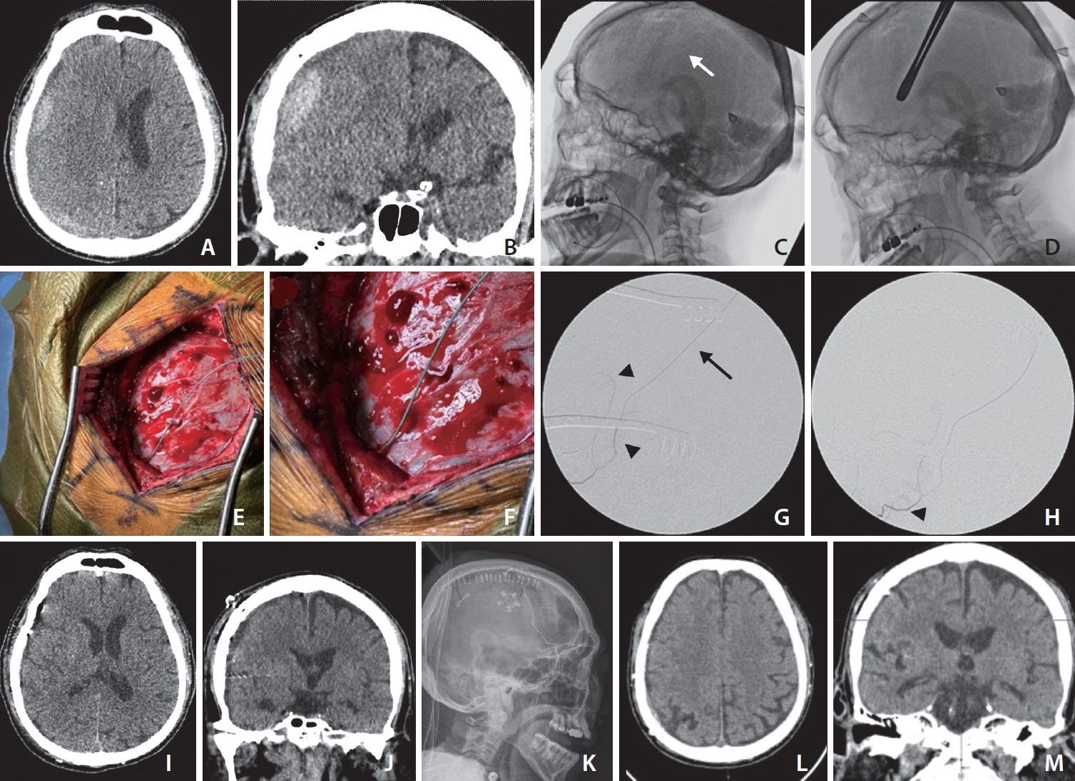
- This report introduces a novel surgical technique for middle meningeal artery embolization (MMAE) during a mini-craniotomy for subdural hematoma (SDH) evacuation. A patient with multiple health issues presented with a 14 mm right subacute SDH. During surgery, the MMA was retrogradely catheterized and embolized using Onyx 18. This approach, combining MMAE with hematoma evacuation, resulted in successful resolution of the SDH without complications. The procedure offers a more efficient workflow by integrating 2 interventions into 1, potentially reducing recurrence rates of SDH.
Keyword
Figure
Reference
-
1. Levitt MR, Hirsch JA, Chen M. Middle meningeal artery embolization for chronic subdural hematoma: an effective treatment with a bright future. J Neurointerv Surg. 2024; 16:329–330.2. Zuo Q, Ni W, Yang P, Gu Y, Yu Y, Yang H, MAGIC-MT investigators, et al. Managing non-acute subdural hematoma using liquid materials: a Chinese randomized trial of middle meningeal artery treatment (MAGIC-MT)-protocol. Trials. 2023; 24:586.3. Edlmann E, Giorgi-Coll S, Whitfield PC, Carpenter KLH, Hutchinson PJ. Pathophysiology of chronic subdural haematoma: inflammation, angiogenesis and implications for pharmacotherapy. J Neuroinflammation. 2017; 14:108.4. Mandai S, Sakurai M, Matsumoto Y. Middle meningeal artery embolization for refractory chronic subdural hematoma. Case report. J Neurosurg. 2000; 93:686–688.5. Link TW, Rapoport BI, Paine SM, Kamel H, Knopman J. Middle meningeal artery embolization for chronic subdural hematoma: Endovascular technique and radiographic findings. Interv Neuroradiol. 2018; 24:455–462.6. Lin N, Brouillard AM, Mokin M, Natarajan SK, Snyder KV, Levy EI, et al. Direct access to the middle meningeal artery for embolization of complex dural arteriovenous fistula: a hybrid treatment approach. J Neurointerv Surg. 2015; 7:e24.7. Oh JS, Yoon SM, Shim JJ, Bae HG. Transcranial direct middle meningeal artery puncture for the onyx embolization of dural arteriovenous fistula involving the superior sagittal sinus. J Korean Neurosurg Soc. 2015; 57:54–57.8. Qiao Y, Zhang YJ, Tsappidi S, Mehta TI, Hui FK. Direct superficial temporal artery access for middle meningeal artery embolization. Interv Neuroradiol. 20249:15910199231225832.9. Khorasanizadeh M, Shutran M, Garcia A, Enriquez-Marulanda A, Moore J, Ogilvy CS, et al. Middle meningeal artery embolization for treatment of chronic subdural hematomas: does selection of embolized branches affect outcomes? J Neurosurg. 2022; 138:1494–1502.10. Duerinck J, Van Der Veken J, Schuind S, Van Calenbergh F, van Loon J, Du Four S, et al. Randomized trial comparing burr hole craniostomy, minicraniotomy, and twist drill craniostomy for treatment of chronic subdural hematoma. Neurosurgery. 2022; 91:304–311.11. Raghavan A, Smith G, Onyewadume L, Peck MR, Herring E, Pace J, et al. Morbidity and mortality after burr hole craniostomy versus craniotomy for chronic subdural hematoma evacuation: a single-center experience. World Neurosurg. 2020; 134:e196–e203.
- Full Text Links
- Actions
-
Cited
- CITED
-
- Close
- Share
- Similar articles
-
- Traumatic middle meningeal artery pseudoaneurysm: Case report and review of literature
- Helical coils augment embolization of the middle meningeal artery for treatment of chronic subdural hematoma: A technical note
- Middle meningeal artery embolizationto treat progressive epidural hematoma:a case report
- Neovascularization in Outer Membrane of Chronic Subdural Hematoma : A Rationale for Middle Meningeal Artery Embolization
- Neovascularization in Outer Membrane of Chronic Subdural Hematoma : A Rationale for Middle Meningeal Artery Embolization


