Anat Cell Biol.
2024 Sep;57(3):463-467. 10.5115/acb.24.023.
Middle meatal nasal recesses of the maxillary sinuses and dangerously modified nasal anatomy
- Affiliations
-
- 1Division of Anatomy, Department 1, Faculty of Dentistry, Carol Davila University of Medicine and Pharmacy, Bucharest, Romania
- KMID: 2559767
- DOI: http://doi.org/10.5115/acb.24.023
Abstract
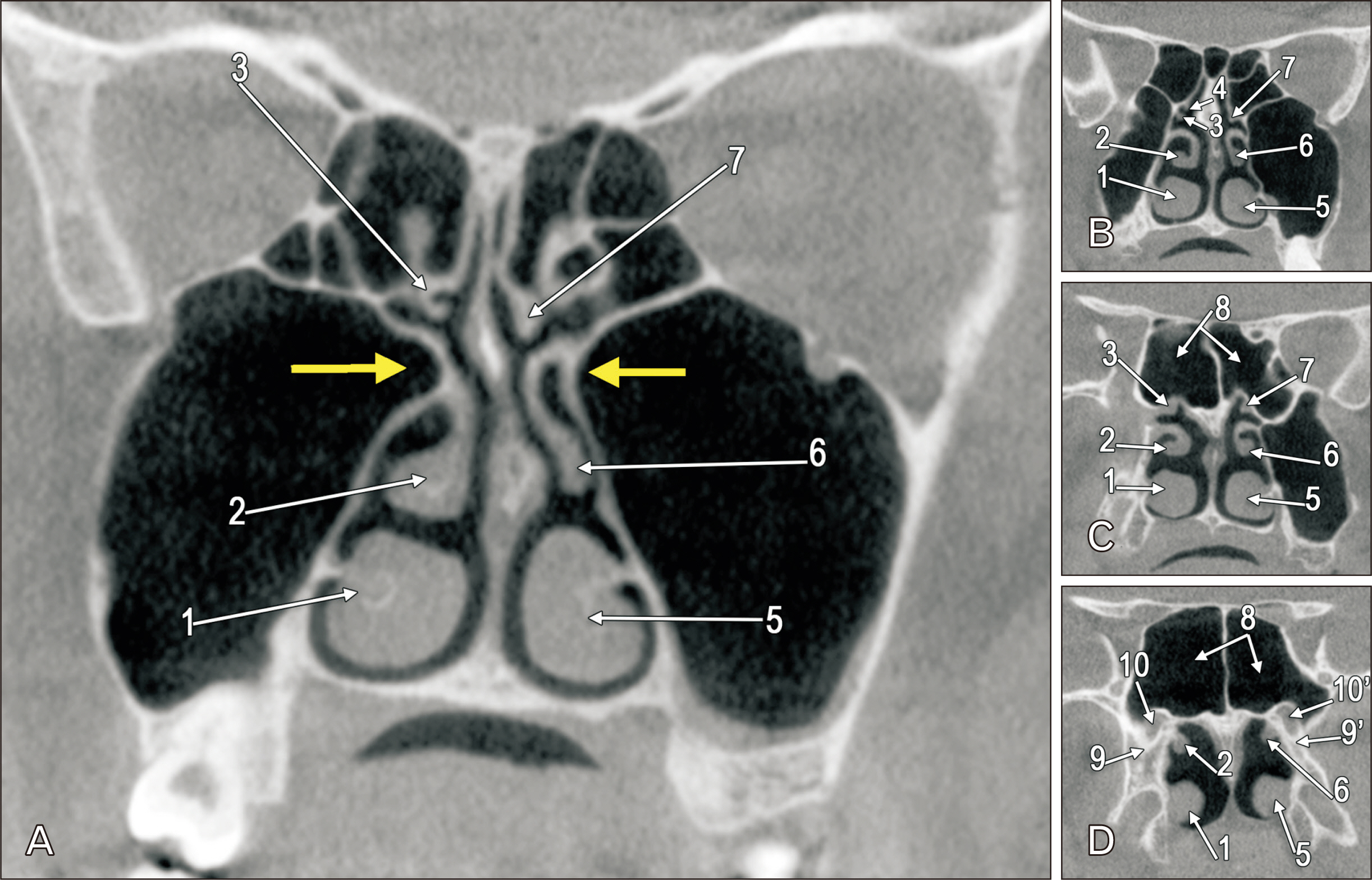
- Pneumatisation of the maxillary sinus (MS) is variable. The archived cone-beam computed tomography file of a 54-year-old female was retrospectively evaluated anatomically. Nasal or retrobullar recesses of the MSs (NRMS) were found. The MSs were bicameral. NRMSs extended from the postero-lateral chambers of the MSs into the lateral nasal walls. The right NRMS was reached superior to the middle turbinate and the ethmoidal bulla was applied on its anterior side. The left NRMS had two medial pouch-like ends, one beneath the ethmoidal bulla and the other on the anterior side of the basal lamella of the middle turbinate. Additional anatomical findings were the uncinate bulla, infraorbital recesses of the MS, maxillary recess of the sphenoidal sinus, and atypical posterior insertions of the superior nasal turbinates, maxillo-ethmoidosphenoidal and ethmoido-sphenoidal. The NRMS is a novel finding and could lead to erroneous endoscopic corridors if not documented before the interventions.
Keyword
Figure
Reference
-
References
1. Craiu C, Rusu MC, Hostiuc S, Săndulescu M, Derjac-Aramă AI. 2017; Anatomic variation in the pterygopalatine angle of the maxillary sinus and the maxillary bulla. Anat Sci Int. 92:98–106. DOI: 10.1007/s12565-015-0320-z. PMID: 26663153.2. Chan HL, Monje A, Suarez F, Benavides E, Wang HL. 2013; Palatonasal recess on medial wall of the maxillary sinus and clinical implications for sinus augmentation via lateral window approach. J Periodontol. 84:1087–93. DOI: 10.1902/jop.2012.120371. PMID: 23106503.3. Günaçar DN, Köse TE, Arsan B, Aydın EZ. 2022; Radioanatomic study of maxillary sinus palatal process pneumatization. Oral Radiol. 38:398–404. DOI: 10.1007/s11282-021-00569-9. PMID: 34554390.4. Niu L, Wang J, Yu H, Qiu L. 2018; New classification of maxillary sinus contours and its relation to sinus floor elevation surgery. Clin Implant Dent Relat Res. 20:493–500. DOI: 10.1111/cid.12606. PMID: 29691967.5. Serindere G, Serindere M, Gunduz K. 2023; Evaluation of maxillary palatal process pneumatization by cone-beam computed tomography. J Stomatol Oral Maxillofac Surg. 124:101432. DOI: 10.1016/j.jormas.2023.101432. PMID: 36921841.6. Cârstocea L, Rusu MC, Mateşică DŞ, Săndulescu M. 2020; Air spaces neighbouring the infraorbital canal. Morphologie. 104:44–50. DOI: 10.1016/j.morpho.2019.07.002. PMID: 31492524.7. Măru N, Rusu M, Săndulescu M. 2013; The conchal recess of the maxillary sinus: a 100 cases CB CT study. Rom J Funct Clin Macro Microsc Anat Anthropol. 12:271–5.8. Wang RG, Jiang SC. 1997; The embryonic development of the human ethmoid labyrinth from 8-40 weeks. Acta Otolaryngol. 117:118–22. DOI: 10.3109/00016489709118002. PMID: 9039492.9. Márquez S, Tessema B, Clement PA, Schaefer SD. 2008; Development of the ethmoid sinus and extramural migration: the anatomical basis of this paranasal sinus. Anat Rec (Hoboken). 291:1535–53. DOI: 10.1002/ar.20775. PMID: 18951481.10. R SSS, Khan N, Parameswaran R, Boovaraghavan S, Nagi M. 2024; Evaluation of dimensional changes in maxillary and frontal sinus in adult patients with anterior open bite and normal overbite: a retrospective cone beam computed tomography (CBCT) study. Cureus. 16:e53710. DOI: 10.7759/cureus.53710. PMID: 38455800. PMCID: PMC10919753.11. Aşantoğrol F, Coşgunarslan A. 2022; The effect of anatomical variations of the sinonasal region on maxillary sinus volume and dimensions: a three-dimensional study. Braz J Otorhinolaryngol. 88(Suppl 1):S118–27. DOI: 10.1016/j.bjorl.2021.05.001. PMID: 34053909. PMCID: PMC9734263.12. Glass M. 1952; Duplication of the maxillary antrum symptomatology, diagnosis and treatment. S Afr Med J. 26:895–902. PMID: 13028765.13. Yue V, Bleach NR, van Hasselt CA. 1994; Double maxillary antrum as a cause of maxillary sinus mucocele. Ear Nose Throat J. 73:839–41. DOI: 10.1177/014556139407301109. PMID: 7828478.14. Bolger WE, Mawn CB. 2001; Analysis of the suprabullar and retrobullar recesses for endoscopic sinus surgery. Ann Otol Rhinol Laryngol Suppl. 186:3–14. DOI: 10.1177/00034894011100S501. PMID: 11372938.15. Rusu MC, Hostiuc S, Motoc AGM, Mogoantă CA, Sava JC, Săndulescu M. 2020; The sphenoethmoidal sinus and the modified anatomy of the related structures. Rom J Morphol Embryol. 61:143–8. DOI: 10.47162/RJME.61.1.16. PMID: 32747905. PMCID: PMC7728111.
- Full Text Links
- Actions
-
Cited
- CITED
-
- Close
- Share
- Similar articles
-
- A Case of Osteoma in the Nasal Cavity
- What is the Relationship between the Localization of Maxillary Fungal Balls and Intranasal Anatomic Variations?
- Evolution of the paranasal sinuses' anatomy through the ages
- Advantage of Middle Meatal Antrostomy in Transnasal Endoscopic Reconstruction of Medial Orbital Blow-out Fracture
- A Case of Huge Fungus Ball in Nasal Cavity Misdiagnosed as Rhinolith on Nasal Septum


