Anat Cell Biol.
2024 Sep;57(3):370-377. 10.5115/acb.24.078.
Challenging arterial pattern of foregut and its potential impact on surgery
- Affiliations
-
- 1Department of Anatomy, Calcutta National Medical College, West Bengal, India
- 2Department of Anatomy, R.G. Kar Medical College, West Bengal, India
- KMID: 2559750
- DOI: http://doi.org/10.5115/acb.24.078
Abstract
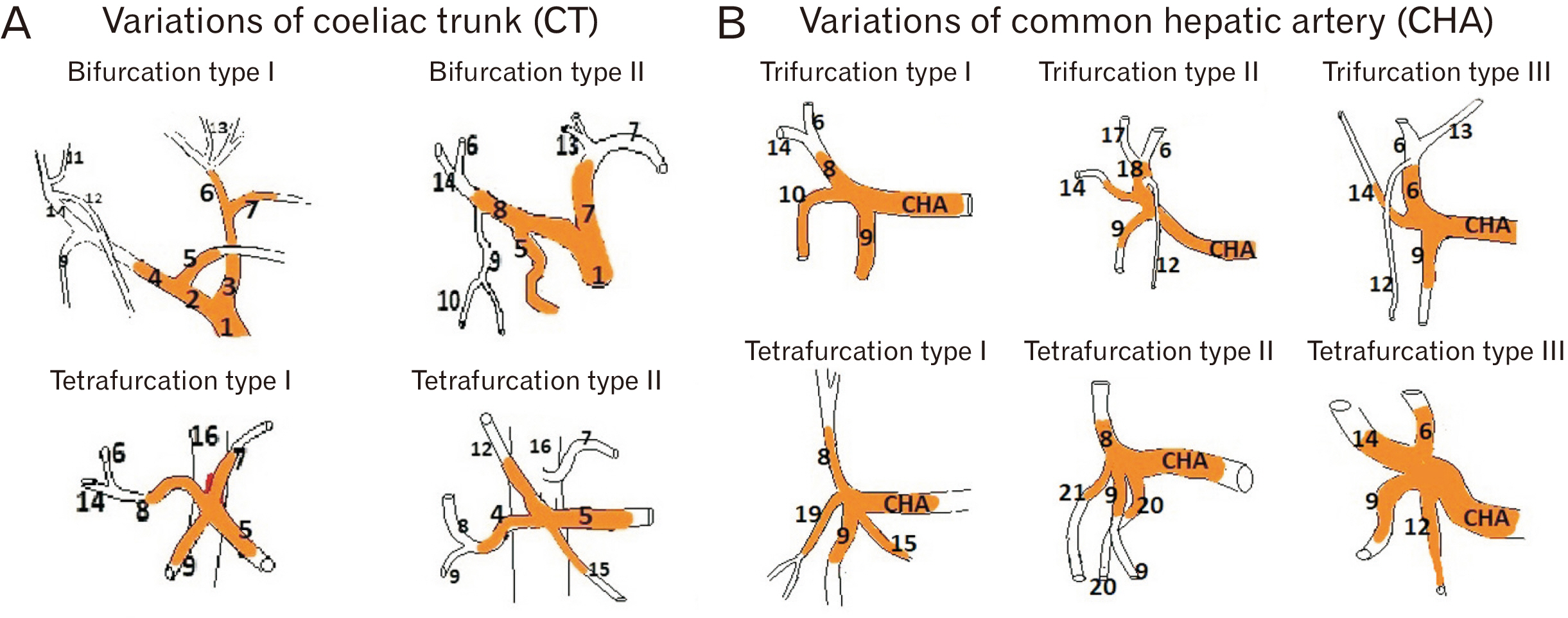
- Anticipating a wide range of morphological variations of arterial anatomy of foregut derivatives beyond the classical pattern, a precise understanding is pertinent to preoperative diagnosis, operative procedure and to avoid potentially devastating post-operative outcome during various traumatic and non-traumatic vascular insult of foregut. The study aimed to revisit the morphological details and update unusual configurations of arteries of foregut to establish clinico-anatomical correlations. This study described the detailed branching pattern of coeliac trunk (CT) as principal artery of foregut with source & course of hepatic, gastric, duodenal and pancreatic branches in 58 cadaveric dissections. Based on morphology, different types and subtypes were made. The descriptions were explained using figures and pertinent tables. Among classical branches of CT, splenic artery was found as most stable whereas other two branches were found to be most variable with missing common hepatic artery in 11 cases. In addition to classical trifurcation (65.52%), different types of bifurcation (12.07%) and tetrafurcations (22.41%) of CT were observed. Regarding variations of hepatic arteries (27.59%), both non-classical origin and accessory hepatic branches were found. In case of gastric branches, more variant origins were seen with right gastric (50%) as compared to left gastric artery (34.48%). Other morphological variations included non-classical origin of gastro-duodenal artery (18.96%) along with presence of accessory pancreatic (17.13%) and duodenal arteries (6.38%). Awareness of anatomical variations regarding circulatory dynamics of foregut is worth knowing in order to facilitate successful planning of surgery involving upper abdominal organs with least complications.
Keyword
Figure
Reference
-
References
1. Hemamalini . 2018; Variations in the branching pattern of the celiac trunk and its clinical significance. Anat Cell Biol. 51:143–9. DOI: 10.5115/acb.2018.51.3.143. PMID: 30310705. PMCID: PMC6172596.2. Standring S, Gray H. Gray's anatomy: the anatomical basis of clinical practice. 40th ed. Churchill Livingstone/Elsevier;2008.3. Walker TG. 2009; Mesenteric vasculature and collateral pathways. Semin Intervent Radiol. 26:167–74. DOI: 10.1055/s-0029-1225663. PMID: 21326561. PMCID: PMC3036491.4. Ochoa JE, Pointer DT Jr, Hamner JB. 2016; Vascular anomalies in pancreaticoduodenectomy: a lesson learned. Case Rep Surg. 2016:5792980. DOI: 10.1155/2016/5792980. PMID: 27200204. PMCID: PMC4856910.5. Malviya KK, Verma A, Nayak AK, Mishra A, More RS. 2021; Unraveling variations in celiac trunk and hepatic artery by CT angiography to aid in surgeries of upper abdominal region. Diagnostics (Basel). 11:2262. DOI: 10.3390/diagnostics11122262. PMID: 34943499. PMCID: PMC8700197.6. Jalamneh B, Nassar IJ, Sabbooba L, Ghanem R, Nazzal Z, Kiwan R, Awadghanem A, Maree M. 2023; Exploring anatomical variations of abdominal arteries through computed tomography: classification, prevalence and implications. Cureus. 15:e41380. DOI: 10.7759/cureus.41380. PMID: 37546145. PMCID: PMC10400811.7. Zaki SM, Abdelmaksoud AHK, Khaled BEA, Abdel Kader IA. 2020; Anatomical variations of hepatic artery using the multidetector computed tomography angiography. Folia Morphol (Warsz). 79:247–54. DOI: 10.5603/FM.a2019.0090. PMID: 31436302.8. Hiatt JR, Gabbay J, Busuttil RW. 1994; Surgical anatomy of the hepatic arteries in 1000 cases. Ann Surg. 220:50–2. DOI: 10.1097/00000658-199407000-00008. PMID: 8024358. PMCID: PMC1234286.9. Michels NA. 1966; Newer anatomy of the liver and its variant blood supply and collateral circulation. Am J Surg. 112:337–47. DOI: 10.1016/0002-9610(66)90201-7. PMID: 5917302.10. Ande T, Makani TK, Nannam K, Velichety SD, Kumar JA. 2023; Left gastric artery variants: a cadaveric, postmortem and radiological investigation. Scr Med. 54:157–61. DOI: 10.5937/scriptamed54-44773.11. Mazurek A, Juszczak A, Walocha JA, Pasternak A. 2021; Rare combined variations of the coeliac trunk, accessory hepatic and gastric arteries with co-occurrence of double cystic arteries. Folia Morphol (Warsz). 80:460–6. DOI: 10.5603/FM.a2020.0052. PMID: 32459367.12. Wu X, Kang J, Liu Y, Sun G, Shi Y, Niu J. 2022; A rare hepatic artery variant reporting and a new classification. Front Surg. 9:1003350. DOI: 10.3389/fsurg.2022.1003350. PMID: 36105121. PMCID: PMC9465518.13. Ugurel MS, Battal B, Bozlar U, Nural MS, Tasar M, Ors F, Saglam M, Karademir I. 2010; Anatomical variations of hepatic arterial system, coeliac trunk and renal arteries: an analysis with multidetector CT angiography. Br J Radiol. 83:661–7. DOI: 10.1259/bjr/21236482. PMID: 20551256. PMCID: PMC3473504.14. Haller AV. [Anatomical icons with which some of the main parts of the human body are outlined are presented & the arteries especially the history is contained]. Vandenhoeck;1756. Latin.15. Adachi B. [The Arterial System of the Japanese]. Kaiserlich-Japanischen Universitat zu Kyoto, Maruzen Publishing Co;1928. Japanese.16. Agarwal S, Pangtey B, Vasudeva N. 2016; Unusual variation in the branching pattern of the celiac trunk and its embryological and clinical perspective. J Clin Diagn Res. 10:AD05–7. DOI: 10.7860/JCDR/2016/19527.8064. PMID: 27504274. PMCID: PMC4963634.17. Sadler TW. Sadler TW, editor. Cardiovascular system. Langman's Medical Embryology. 12th ed. Wolters Kluwer;2012. p. 189.18. Chitra R. 2010; Clinically relevant variations of the coeliac trunk. Singapore Med J. 51:216–9. PMID: 20428743.19. Juszczak A, Mazurek A, Walocha JA, Pasternak A. 2021; Coeliac trunk and its anatomic variations: a cadaveric study. Folia Morphol (Warsz). 80:114–21. DOI: 10.5603/FM.a2020.0042. PMID: 32301103.20. Mburu KS, Alexander OJ, Hassan S, Bernard N. 2010; Variations in the branching pattern of the celiac trunk in a Kenyan population. Int J Morphol. 28:199–204. DOI: 10.4067/S0717-95022010000100028.21. Panagouli E, Venieratos D, Lolis E, Skandalakis P. 2013; Variations in the anatomy of the celiac trunk: a systematic review and clinical implications. Ann Anat. 195:501–11. DOI: 10.1016/j.aanat.2013.06.003. PMID: 23972701.22. Pinal-Garcia DF, Nuno-Guzman CM, Gonzalez-Gonzalez ME, Ibarra-Hurtado TR. 2018; The celiac trunk and its anatomical variations: a cadaveric study. J Clin Med Res. 10:321–9. DOI: 10.14740/jocmr3356w. PMID: 29511421. PMCID: PMC5827917.23. Santos PVD, Barbosa ABM, Targino VA, Silva NA, Silva YCM, Barbosa F, Oliveira ASB, Assis TO. 2018; Anatomical variations of the celiac trunk: a systematic review. Arq Bras Cir Dig. 31:e1403. DOI: 10.1590/0102-672020180001e1403. PMID: 30539978. PMCID: PMC6284376.24. Srivastava AK, Sehgal G, Sharma PK, Kumar N, Singh R, Parihar A, Aga P. 2012; Various types of branching patterns of celiac trunk. FASEB J. 26(Suppl 1):722.5. DOI: 10.1096/fasebj.26.1_supplement.722.5.25. Sureka B, Mittal MK, Mittal A, Sinha M, Bhambri NK, Thukral BB. 2013; Variations of celiac axis, common hepatic artery and its branches in 600 patients. Indian J Radiol Imaging. 23:223–33. DOI: 10.4103/0971-3026.120273. PMID: 24347852. PMCID: PMC3843330.26. Torres K, Staśkiewicz G, Denisow M, Pietrzyk Ł, Torres A, Szukała M, Czekajska-Chehab E, Drop A. 2015; Anatomical variations of the coeliac trunk in the homogeneous Polish population. Folia Morphol (Warsz). 74:93–9. DOI: 10.5603/FM.2014.0059. PMID: 25792402.27. Venieratos D, Panagouli E, Lolis E, Tsaraklis A, Skandalakis P. 2013; A morphometric study of the celiac trunk and review of the literature. Clin Anat. 26:741–50. DOI: 10.1002/ca.22136. PMID: 22886953.28. Huang Y, Mu GC, Qin XG, Chen ZB, Lin JL, Zeng YJ. 2015; Study of celiac artery variations and related surgical techniques in gastric cancer. World J Gastroenterol. 21:6944–51. DOI: 10.3748/wjg.v21.i22.6944. PMID: 26078572. PMCID: PMC4462736.29. Cirocchi R, D'Andrea V, Amato B, Renzi C, Henry BM, Tomaszewski KA, Gioia S, Lancia M, Artico M, Randolph J. 2020; Aberrant left hepatic arteries arising from left gastric arteries and their clinical importance. Surgeon. 18:100–12. DOI: 10.1016/j.surge.2019.06.002. PMID: 31337536.30. van den Hoven AF, Smits ML, de Keizer B, van Leeuwen MS, van den Bosch MA, Lam MG. 2014; Identifying aberrant hepatic arteries prior to intra-arterial radioembolization. Cardiovasc Intervent Radiol. 37:1482–93. DOI: 10.1007/s00270-014-0845-x. PMID: 24469409.
- Full Text Links
- Actions
-
Cited
- CITED
-
- Close
- Share
- Similar articles
-
- Endoscopic Treatment for Early Foregut Neuroendocrine Tumors
- Oral foregut cyst in the ventral tongue: a case report
- A case of ciliated hepatic foregut cyst treated by laparoscopic excision
- Restoration for the foregut surgery: bridging gaps between foregut surgery practice and academia
- Ciliated Foregut Cyst of the Liver: Report of a case


