J Korean Foot Ankle Soc.
2024 Sep;28(3):114-118. 10.14193/jkfas.2024.28.3.114.
Minimally Invasive Distal Transverse Metatarsal Osteotomy – Akin Osteotomy (MITA) for Recurrent Hallux Valgus: A Report of Four Cases
- Affiliations
-
- 1SNU Seoul Hospital, Seoul, Korea
- KMID: 2559642
- DOI: http://doi.org/10.14193/jkfas.2024.28.3.114
Abstract
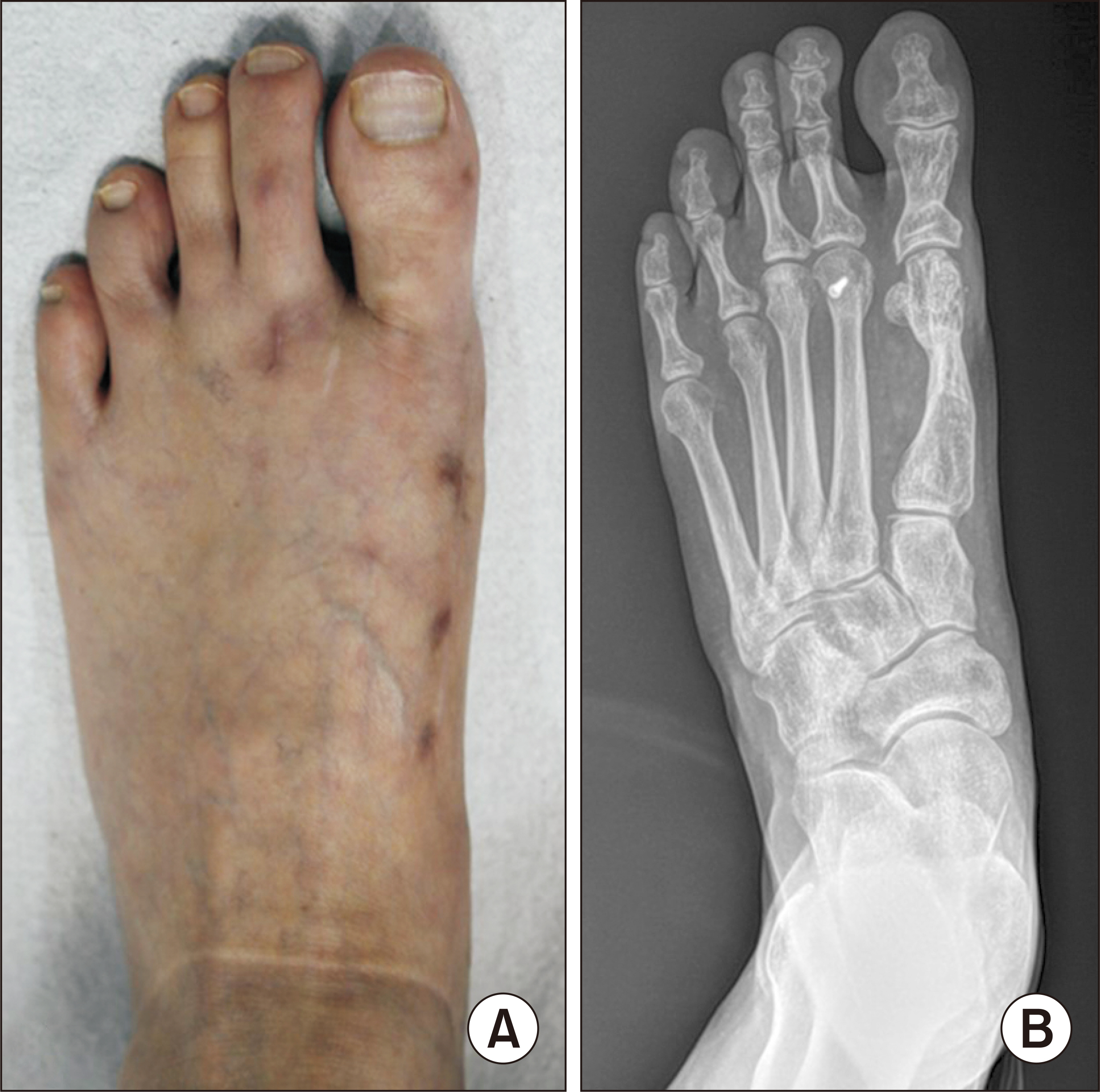
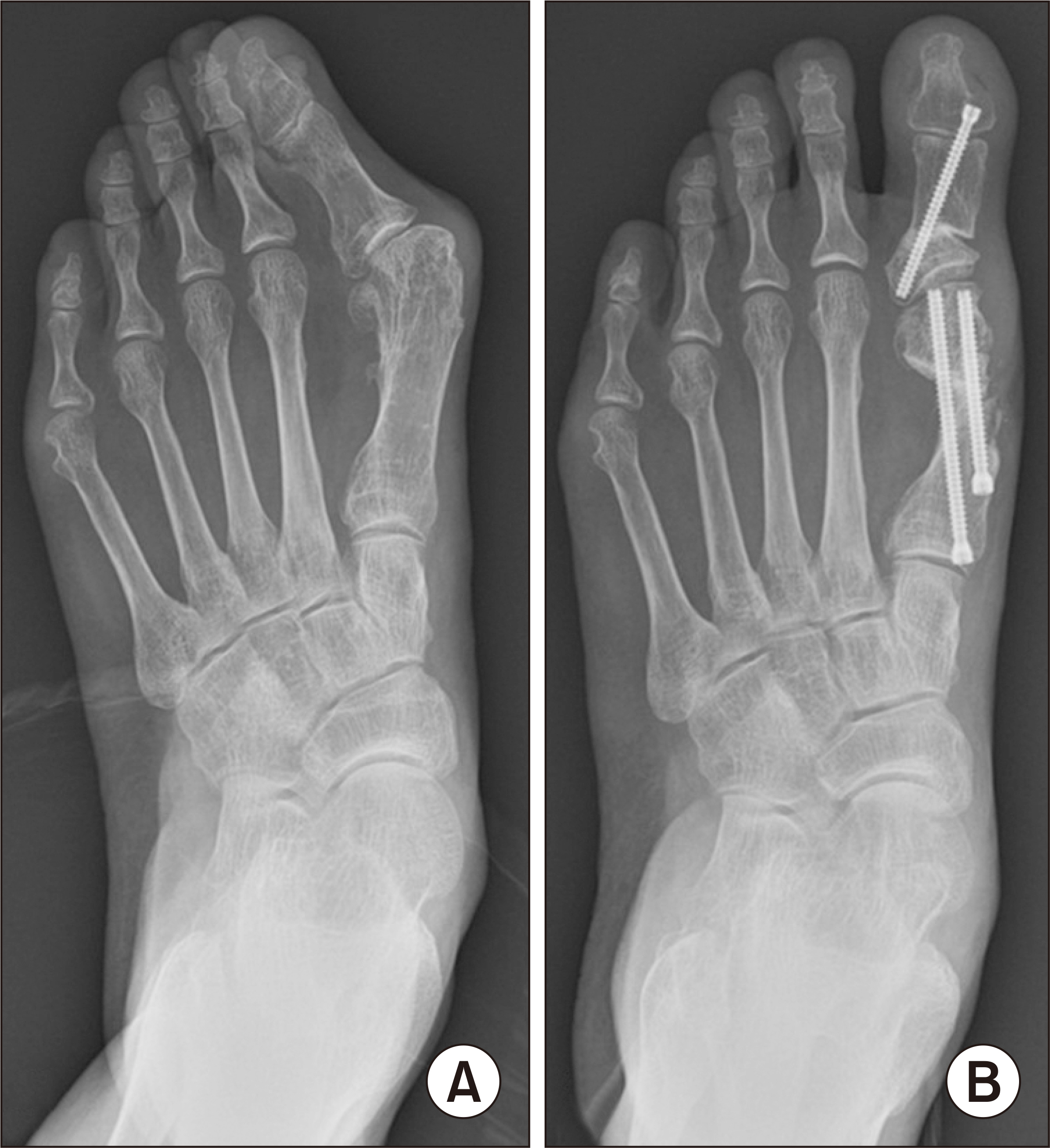
- Recurrent deformity following hallux valgus surgery can be technically challenging to treat. In cases of revision surgery, a surgical technique with greater corrective power is often chosen compared to the primary surgery. Therefore, minimally invasive surgery is not commonly performed. On the other hand, minimally invasive surgery minimizes soft tissue damage and allows for greater correction of deformity compared to traditional open approaches. This paper reports four cases of recurrent hallux valgus treated with a minimally invasive distal transverse metatarsal osteotomy – Akin osteotomy (MITA), resulting in significant improvements in the clinical and radiographic outcomes.
Figure
Reference
-
1. Thompson FM. 1990; Complications of hallux valgus surgery and salvage. Orthopedics. 13:1059–67. doi: 10.3928/0147-7447-19900901-19. DOI: 10.3928/0147-7447-19900901-19.2. Raikin SM, Miller AG, Daniel J. 2014; Recurrence of hallux valgus: a review. Foot Ankle Clin. 19:259–74. doi: 10.1016/j.fcl.2014.02.008. DOI: 10.1016/j.fcl.2014.02.008.3. Isham SA. 1991; The Reverdin-Isham procedure for the correction of hallux abducto valgus. A distal metatarsal osteotomy procedure. Clin Podiatr Med Surg. 8:81–94. DOI: 10.1016/S0891-8422(23)00420-2.4. Bösch P, Wanke S, Legenstein R. 2000; Hallux valgus correction by the method of Bösch: a new technique with a seven-to-ten-year follow-up. Foot Ankle Clin. 5:485–98. v–vi.5. Redfern D, Vernois J. 2016; Minimally invasive chevron akin (MICA) for correction of hallux valgus. Tech Foot Ankle Surg. 15:3–11. doi: 10.1097/BTF.0000000000000102. DOI: 10.1097/BTF.0000000000000102.6. Kwon KB, Lee KM. 2019; Treatment of recurrent hallux valgus after surgery. J Korean Foot Ankle Soc. 23:149–53. doi: 10.14193/jkfas.2019.23.4.149. DOI: 10.14193/jkfas.2019.23.4.149.7. Scala A, Cipolla M, Giannini S, Oliva G. 2020; The modified subcapital metatarsal osteotomy in the treatment of hallux valgus recurrence. Foot Ankle Spec. 13:404–14. doi: 10.1177/1938640019875322. DOI: 10.1177/1938640019875322.8. Magnan B, Negri S, Maluta T, Dall'Oca C, Samaila E. 2019; Minimally invasive distal first metatarsal osteotomy can be an option for recurrent hallux valgus. Foot Ankle Surg. 25:332–9. doi: 10.1016/j.fas.2017.12.010. DOI: 10.1016/j.fas.2017.12.010.9. Lewis TL, Ray R, Lam P. Revision of recurrent hallux valgus deformity using a percutaneous distal transverse osteotomy: surgical considerations and early results. Foot Ankle Clin. Published online May 30, 2024; doi: 10.1016/j.fcl.2024.04.010. DOI: 10.1016/j.fcl.2024.04.010.10. Lewis TL, Lau B, Alkhalfan Y, Trowbridge S, Gordon D, Vernois J, et al. 2023; Fourth-generation minimally invasive hallux valgus surgery with metaphyseal extra-articular transverse and akin osteotomy (META): 12 month clinical and radiologic results. Foot Ankle Int. 44:178–91. doi: 10.1177/10711007231152491. DOI: 10.1177/10711007231152491.11. Aiyer A, Massel DH, Siddiqui N, Acevedo JI. 2021; Biomechanical comparison of 2 common techniques of minimally invasive hallux valgus correction. Foot Ankle Int. 42:373–80. doi: 10.1177/1071100720959029. DOI: 10.1177/1071100720959029.12. Najefi AA, Katmeh R, Zaveri AK, Alsafi MK, Garrick F, Malhotra K, et al. 2022; Imaging findings and first metatarsal rotation in hallux valgus. Foot Ankle Int. 43:665–75. doi: 10.1177/10711007211064609. DOI: 10.1177/10711007211064609.
- Full Text Links
- Actions
-
Cited
- CITED
-
- Close
- Share
- Similar articles
-
- Minimally Invasive Proximal Transverse Metatarsal Osteotomy Followed by Intramedullary Plate Fixation for Hallux Valgus Deformity: A Case Report
- The Effect of Derotational Closing Wedge Akin Osteotomy for the Treatment of Hallux Valgus with the Pronation of Great Toe
- Comparison of Operative Results of Distal Chevron Osteotomy with and without Akin Osteotomy for Moderate to Severe Hallux Valgus
- Treatment of Severe Hallux Valgus Deformity with Proximal Reverse Chevron Metatarsal Osteotomy and Akin Osteotomy
- Corrective Osteotomies in Hallux Valgus

