J Korean Foot Ankle Soc.
2024 Sep;28(3):102-106. 10.14193/jkfas.2024.28.3.102.
Successful Treatment of Chronic Ulcerative Lesion on the Heel with a Half-Width Reverse Sural Flap in a Patient Who Underwent Achilles Tendon Repair Three Years Ago: A Case Report
- Affiliations
-
- 1Department of Plastic and Reconstructive Surgery, Kangwon National University Hospital, Chuncheon, Korea
- 2Department of Plastic and Reconstructive Surgery, Kangwon National University School of Medicine, Chuncheon, Korea
- 3Department of Orthopedic Surgery, Kangwon National University Hospital, Chuncheon, Korea
- KMID: 2559639
- DOI: http://doi.org/10.14193/jkfas.2024.28.3.102
Abstract
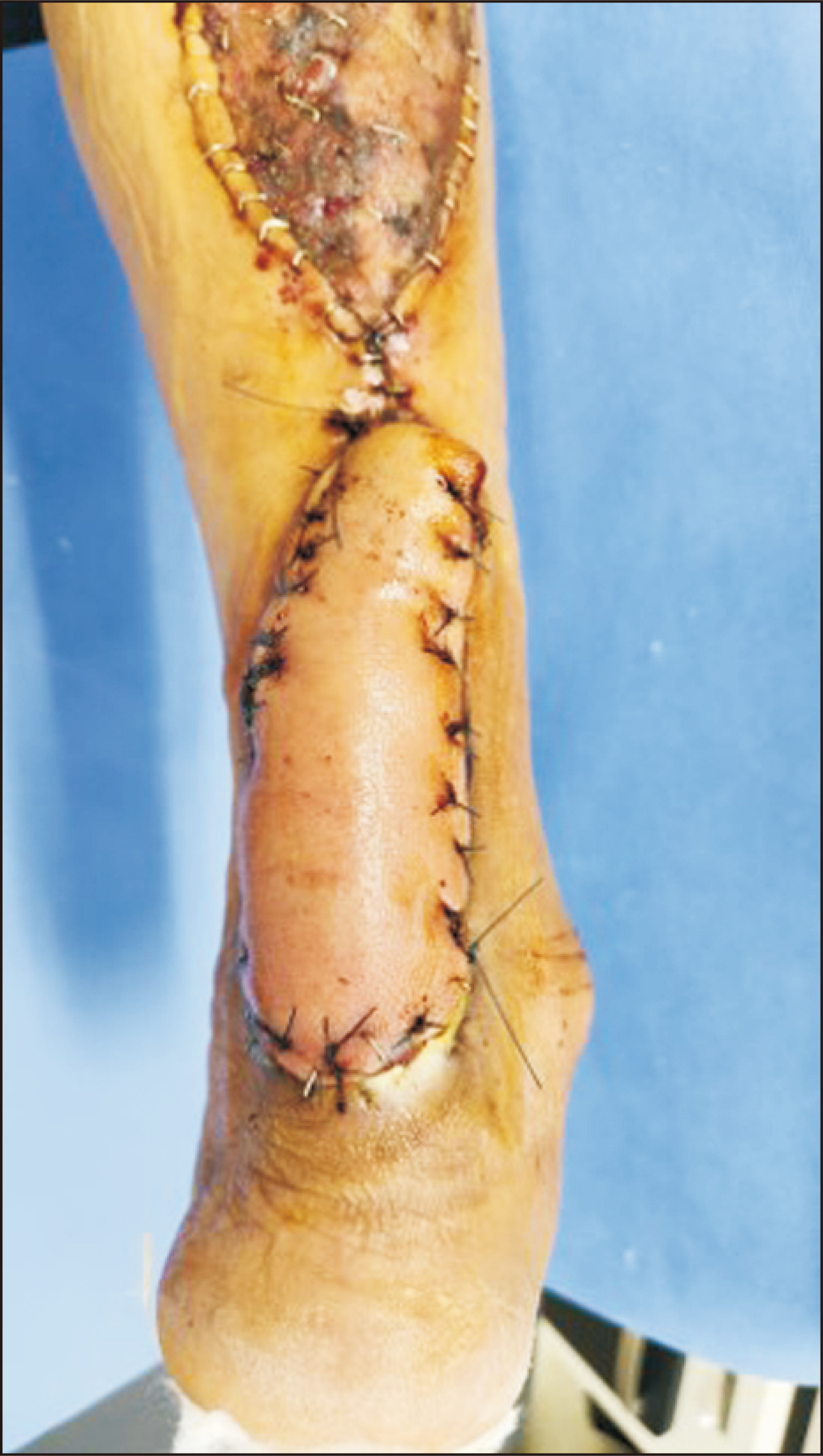
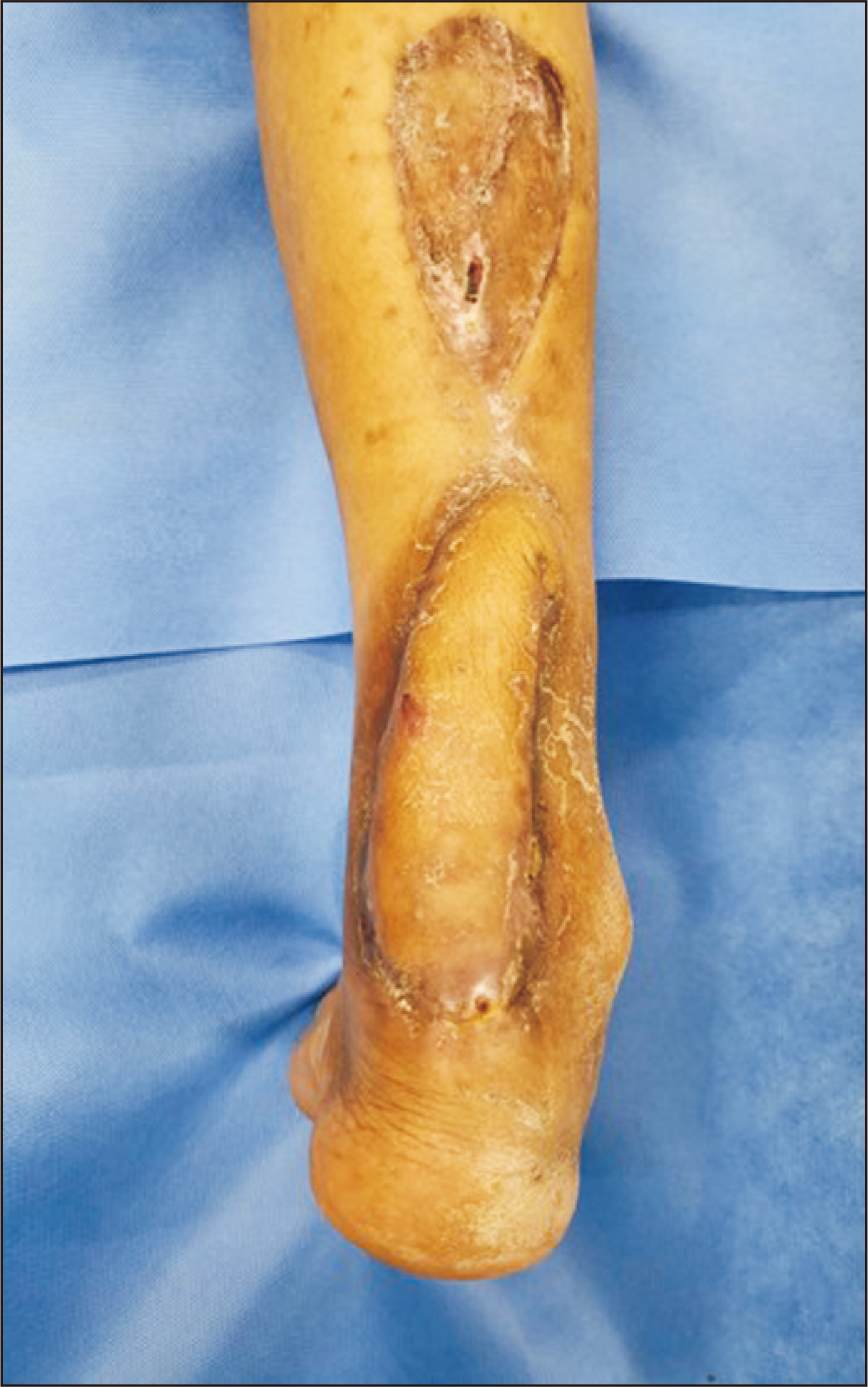
- A reverse sural flap is a surgical procedure to repair soft tissue defects, usually in the ankle region. This procedure involves moving a tissue flap from the calf to cover a defect in the ankle. The flap is turned 180° so that the tissue around the wound is supplied with blood by the vessels at the base of the flap, typically preserving the sural nerve and artery. This method is particularly valuable when thick and robust tissue is required to cover defects resulting from traumatic injuries, chronic wounds, or post-skin tumor removal when the local tissue is insufficient for direct closure. In this case, a patient who had undergone surgery for a chronic ulcerative lesion on the Achilles tendon three years prior to presentation at the authors’ hospital was treated using a half-width reverse sural flap. Modifications to the sural flap design may be crucial considering the surgical history, blood supply, and defect size around the lower leg. In particular, previous surgeries for lower leg fractures or ligament damage may limit blood supply and require flap design modifications.
Keyword
Figure
Reference
-
1. Dhamangaonkar AC, Patankar HS. 2014; Reverse sural fasciocutaneous flap with a cutaneous pedicle to cover distal lower limb soft tissue defects: experience of 109 clinical cases. J Orthop Traumatol. 15:225–9. doi: 10.1007/s10195-014-0304-0. DOI: 10.1007/s10195-014-0304-0.2. Chang SM, Li XH, Gu YD. 2015; Distally based perforator sural flaps for foot and ankle reconstruction. World J Orthop. 6:322–30. doi: 10.5312/wjo.v6.i3.322. DOI: 10.5312/wjo.v6.i3.322.3. Altinkaya A, Yazar S, Bengur FB. 2021; Reconstruction of soft tissue defects around the Achilles region with distally based extended peroneal artery perforator flap. Injury. 52:1985–92. doi: 10.1016/j.injury.2021.04.015. DOI: 10.1016/j.injury.2021.04.015.4. Krishna D, Chaturvedi G, Khan MM, Cheruvu VPR, Laitonjam M, Minz R. 2021; Reconstruction of heel soft tissue defects: an algorithm based on our experience. World J Plast Surg. 10:63–72. doi: 10.29252/wjps.10.3.63. DOI: 10.52547/wjps.10.3.63.5. Hashmi DPM, Musaddiq A, Ali DM, Hashmi A, Zahid DM, Nawaz DZ. 2021; Long-term clinical and functional outcomes of distally based sural artery flap: a retrospective case series. JPRAS Open. 30:61–73. doi: 10.1016/j.jpra.2021.01.013. DOI: 10.1016/j.jpra.2021.01.013.6. Dastagir K, Gamrekelashvili J, Dastagir N, Limbourg A, Kijas D, Kapanadze T, et al. 2023; A new fasciocutaneous flap model identifies a critical role for endothelial Notch signaling in wound healing and flap survival. Sci Rep. 13:12542. doi: 10.1038/s41598-023-39722-1. DOI: 10.1038/s41598-023-39722-1.7. Novriansyah R, Prabowo I, Laras S. 2021; Non-microsurgical bipedicled reverse sural fasciocutaneous flap with preservation of medial and lateral sural cutaneous nerve: current surgical management of skin defect after traumatic Achilles tendon rupture - a case report. Int J Surg Case Rep. 78:259–64. doi: 10.1016/j.ijscr.2020.12.027. DOI: 10.1016/j.ijscr.2020.12.027.8. Talukdar A, Yadav J, Purkayastha J, Pegu N, Singh PR, Kodali RK, et al. 2019; Reverse sural flap - a feasible option for oncological defects of the lower extremity, ankle, and foot: our experience from Northeast India. South Asian J Cancer. 8:255–7. doi: 10.4103/sajc.sajc_11_19. DOI: 10.4103/sajc.sajc_11_19.9. de Rezende MR, Saito M, Paulos RG, Ribak S, Abarca Herrera AK, Cho ÁB, et al. 2018; Reduction of morbidity with a reverse-flow sural flap: a two-stage technique. J Foot Ankle Surg. 57:821–5. doi: 10.1053/j.jfas.2017.11.020. DOI: 10.1053/j.jfas.2017.11.020.10. Mohamed AY, Ibrahim YB, Taşkoparan H, Çi Çek Eİ, May H. 2022; Reverse sural flap for anteromedial ankle and dorsal foot soft-tissue defect following an injury: a case report. Ann Med Surg (Lond). 84:104935. doi: 10.1016/j.amsu.2022.104935. DOI: 10.1016/j.amsu.2022.104935.11. Johnson L, Liette MD, Green C, Rodriguez P, Masadeh S. 2020; The reverse sural artery flap: a reliable and versatile flap for wound coverage of the distal lower extremity and hindfoot. Clin Podiatr Med Surg. 37:699–726. doi: 10.1016/j.cpm.2020.05.004. DOI: 10.1016/j.cpm.2020.05.004.
- Full Text Links
- Actions
-
Cited
- CITED
-
- Close
- Share
- Similar articles
-
- Reverse Superficial Sural artery flap for the Reconstruction of Soft Tissue Defect on Posterior side of heel exposing Achilles tendon
- Treatment of Deep Infection Following Repair of Achilles Tendon Rupture
- Anterior Lateral Thigh Free Flap and Achilles Tendon Reconstruction Surgery for Contact Dermal Burn of Heel Including Achilles Tendon: A Case Report -Surgical Treatment for Functional Recovery-
- Intraoperative Ultrasound-Guided Percutaneous Repair of a Ruptured Achilles Tendon: A Comparative Study with Open Repair
- Treatment of Massive Defect in Achilles Tendon with Tendon Allograft: A Case Report


