Anterior Lateral Thigh Free Flap and Achilles Tendon Reconstruction Surgery for Contact Dermal Burn of Heel Including Achilles Tendon: A Case Report -Surgical Treatment for Functional Recovery-
- Affiliations
-
- 1Department of Orthopedic Surgery, Kangdong Sacred Heart Hospital, Seoul, Korea. kiga9@hanmail.net
- KMID: 2428671
- DOI: http://doi.org/10.14193/jkfas.2018.22.3.127
Abstract
- A 3rd degree burn on the heel including the Achilles tendon is vulnerable and requires active treatment to improve the functional outcomes. Previously, there have been a few treatments on severe burns, such as amputation, debridement or simple skin graft. The cooperative technique of an anterior lateral thigh flap with Achilles tendon reconstruction can be an innovative procedure that preserves the major arteries. The authors review a case and report the clinical outcome.
MeSH Terms
Figure
-
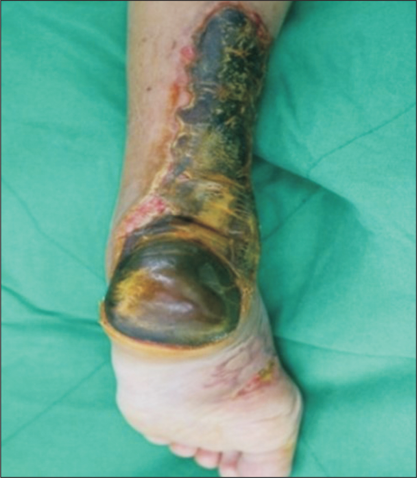
Figure 1. Photograph taken before the surgery shows a large soft tissue defect of posteromedial side of ankle and heel area.
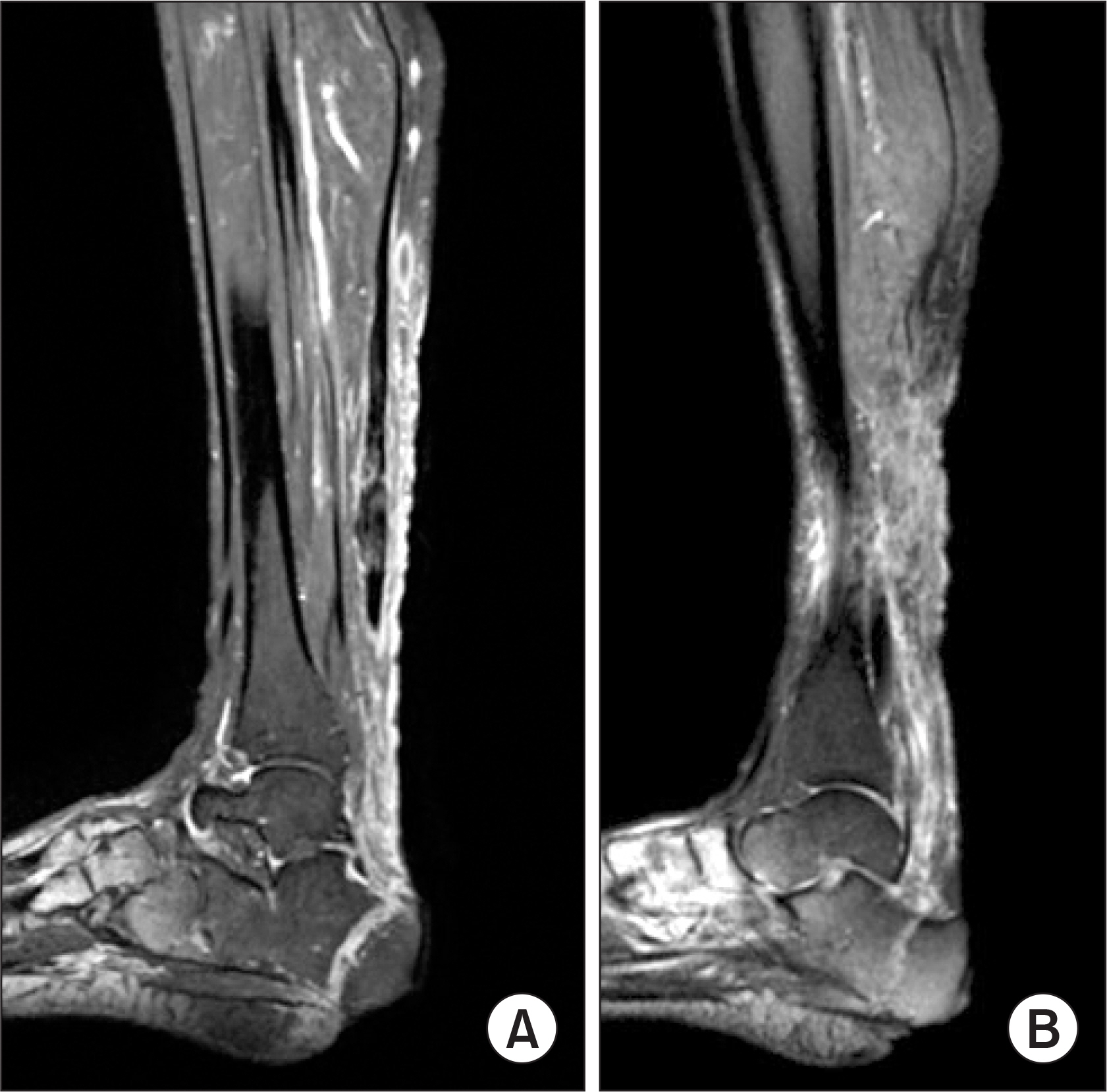
Figure 2. The sagittal images of magnetic resonance imaging show Achilles tendon defect (A) and damaged soft tissue (B).

Figure 3. (A) Photograph of necrotic tissues and nonfunction of Achilles tendon. (B) The damaged Achilles tendon was removed from the calcaneus to one third of distal lower leg.
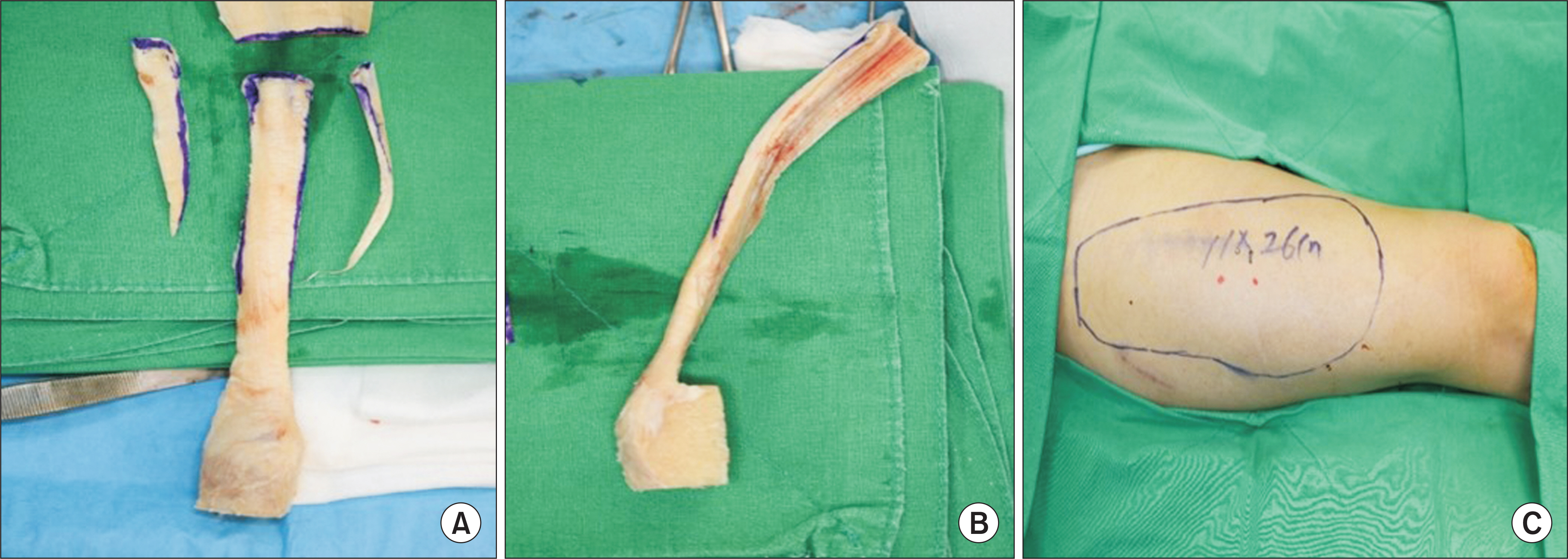
Figure 4. (A) An allotendon of Achilles tendon and allobone used in reconstruction. (B) An intraoperative lateral image of allotendon and allobone. (C) Flap donor site of anterior surface of thigh.
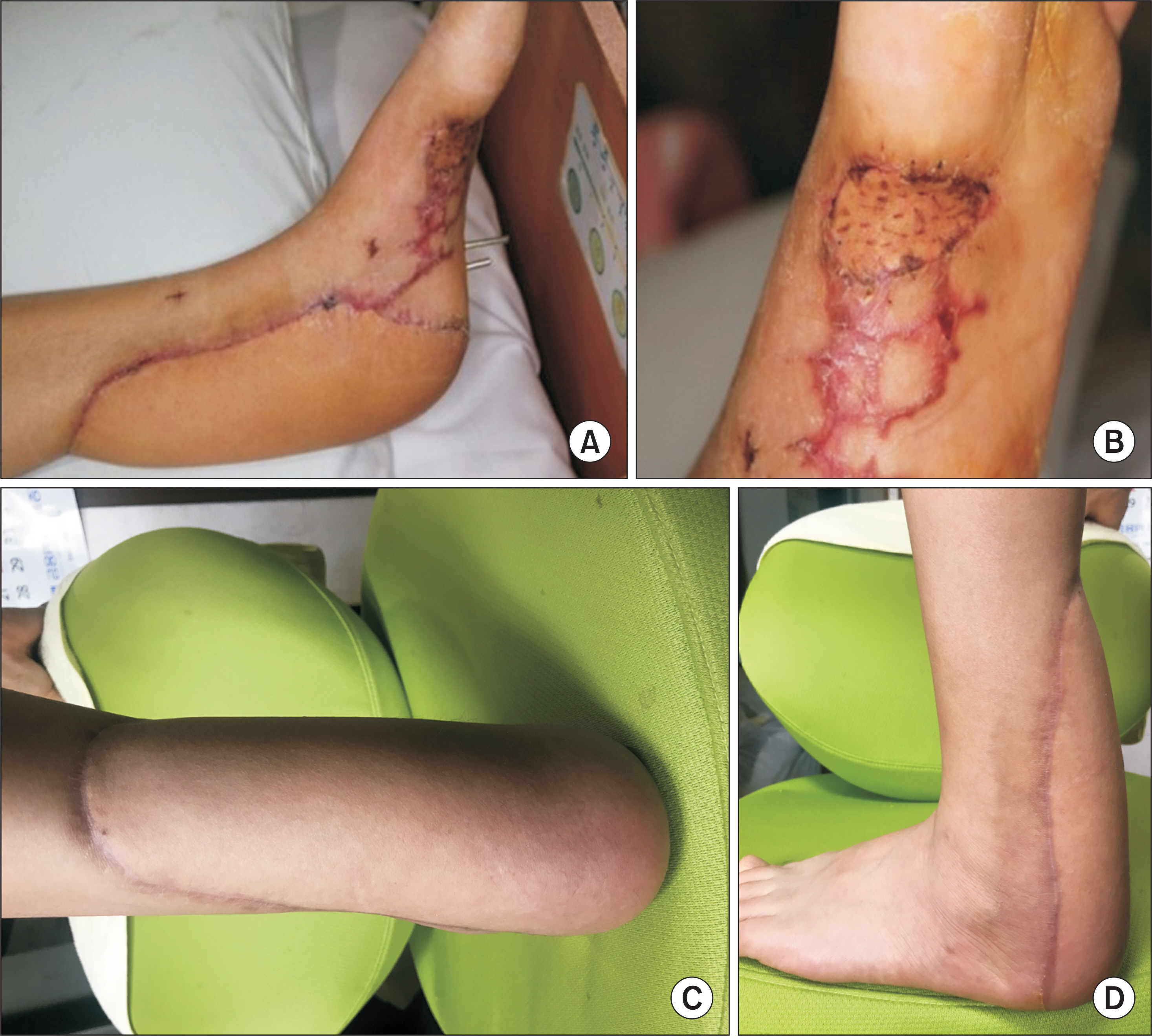
Figure 5. Gross photography (A) and plantar area (B) of foot flap site of 1 month after the surgery shows no complications. Posteroanterior view (C) and laterial view (D) of lower leg gross photography of 3 months after the surgery shows good healing and no complications.

Figure 6. (A) Preoperative simple lateral radiography shows soft tissue damage. (B) Postoperative simple lateral radiography shows fixation of allobone to calcaneus with screws. And ankle joint was immobilized with Steinmann pins.
Reference
-
References
1. Leppilahti J, Orava S. Total Achilles tendon rupture. Sports Med. 1998; 25:79–100.
Article2. Marchesi A, Parodi PC, Brioschi M, Riccio M, Perrotta RE, Colombo M, et al. Soft-tissue defects of the Achilles tendon region: management and reconstructive ladder: review of the literature. Injury. 2016; 47(Suppl 4):147–53.
Article3. Bullocks JM, Hickey RM, Basu CB, Hollier LH, Kim JY. Singlestage reconstruction of Achilles tendon injuries and distal lower extremity soft tissue defects with the reverse sural fasciocutaneous flap. J Plast Reconstr Aesthet Surg. 2008; 61:566–72.
Article4. Deiler S, Pfadenhauer A, Widmann J, Stützle H, Kanz KG, Stock W. Tensor fasciae latae perforator flap for reconstruction of composite Achilles tendon defects with skin and vascularized fascia. Plast Reconstr Surg. 2000; 106:342–9.
Article5. Lee JW, Yu JC, Shieh SJ, Liu C, Pai JJ. Reconstruction of the Achilles tendon and overlying soft tissue using anterolateral thigh free flap. Br J Plast Surg. 2000; 53:574–7.
Article6. Smith PJ, Foley B, McGregor IA, Jackson IT. The anatomical basis of the groin flap. Plast Reconstr Surg. 1972; 49:41–7.
Article7. Baumeister SP, Spierer R, Erdmann D, Sweis R, Levin LS, Germann GK. A realistic complication analysis of 70 sural artery flaps in a multimorbid patient group. Plast Reconstr Surg. 2003; 112:129–40. ; discussion 141–2.
Article8. Kim GC, Chung CI, Kim SE, Kim HS, Rhyou IH. Reconstruction of soft tissue defect of lower extremity with anterolateral thigh perforator flap. J Korean Soc Micorsurg. 2006; 15:70–6.9. Clark N, Sherman R. Soft-tissue reconstruction of the foot and ankle. Orthop Clin North Am. 1993; 24:489–503.
Article10. Choi YR, Lee SY, Lee SC, Lee HJ, Han SH. Reverse superficial sural artery flap for the reconstruction of soft tissue defect on posterior side of heel exposing Achilles tendon. J Korean Soc Micorsurg. 2012; 21:159–64.
- Full Text Links
-
- Actions
-
Cited
- CITED
-
- Close
- Share
-
- Similar articles
-
- Heterotopic Ossification of a Partially Ruptured Achilles Tendon (A Case Report)
- Repair of Neglected Rupture of the Achilles Tendon using V-Y Tendinous Flap
- Long Term Result of Four Cases without a Staged Reconstruction of an Infected Achilles Tendon Following Repair
- Management of Postoperative Complications Following Surgical Repair of Achilles Tendon Rupture
- One-stage Reconstruction of Soft-tissue Defect including Achilles Tendon: A Case Report
