J Cerebrovasc Endovasc Neurosurg.
2024 Sep;26(3):318-323. 10.7461/jcen.2024.E2023.07.002.
Isolated ipsilateral abducens nerve palsy and contralateral homonymous hemianopsia associated with unruptured posterior cerebral artery aneurysm: A rare neurological finding
- Affiliations
-
- 1Department of Neurosurgery, All India Institute of Medical Sciences, New Delhi, India
- 2Department of Radiology, Postgraduate Institute of Medical Education and Research, Chandigarh, India
- 3Division of Neuroanesthesia, Department of Anaesthesia and Intensive Care, Postgraduate Institute of Medical Education and Research, Chandigarh, India
- 4Department of Neuroimaging and Interventional Neuroradiology, All India Institute of Medical Sciences, New Delhi, India
- KMID: 2559443
- DOI: http://doi.org/10.7461/jcen.2024.E2023.07.002
Abstract
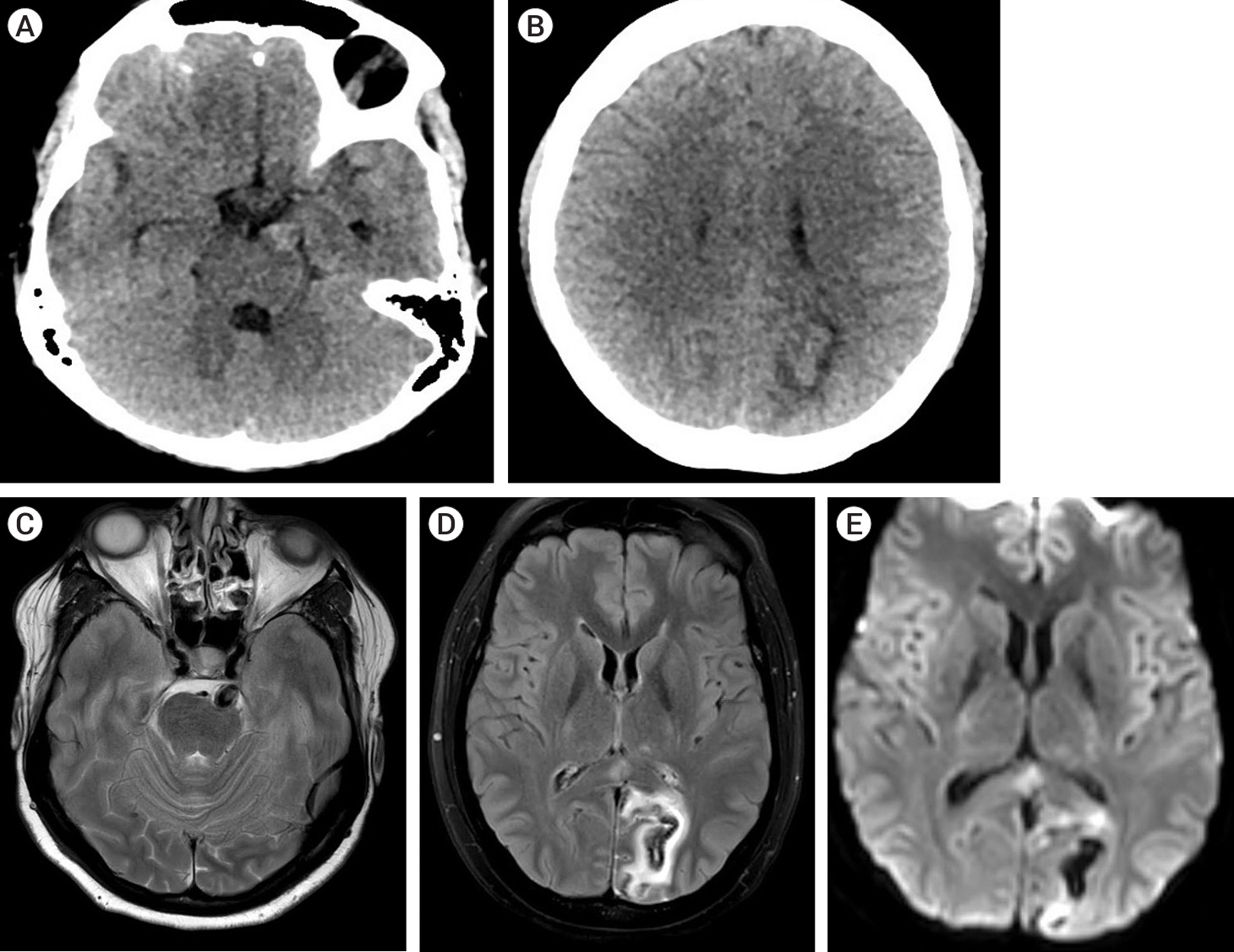
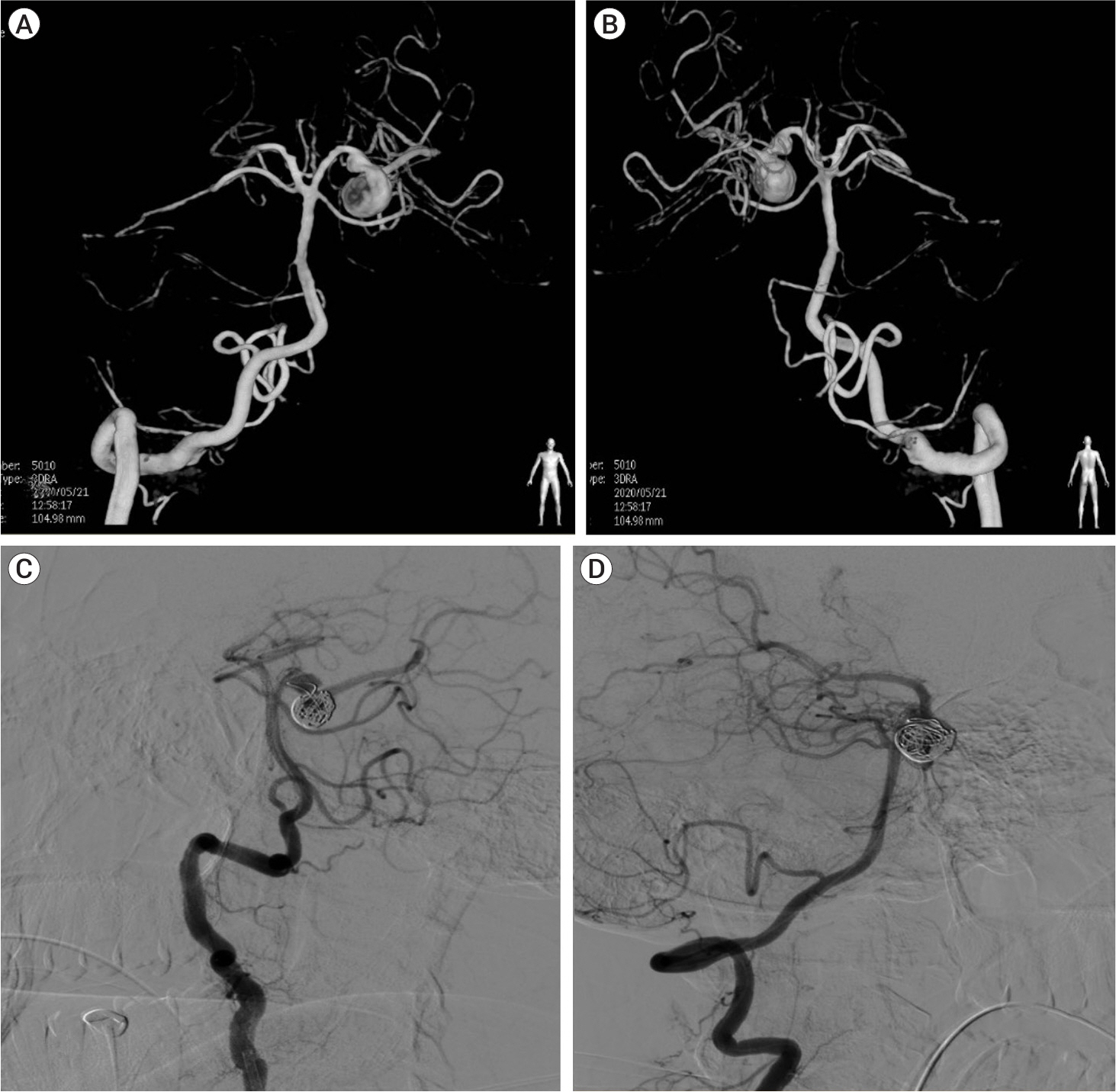
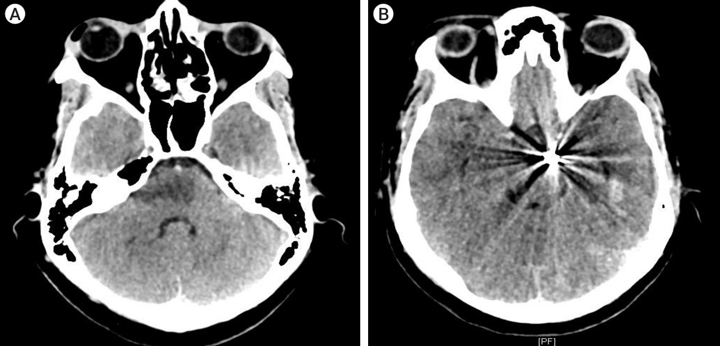
- Cranial nerve palsies can be presenting signs of intracranial aneurysms. There is a classic pairing between an aneurysmal vessel and adjacent nerves leading to cranial neuropathy. Isolated abducens nerve palsy can be a localizing sign of an unruptured vertebrobasilar circulation aneurysm. Aneurysms involving Anterior Inferior Cerebellar Artery (AICA) and Posterior Inferior Cerebellar Artery (PICA) have been reported to be associated with abducens nerve palsy. The symptoms in unruptured aneurysms are due to the mass effect on adjacent neurovascular structures. Most of the abducens nerve palsy resolves following microsurgical clipping. Here, we present a rare case of an unruptured Posterior Cerebral Artery (PCA) aneurysm presenting with abducens nerve palsy and diplopia associated with contralateral hemianopsia which markedly improved following endovascular coil embolization.
Keyword
Figure
Reference
-
1. Ciceri EF, Klucznik RP, Grossman RG, Rose JE, Mawad ME. Aneurysms of the posterior cerebral artery: Classification and endovascular treatment. AJNR Am J Neuroradiol. 2001; Jan. 22(1):27–34.2. Drake CG, Peerless SJ, Hernesniemi JA. Surgery of Vertebrobasilar Aneurysms: London, Ontario Experience on 1767 Patients. Vienna: Springer Vienna;1996.3. Gerber CJ, Neil-Dwyer G. A review of the management of 15 cases of aneurysms of the posterior cerebral artery. Br J Neurosurg. 1992; 6(6):521–7.
Article4. Oran I, Cinar C, Yağci B, Tarhan S, Kiroğlu Y, Serter S. Ruptured dissecting aneurysms arising from non-vertebral arteries of the posterior circulation: Endovascular treatment perspective. Diagn Interv Radiol. 2009; Sep. 15(3):159–65.5. Parr M, Carminucci A, Al-Mufti F, Roychowdhury S, Gupta G. Isolated abducens nerve palsy associated with ruptured posterior inferior cerebellar artery aneurysm: Rare neurologic finding. World Neurosurg. 2019; Jan. 121:97–9.6. Pia HW, Fontana H. Aneurysms of the posterior cerebral artery. Locations and clinical pictures. Acta Neurochir (Wien). 1977; 38(1-2):13–35.
Article7. Seoane ER, Tedeschi H, de Oliveira E, Siqueira MG, Calderón GA, Rhoton AL Jr. Management strategies for posterior cerebral artery aneurysms: A proposed new surgical classification. Acta Neurochir (Wien). 1997; 139(4):325–31.
Article8. Sherman P, Oka M, Aldrich E, Jordan L, Gailloud P. Isolated posterior cerebral artery dissection: Report of three cases. AJNR Am J Neuroradiol. 2006; Mar. 27(3):648–52.
Article9. Suzuki O, Miyachi S, Negoro M, Okamoto T, Sahara Y, Hattori K, et al. Treatment strategy for aneurysms of the posterior cerebral artery. Interv Neuroradiol. 2003; May. 9(Suppl 1):83–8.
Article10. Taqi MA, Lazzaro MA, Pandya DJ, Badruddin A, Zaidat OO. Dissecting aneurysms of posterior cerebral artery: Clinical presentation, angiographic findings, treatment, and outcome. Front Neurol. 2011; Jun. 2:38.11. Walter E, Liao EA, De Lott LB, Trobe JD. Acute isolated sixth nerve palsy caused by unruptured intradural saccular aneurysm. J Neuroophthalmol. 2019; Dec. 39(4):458–61.
Article12. Zelman S, Goebel MC, Manthey DE, Hawkins S. Large posterior communicating artery aneurysm: Initial presentation with reproducible facial pain without cranial nerve deficit. West J Emerg Med. 2016; Nov. 17(6):808–10.
Article
- Full Text Links
- Actions
-
Cited
- CITED
-
- Close
- Share
- Similar articles
-
- Isolated Bilateral Abducens Nerve Palsy Caused by Basilar Artery Dissecting Aneurysm
- An Unruptured Anterior Communicating Artery Aneurysm Presenting with Left Homonymous Hemianopsia: A Case Report
- Unilateral Abducens Nerve Palsy Associated with Ruptured Anterior Communicating Artery Aneurysm
- Combined Facial and Abducens Nerve Palsy in Pontine Infarction
- Dissecting Aneurysm of Vertebral Artery Manifestating as Contralateral Abducens Nerve Palsy


