J Cerebrovasc Endovasc Neurosurg.
2024 Sep;26(3):304-310. 10.7461/jcen.2024.E2023.04.006.
Giant cerebellar cavernous malformation in children: A case report and literature review
- Affiliations
-
- 1Department of Neurosurgery, National Children’s Medical Center, Ministry of Healthcare of Republic of Uzbekistan, Tashkent, Uzbekistan
- 2Department of Neurosurgery, Tashkent Pediatric Medical Institute, Tashkent, Uzbekistan
- 3Department of Neurosurgery, Neurosurgery Clinic, Birgunj, Nepal
- KMID: 2559441
- DOI: http://doi.org/10.7461/jcen.2024.E2023.04.006
Abstract
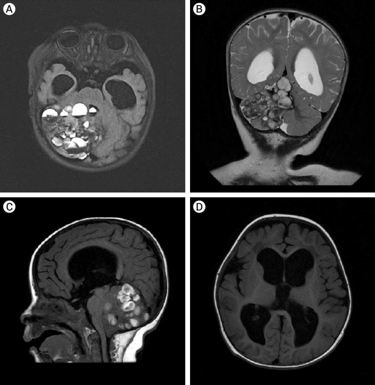
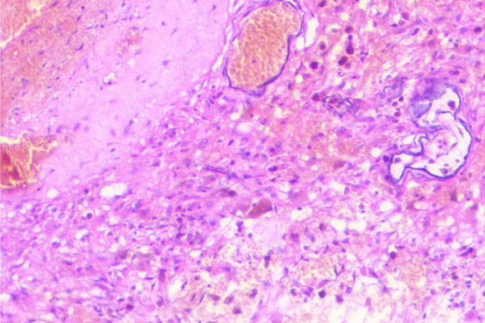
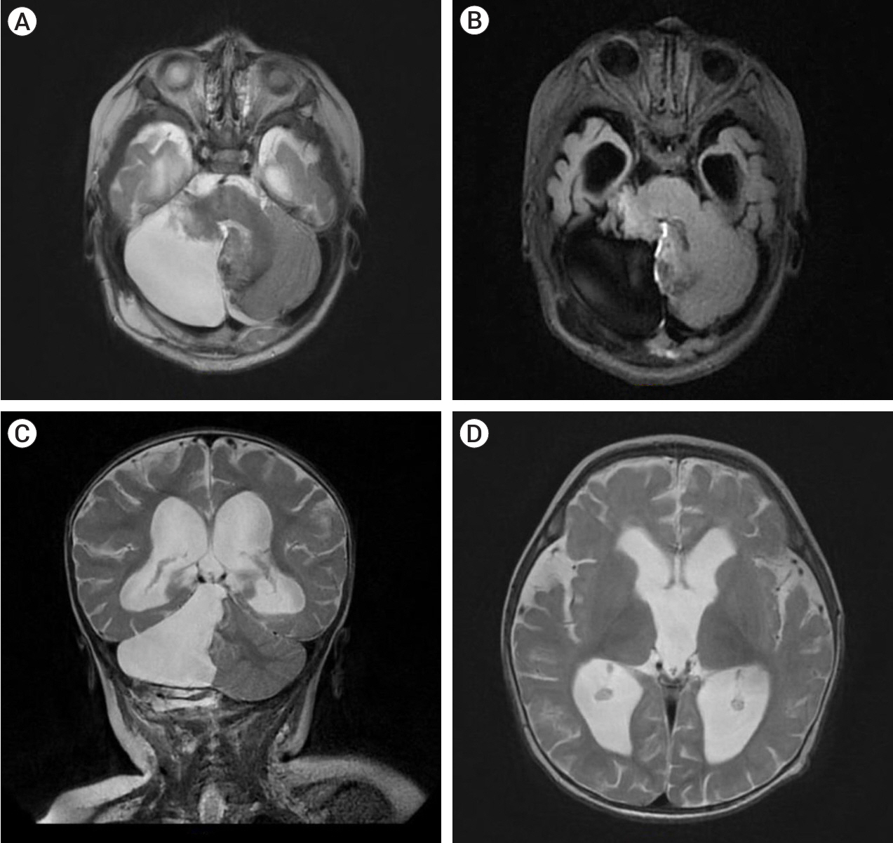
- Giant cerebellar cavernomas in children are rare and must be differentiated from hemorrhagic cerebellar tumors. The diagnosis and treatment of giant cerebellar cavernomas is challenging, but complete surgical resection can lead to favorable outcomes and complete neurological recovery in most cases. We present a case of eight months old baby who was diagnosed with a giant cavernoma resulting in secondary obstructive hydrocephalus with neuropsychiatric presentations. The patient underwent a paramedian craniotomy surgery with a suboccipital approach and complete surgical resection of the cavernoma was done. Over nine months of observation, the child showed improvement in their ability to walk and fully recovered from a neurological perspective. We also conducted a literature review to identify eleven cases of giant cerebellar cavernomas in children, including our case. The data were analyzed to determine the clinical features, treatment, and outcomes of giant cerebellar cavernomas in children.
Figure
Reference
-
1. Atalar M, Kars Z, Egilmez R, Egilmez H. Giant cavernous angioma mimicking cerebellar neoplasm with major bleed: A case report. Tip Arastirmalari Dergisi. 2007; 5(3):153–6.2. Avci E, Oztürk A, Baba F, Karabağ H, Cakir A. Huge cavernoma with massive intracerebral hemorrhage in a child. Turk Neurosurg. 2007; 17(1):23–6.3. Del Curling O Jr, Kelly DL Jr, Elster AD, Craven TE. An analysis of the natural history of cavernous angiomas. J Neurosurg. 1991; Nov. 75(5):702–8.
Article4. Gaddi MJS, Pascual JSG, Legaspi EDC, Rivera PP, Omar AT 2nd. Giant cerebellar cavernomas in pediatric patients: Systematic review with illustrative case. J Stroke Cerebrovasc Dis. 2020; Nov. 29(11):105264.5. Gezen F, Karatas A, Is M, Yildirim U, Aytekin H. Giant cavernous haemangioma in an infant. Br J Neurosurg. 2008; Dec. 22(6):787–9.
Article6. Gross BA, Lin N, Du R, Day AL. The natural history of intracranial cavernous malformations. Neurosurg Focus. 2011; Jun. 30(6):e24.
Article7. Grujić J, Jovanović V, Tasić G, Savić A, Stojiljković A, Matić S, et al. Giant cavernous malformation with unusually aggressive clinical course: A case report. Acta Clin Croat. 2020; Mar. 59(1):183–87.
Article8. Hayashi T, Fukui M, Shyojima K, Utsunomiya H, Kawasaki K. Giant cerebellar hemangioma in an infant. Childs Nerv Syst. 1985; 1(4):230–3.
Article9. Jurkiewicz E, Marcinska B, Malczyk K, Grajkowska W, Daszkiewicz P, Roszkowski M. Giant cerebellar cavernous malformation in 4-month-old boy. Case report and review of the literature. Neurol Neurochir Pol. 2013; Nov-Dec. 47(6):596–600.10. Kan P, Tubay M, Osborn A, Blaser S, Couldwell WT. Radiographic features of tumefactive giant cavernous angiomas. Acta Neurochir (Wien). 2008; Jan. 150(1):49–55. discussion 55.
Article11. Kim DS, Park YG, Choi JU, Chung SS, Lee KC. An analysis of the natural history of cavernous malformations. Surg Neurol. 1997; Jul. 48(1):9–17. discussion 17.
Article12. Konovalov A, Saripov O, Gadzhiagaev V, Titov O, Lasunin N, Zhumabekov A, et al. Optochiasmatic cavernoma: Surgical treatment and outcomes. J Cerebrovasc Endovasc Neurosurg. 2023; Dec. 25(4):411–9.
Article13. Lawton MT, Vates GE, Quinones-Hinojosa A, McDonald WC, Marchuk DA, Young WL. Giant infiltrative cavernous malformation: Clinical presentation, intervention, and genetic analysis: Case report. Neurosurgery. 2004; Oct. 55(4):979–80.
Article14. Lew SM. Giant posterior fossa cavernous malformations in 2 infants with familial cerebral cavernomatosis: The case for early screening. Neurosurg Focus. 2010; Sep. 29(3):e18.15. McCormick WF, Hardman JM, Boulter TR. Vascular malformations (“angiomas”) of the brain, with special reference to those occurring in the posterior fossa. J Neurosurg. 1968; Mar. 28(3):241–51.
Article16. Otten P, Pizzolato GP, Rilliet B, Berney J. [131 cases of cavernous angioma (cavernomas) of the CNS, discovered by retrospective analysis of 24,535 autopsies]. Neurochirurgie. 1989; 35(2):82–31. 82-3, 128-31. Article in French.17. Ozgen B, Senocak E, Oguz KK, Soylemezoglu F, Akalan N. Radiological features of childhood giant cavernous malformations. Neuroradiology. 2011; 53(4):283–9.
Article18. Ozsoy KM, Oktay K, Gezercan Y, Cetinalp NE, Olguner SK, Erman T. Giant cavernous malformations in childhood: A case report and review of the literature. Pediatr Neurosurg. 2017; 52(1):30–5.
Article19. Parizel MR, Menovsky T, Van Marck V, Lammens M, Parizel PM. Giant cavernous malformations in young adults: Report of two cases, radiological findings and surgical consequences. JBR-BTR. 2014; Sep-Oct. 97(5):274–8.20. Porter PJ, Willinsky RA, Harper W, Wallace MC. Cerebral cavernous malformations: Natural history and prognosis after clinical deterioration with or without hemorrhage. J Neurosurg. 1997; Aug. 87(2):190–7.
Article21. Robinson JR, Awad IA, Little JR. Natural history of the cavernous angioma. J Neurosurg. 1991; Nov. 75(5):709–14.
Article22. Shroff K, Deopujari C, Karmarkar V, Mohanty C. Paediatric giant cavernomas: Report of three cases with a review of the literature. Childs Nerv Syst. 2021; Dec. 37(12):3835–45.
Article23. Son DW, Lee SW, Choi CH. Giant cavernous malformation: A case report and review of the literature. J Korean Neurosurg Soc. 2008; Apr. 43(4):198–200.
Article24. Villaseñor-Ledezma J, Budke M, Alvarez-Salgado JA, Cañizares MA, Moreno L, Villarejo F. Pediatric cerebellar giant cavernous malformation: Case report and review of literature. Childs Nerv Syst. 2017; 33(12):2187–91.
- Full Text Links
- Actions
-
Cited
- CITED
-
- Close
- Share
- Similar articles
-
- Giant Cystic Cerebral Cavernous Malformation with Multiple Calcification: Case Report
- A Case of Cavernous Angioma of the Cerebellar Vermis
- An Unusual Case of a Thrombosed Giant Distal PICA Aneurysm Simulating a Large Cavernous Angioma
- A case involving anesthesia for cesarean section followed by resection of ruptured cavernous malformation of pons :A case report
- Giant Cavernous Malformation : A Case Report and Review of the Literature


