Clin Endosc.
2024 Sep;57(5):688-689. 10.5946/ce.2024.052.
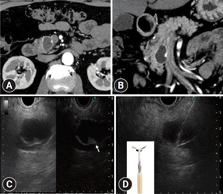
Usefulness of micro forceps biopsy for cystic degenerated pancreatic neuroendocrine neoplasm
- Affiliations
-
- 1Division of Endoscopy, Shizuoka Cancer Center, Shizuoka, Japan
- KMID: 2559221
- DOI: http://doi.org/10.5946/ce.2024.052
Figure
Reference
-
1. Nakai Y, Isayama H, Chang KJ, et al. A pilot study of EUS-guided through-the-needle forceps biopsy (with video). Gastrointest Endosc. 2016; 84:158–162.2. Barresi L, Crinò SF, Fabbri C, et al. Endoscopic ultrasound-through-the-needle biopsy in pancreatic cystic lesions: a multicenter study. Dig Endosc. 2018; 30:760–770.3. Yang D, Trindade AJ, Yachimski P, et al. Histologic analysis of endoscopic ultrasound-guided through the needle microforceps biopsies accurately identifies mucinous pancreas cysts. Clin Gastroenterol Hepatol. 2019; 17:1587–1596.
- Full Text Links
- Actions
-
Cited
- CITED
-
- Close
- Share
- Similar articles
-
- Pancreatic Collision Tumor of Desmoid-Type Fibromatosis and Mucinous Cystic Neoplasm: A Case Report
- Well-Differentiated Pancreatic Neuroendocrine Tumor with Solitary Hepatic Metastasis Presenting as a Benign Cystic Mass: A Case Report
- Surgical Indications and Postsurgical Follow-up Strategy for Pancreatic Cystic Neoplasm
- Pathologic Features of Pancreatic Cystic Neoplasms
- Pancreatic Cystic Neoplasm: Radiologic Evaluation and Differential Diagnosis


