J Pathol Transl Med.
2024 Sep;58(5):241-248. 10.4132/jptm.2024.07.14.
International Academy of Cytology standardized reporting of breast fine-needle aspiration cytology with cyto-histopathological correlation of breast carcinoma
- Affiliations
-
- 1University Hospital Lewisham, London, UK
- 2Department of Pathology, SCMCH & RI, Karnataka, India
- KMID: 2559069
- DOI: http://doi.org/10.4132/jptm.2024.07.14
Abstract
- Background
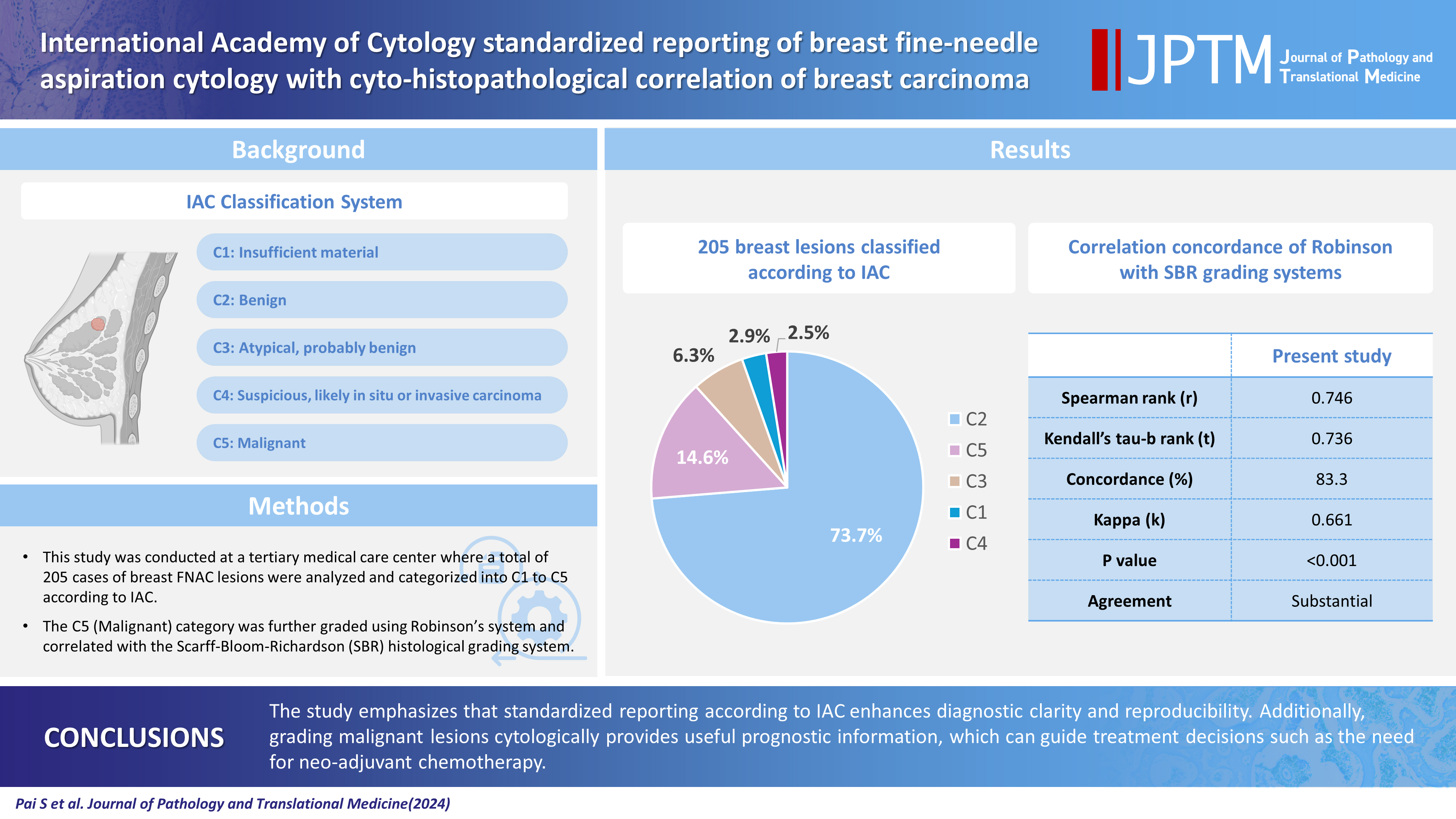
The International Academy of Cytology (IAC) has developed a standardized approach for reporting the findings of breast fine-needle aspiration cytology (FNAC). Accordingly, there are five chief categories of breast lesions, C1 (insufficient material), C2 (benign), C3 (atypical), C4 (suspicious), and C5 (malignant). The prognostication and management of breast carcinoma can be performed readily on the basis of this classification system. The aim of this study was to classify various breast lesions into one of the above-named categories and to further grade the C5 lesions specifically using the Robinson system. The latter grades were then correlated with modified Scarff-Bloom-Richardson (SBR) grades.
Methods
This retrospective study was undertaken in the pathology department of a hospital located in the urban part of the city of Bangalore. All FNAC procedures performed on breast lumps spanning the year 2020 were included in the study.
Results
A total of 205 breast lesions was classified according to the IAC guidelines into C1 (6 cases, 2.9%), C2 (151 cases, 73.7%), C3 (13 cases, 6.3%), C4 (5 cases, 2.5%), and C5 (30 cases, 14.6%) groups. The C5 cases were further graded using Robinson’s system. The latter showed a significant correlation with the SBR system (concordance=83.3%, Spearman correlation=0.746, Kendall’s tau-b=0.736, kappa=0.661, standard error=0.095, p≤.001).
Conclusions
A standardized approach for FNAC reporting of breast lesions, as advocated for by the IAC, improves the quality and clarity of the reports and assures diagnostic reproducibility on a global scale. Further, the cytological grading of C5 lesions provides reliable cyto-prognostic scores that can help assess a tumor’s aggressiveness and predict its histological grade.
Keyword
Figure
Reference
-
References
1. Panwar H, Ingle P, Santosh T, Singh V, Bugalia A, Hussain N. FNAC of breast lesions with special reference to IAC standardized reporting and comparative study of cytohistological grading of breast carcinoma. J Cytol. 2020; 37:34–9.2. Field AS, Schmitt F, Vielh P. IAC standardized reporting of breast fine-needle aspiration biopsy cytology. Acta Cytol. 2017; 61:3–6.3. Elston CW, Ellis IO. Pathological prognostic factors in breast cancer. I. The value of histological grade in breast cancer: experience from a large study with long-term follow-up. Histopathology. 1991; 19:403–10.4. Robinson IA, McKee G, Nicholson A, et al. Prognostic value of cytological grading of fine-needle aspirates from breast carcinomas. Lancet. 1994; 343:947–9.5. Hunt CM, Ellis IO, Elston CW, Locker A, Pearson D, Blamey RW. Cytological grading of breast carcinoma: a feasible proposition? Cytopathology. 1990; 1:287–95.6. Mouriquand J, Gozlan-Fior M, Villemain D, et al. Value of cytoprognostic classification in breast carcinomas. J Clin Pathol. 1986; 39:489–96.7. Bansal C, Pujani M, Sharma KL, Srivastava AN, Singh US. Grading systems in the cytological diagnosis of breast cancer: a review. J Cancer Res Ther. 2014; 10:839–45.8. Robinson IA, McKee G, Kissin MW. Typing and grading breast carcinoma on fine-needle aspiration: is this clinically useful information? Diagn Cytopathol. 1995; 13:260–5.9. Wani FA, Bhardwaj S, Kumar D, Katoch P. Cytological grading of breast cancers and comparative evaluation of two grading systems. J Cytol. 2010; 27:55–8.10. Das AK, Kapila K, Dinda AK, Verma K. Comparative evaluation of grading of breast carcinomas in fine needle aspirates by two methods. Indian J Med Res. 2003; 118:247–50.11. Phukan JP, Sinha A, Deka JP. Cytological grading of breast carcinoma on fine needle aspirates and its relation with histological grading. South Asian J Cancer. 2015; 4:32–4.12. Skrbinc B, Babic A, Cufer T, Us-Krasovec M. Cytological grading of breast cancer in Giemsa-stained fine needle aspiration smears. Cytopathology. 2001; 12:15–25.13. Khan N, Afroz N, Rana F, Khan M. Role of cytologic grading in prognostication of invasive breast carcinoma. J Cytol. 2009; 26:65–8.14. Sneige N. Should specimen adequacy be determined by the opinion of the aspirator or by the cells on the slides? Cancer. 1997; 81:3–5.15. Hussain MT. Comparison of fine needle aspiration cytology with excision biopsy of breast lump. J Coll Physicians Surg Pak. 2005; 15:211–4.16. McManus DT, Anderson NH. Fine needle aspiration cytology of the breast. Curr Diagn Pathol. 2001; 7:262–71.17. Tariq GR, Haleem A, Zaidi AH, Afzal M, Abbasi S. Role of FNA cytology in the management of carcinoma breast. J Coll Physicians Surg Pak. 2005; 15:207–10.18. Montezuma D, Malheiros D, Schmitt FC. Breast fine needle aspiration biopsy cytology using the newly proposed IAC Yokohama System for Reporting Breast Cytopathology: the experience of a single institution. Acta Cytol. 2019; 1–6.19. Haobam S, Thiyam U, Sharma DC. Cytomorphological study of breast lesions with sonomammographic correlation. J Evol Med Dent Sci. 2015; 4:14137–42.20. Georgieva RD, Obdeijn IM, Jager A, Hooning MJ, Tilanus-Linthorst MM, van Deurzen CH. Breast fine-needle aspiration cytology performance in the high-risk screening population: a study of BRCA1/BRCA2 mutation carriers. Cancer Cytopathol. 2013; 121:561–7.21. Bajwa R, Zulfiqar T. Association of fine needle aspiration cytology with tumor size in palpable breast lesions. Biomedica. 2010; 26:124–9.22. Modi P, Oza H, Bhalodia J. FNAC as preoperative diagnostic tool for neoplastic and non-neoplastic breast lesions: a teaching hospital experience. Natl J Med Res. 2014; 4:274–8.23. Arul P, Masilamani S. Comparative evaluation of various cytomorphological grading systems in breast carcinoma. Indian J Med Paediatr Oncol. 2016; 37:79–84.24. Saha K, Raychaudhuri G, Chattopadhyay BK, Das I. Comparative evaluation of six cytological grading systems in breast carcinoma. J Cytol. 2013; 30:87–93.25. Einstien D, Omprakash BO, Ganapathy H, Rahman S. Comparison of 3-tier cytological grading systems for breast carcinoma. ISRN Oncol. 2014; 2014:252103.26. Sinha S, Sinha N, Bandyopadhyay R, Mondal SK. Robinson’s cytological grading on aspirates of breast carcinoma: correlation with Bloom Richardson’s histological grading. J Cytol. 2009; 26:140–3.27. Vasudev V, R R, V G. The cytological grading of malignant neoplasms of the breast and its correlation with the histological grading. J Clin Diagn Res. 2013; 7:1035–9.28. Pal S, Gupta ML. Correlation between cytological and histological grading of breast cancer and its role in prognosis. J Cytol. 2016; 33:182–6.
- Full Text Links
- Actions
-
Cited
- CITED
-
- Close
- Share
- Similar articles
-
- Fine Needle Aspiration Cytology of Medullary Carcinoma of the Breast: A Case Report
- The Usefulness of Ultrasound-Guided Fine Needle Aspiration in Breast Lesions
- Fine Needle Aspiration Cytology of Tubulolobular Carcinoma of the Breast: A Case Report
- Fine Needle Aspiration Cytology of Intracystic Papillary Carcinoma of the Breast
- Fine Needle Aspiration Cytology of Cystic Hypersecretory Intraductal Carcinoma of the Breast: Report of Two Cases


