Salvaging Vision: A Study of Non-Traumatic Optic Neuropathies
- Affiliations
-
- 1Department of ENT and Head-Neck Surgery, Seth G.S. Medical College and King Edward Memorial Hospital, Mumbai, India
- KMID: 2558233
- DOI: http://doi.org/10.18787/jr.2024.00012
Abstract
- Background and Objectives
Various ear, nose, and throat (ENT) conditions can result in vision loss. The purpose of this study is to identify the etiologies, presentations, and radiological findings associated with impaired vision in the context of ENT. Additionally, this article discusses management protocols, including optic nerve decompression and orbital decompression.
Methods
In a retrospective study, we examined the period from 2016 to 2022 at a tertiary care hospital in Mumbai, India. The analysis included 11 patients who presented with progressive diminution of vision. All patients received a regimen of broad-spectrum intravenous antibiotics and high-dose intravenous steroids. This was followed by either endoscopic optic nerve decompression or orbital decompression. Subsequent improvements in vision were documented, and any complications were evaluated.
Results
A total of 11 patients were treated with medical management followed by successful surgery, with 10 patients demonstrating significant vision improvement.
Conclusion
Identifying the etiology of vision loss and managing the condition can present challenges for otorhinolaryngologists. A thorough grasp of the underlying pathophysiology, combined with active surveillance of clinical and radiological indicators, can enable these clinicians to achieve effective and rewarding outcomes.
Figure
-
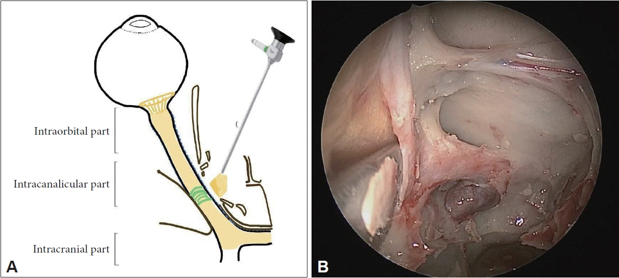
Fig. 1. Endoscopic anatomy of optic nerve. A: Schematic representation of intracanalicular part of optic nerve that can be assessed by transnasal endoscopy. B: Endoscopic anatomical relations of the right optic nerve showing carotid artery, opticocarotid recess and posterior ethmoidal artery in the Onodi cell in cadaveric dissection
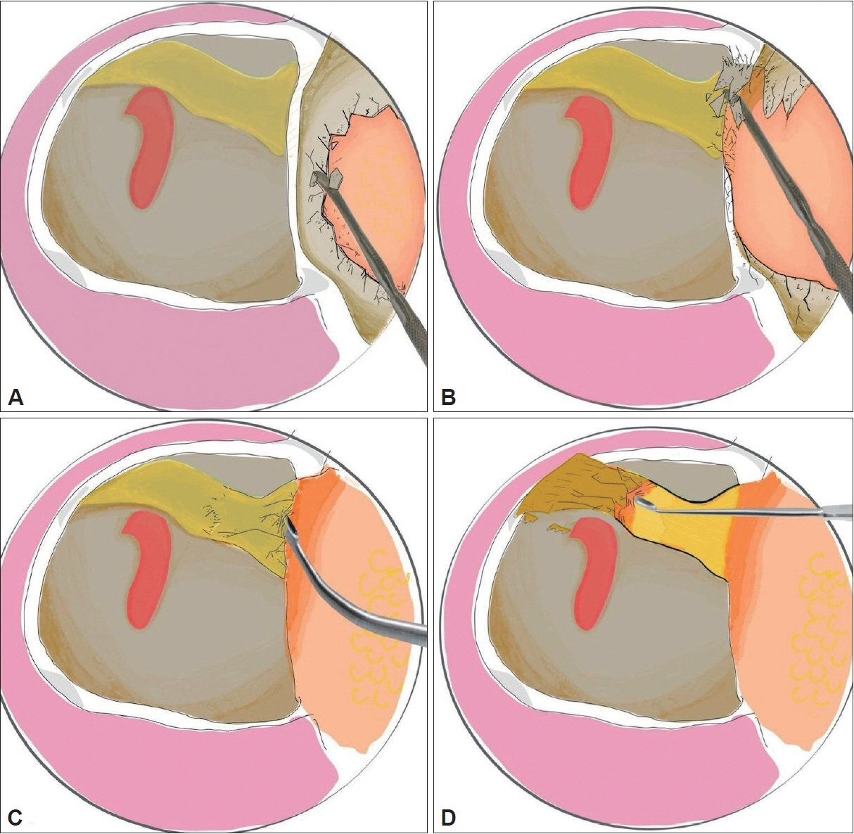
Fig. 2. Schematic diagram showing the steps of left trans-nasal endoscopic optic nerve decompression. A and B: The relationship of the left optic nerve with lamina papyracea and carotid. Removal of the lamina papyracea with a Freer’s elevator. C and D: Removal of the optic canal and annulus of Zinn with curette to decompress the optic nerve.
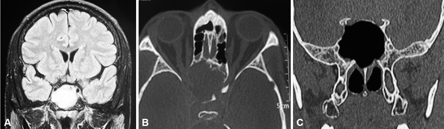
Fig. 3. Contrast-enhanced computed tomography of the paranasal sinuses in a 62-year-old male patient presenting with headache and diminution of vision. A and B: Imaging revealed a sphenoid mucocele causing compression of the optic nerves. C: The patient underwent endoscopic marsupialization of the sphenoid mucocele and a postoperative scan for the same was done.
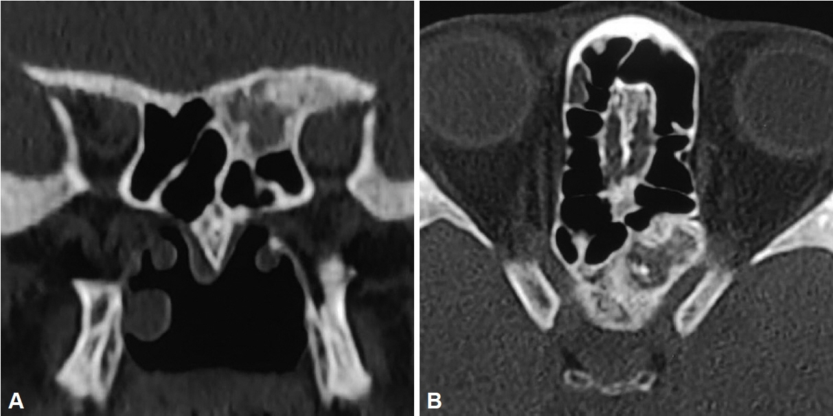
Fig. 4. Coronal (A) and axial (B) computed tomography images of the paranasal sinuses showing ground glass opacity in the Onodi cell. A 25-year-old female patient presented to our outpatient department with left-sided diminution of vision and diplopia and left ophthalmoplegia of insidious onset for 15 days. On starting intravenous steroids, the ophthalmoplegia had improved. CT scan of paranasal sinuses revealed ground glass opacification of the Onodi cell suggestive of fibrous dysplasia with involvement of optic nerve on the left. Fundoscopy was done to check the viability of the optic disc plan endoscopic decompression. The patient required drilling of the dense fibrous dysplasia lesion in order to release the compression from the left optic nerve.
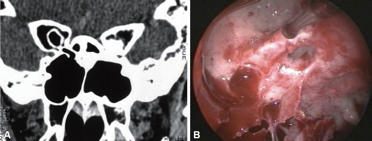
Fig. 5. A rare case of osteopetrosis was identified in our study population (20-year-old male). The patient had presented with a recent onset, progressive diminution of vision on the left side. A: Axial CT scan revealed dense bone around the optic nerve causing optic canal stenosis. B: Endoscopic decompression of the left optic nerve was achievable only with the use of micro-drill. On follow-up, the progression of vision deterioration had halted and the patient was taken for endoscopic decompression of optic nerve on the right side after 3 months.
Reference
-
References
1. Samardzic K, Samardzic J, Janjetovic Z, Samardzic I, Sekelj S, Latic-Hodzic L. Traumatic optic neuropathy - to treat or to observe? Acta Inform Med. 2012; 20(2):131–2.
Article2. Rodriguez-Beato FY, De Jesus O. Compressive optic neuropathy. In: StatPearls [Internet]. Treasure Island: StatPearls Publishing;2023. [cited 2023 Aug 23]. Available from: https://www.ncbi.nlm.nih.gov/books/NBK560583.3. Newton N Jr, Baratham G, Sinniah R, Lim A. Bilateral compressive optic neuropathy secondary to bilateral sphenoethmoidal mucoceles. Ophthalmologica. 1989; 198(1):13–9.
Article4. DeLano MC, Fun FY, Zinreich SJ. Relationship of the optic nerve to the posterior paranasal sinuses: a CT anatomic study. AJNR Am J Neuroradiol. 1996; 17(4):669–75.
Article5. Lal D, Stankiewicz JA. Endoscopic optic nerve decompression. Oper Tech Otolayngol Head Neck Surg. 2009; 20(2):96–100.
Article6. Karimi S, Arabi A, Ansari I, Shahraki T, Safi S. A systematic literature review on traumatic optic neuropathy. J Ophthalmol. 2021; 2021:5553885.
Article7. Li HB, Shi JB, Cheng L, Yun O, Xu G. Salvage optic nerve decompression for traumatic blindness under nasal endoscopy: risk and benefit analysis. Clin Otolaryngol. 2007; 32(6):447–51.8. Loeb HW. XXI. A study of the anatomic relations of the optic nerve to the accessory cavities of the nose. Ann Otol Rhinol Laryngol. 1909; 18(2):243–306.
Article9. Chandler JR, Langenbrunner DJ, Stevens ER. The pathogenesis of orbital complications in acute sinusitis. Laryngoscope. 1970; 80(9):1414–28.
Article10. Kim YH, Kim J, Kang MG, Lee DH, Chin HS, Jang TY, et al. Optic nerve changes in chronic sinusitis patients: correlation with disease severity and relevant sinus location. PLoS One. 2018; 13(7):e0199875.
Article11. Che SA, Lee YW, Yoo YJ. Compressive optic neuropathy due to posterior ethmoid mucocele. BMC Ophthalmol. 2023; 23(1):426.
Article12. Capra GG, Carbone PN, Mullin DP. Paranasal sinus mucocele. Head Neck Pathol. 2012; 6(3):369–72.
Article13. Gupta S, Sethi P, Duvesh R, Sethi HS, Naik M, Rai HK. Optic perineuritis. BMJ Open Ophthalmol. 2021; 6(1):e000745.
Article14. Van Stavern GP. Metabolic, hereditary, traumatic, and neoplastic optic neuropathies. Continuum (Minneap Minn). 2014; 20(4):877–906.
Article15. Bailey JR, Tapscott DC. Osteopetrosis. In: StatPearls [Internet]. Treasure Island: StatPearls Publishing;2023. [cited 2023 Apr 24]. Available from: https://www.ncbi.nlm.nih.gov/books/NBK557529.
Article16. Luxenberger W, Stammberger H, Jebeles JA, Walch C. Endoscopic optic nerve decompression: the Graz experience. Laryngoscope. 1998; 108(6):873–82.
Article
- Full Text Links
-
- Actions
-
Cited
- CITED
-
- Close
- Share
-
- Similar articles
-
- 2 Cases of Optic Nerve Decompression of Two Traumatic Optic Neuropathies Using Intranasal Endoscope
- Correlation between Visual Acuity and Retinal Nerve Fiber Layer Thickness in Optic Neuropathies
- Clinical Evaluation of the Traumatic Optic Neuropathy
- Importance of Preoperative Pupillary Reflex in Traumatic Optic Neuropathy
- Visual Rehabilitation of Optic Atrophy Patients with Low Vision Aids
