Diagnostic Performance of On-Site Automatic Coronary Computed Tomography Angiography-Derived Fractional Flow Reserve
- Affiliations
-
- 1Department of Internal Medicine and Cardiovascular Center, Seoul National University Hospital, Seoul, Korea
- 2Department of Medicine, Keimyung University Dongsan Medical Center, Daegu, Korea
- 3Department of Medicine, Inje University Ilsan Paik Hospital, Goyang, Korea
- 4Chosun University Hospital, University of Chosun College of Medicine, Gwangju, Korea
- 5Division of Cardiology, Department of Internal Medicine, Yonsei University College of Medicine and Cardiovascular Center, Yongin Severance Hospital, Yongin, Korea
- 6Department of Radiology, Seoul National University Bundang Hospital, Seongnam, Korea
- KMID: 2557977
- DOI: http://doi.org/10.4070/kcj.2023.0288
Abstract
- Background and Objectives
Fractional flow reserve (FFR) is an invasive standard method to identify ischemia-causing coronary artery disease (CAD). With the advancement of technology, FFR can be noninvasively computed from coronary computed tomography angiography (CCTA). Recently, a novel simpler method has been developed to calculate onsite CCTA-derived FFR (CT-FFR) with a commercially available workstation.
Methods
A total of 319 CAD patients who underwent CCTA, invasive coronary angiography, and FFR measurement were included. The primary outcome was the accuracy of CT-FFR for defining myocardial ischemia evaluated with an invasive FFR as a reference. The presence of ischemia was defined as FFR ≤0.80. Anatomical obstructive stenosis was defined as diameter stenosis on CCTA ≥50%, and the diagnostic performance of CT-FFR and CCTA stenosis for ischemia was compared.
Results
Among participants (mean age 64.7±9.4 years, male 77.7%), mean FFR was 0.82±0.10, and 126 (39.5%) patients had an invasive FFR value of ≤0.80. The diagnostic accuracy, sensitivity, specificity, positive predictive value, and negative predictive value of CT-FFR were 80.6% (95% confidence interval [CI], 80.5–80.7%), 88.1% (95% CI, 82.4–93.7%), 75.6% (95% CI, 69.6–81.7%), 70.3% (95% CI, 63.1–77.4%), and 90.7% (95% CI, 86.2–95.2%), respectively. CT-FFR had higher diagnostic accuracy (80.6% vs. 59.1%, p<0.001) and discriminant ability (area under the curve from receiver operating characteristic curve 0.86 vs. 0.64, p<0.001), compared with anatomical obstructive stenosis on CCTA.
Conclusions
This novel CT-FFR obtained from an on-site workstation demonstrated clinically acceptable diagnostic performance and provided better diagnostic accuracy and discriminant ability for identifying hemodynamically significant lesions than CCTA alone.
Figure
-
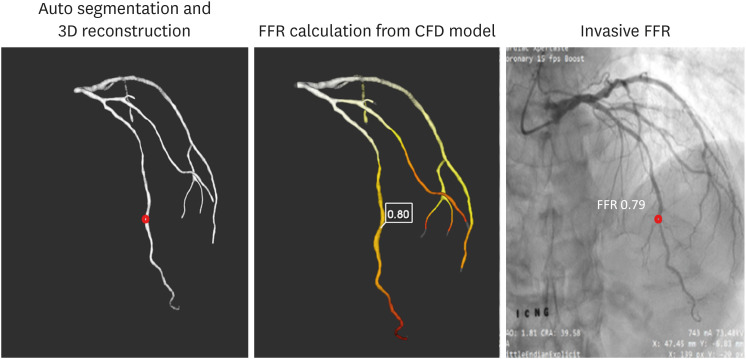
Figure 1 Case example of CT-FFR.The process of calculating CT-FFR is shown. Three-dimensional model of epicardial coronary artery tree was reconstructed by auto-segmentation from CCTA, and the value of CT-FFR was calculated from the CFD model at the selected point (0.80), which was comparable with the invasive FFR value of 0.82.3D = three-dimensional; CCTA = coronary computed tomography angiography; CFD = computational fluid dynamic; CT-FFR = coronary computed tomography angiography-derived fractional flow reserve; FFR = fractional flow reserve.
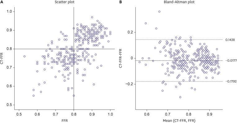
Figure 2 Correlation between CT-FFR and FFR.The correlation between CT-FFR and FFR is shown in (A) the scatter plot and (B) Bland-Altman plot.CT-FFR = coronary computed tomography angiography-derived fractional flow reserve; FFR = fractional flow reserve.
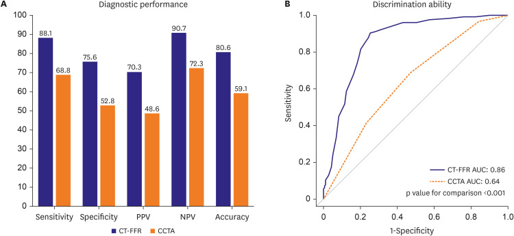
Figure 3 Diagnostic performances and discrimination abilities of CT-FFR and CCTA.(A) Diagnostic performance and (B) receiver operating characteristic curves comparing CT-FFR and CCTA to predict hemodynamically significant CAD are shown.AUC = area under the curve; CAD = coronary artery disease; CCTA = coronary computed tomography angiography; CT-FFR = coronary computed tomography angiography-derived fractional flow reserve; NPV = negative predictive value; PPV = positive predictive value.
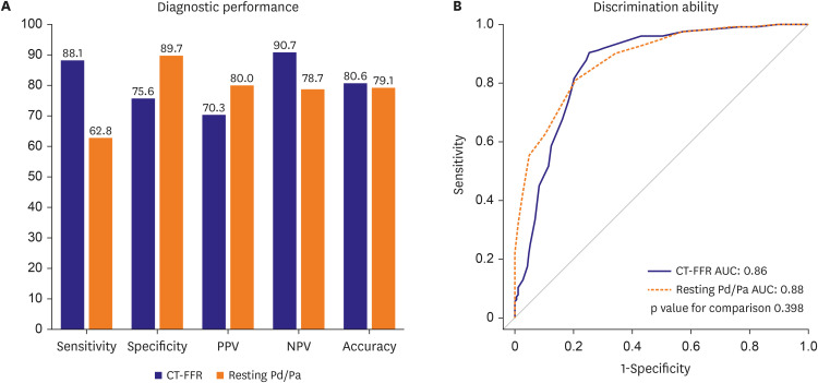
Figure 4 Diagnostic performances and discrimination abilities of CT-FFR and resting Pd/Pa.(A) Diagnostic performance and (B) receiver operating characteristic curves comparing CT-FFR and resting Pd/Pa to predict hemodynamically significant CAD are shown.AUC = area under the curve; CAD = coronary artery disease; CT-FFR = coronary computed tomography angiography-derived fractional flow reserve; NPV = negative predictive value; PPV = positive predictive value; Pd/Pa = distal to aortic coronary pressure.
Reference
-
1. Miller JM, Rochitte CE, Dewey M, et al. Diagnostic performance of coronary angiography by 64-row CT. N Engl J Med. 2008; 359:2324–2336. PMID: 19038879.
Article2. Budoff MJ, Dowe D, Jollis JG, et al. Diagnostic performance of 64-multidetector row coronary computed tomographic angiography for evaluation of coronary artery stenosis in individuals without known coronary artery disease: results from the prospective multicenter ACCURACY (Assessment by Coronary Computed Tomographic Angiography of Individuals Undergoing Invasive Coronary Angiography) trial. J Am Coll Cardiol. 2008; 52:1724–1732. PMID: 19007693.3. Knuuti J, Wijns W, Saraste A, et al. 2019 ESC Guidelines for the diagnosis and management of chronic coronary syndromes. Eur Heart J. 2020; 41:407–477. PMID: 31504439.4. Douglas PS, Hoffmann U, Patel MR, et al. Outcomes of anatomical versus functional testing for coronary artery disease. N Engl J Med. 2015; 372:1291–1300. PMID: 25773919.
Article5. Meijboom WB, Van Mieghem CA, van Pelt N, et al. Comprehensive assessment of coronary artery stenoses: computed tomography coronary angiography versus conventional coronary angiography and correlation with fractional flow reserve in patients with stable angina. J Am Coll Cardiol. 2008; 52:636–643. PMID: 18702967.6. Virani SS, Newby LK, Arnold SV, et al. 2023 AHA/ACC/ACCP/ASPC/NLA/PCNA Guideline for the Management of Patients With Chronic Coronary Disease: A Report of the American Heart Association/American College of Cardiology Joint Committee on Clinical Practice Guidelines. J Am Coll Cardiol. 2023; 82:833–955. PMID: 37480922.7. Koo BK, Erglis A, Doh JH, et al. Diagnosis of ischemia-causing coronary stenoses by noninvasive fractional flow reserve computed from coronary computed tomographic angiograms. Results from the prospective multicenter DISCOVER-FLOW (Diagnosis of Ischemia-Causing Stenoses Obtained Via Noninvasive Fractional Flow Reserve) study. J Am Coll Cardiol. 2011; 58:1989–1997. PMID: 22032711.8. Min JK, Leipsic J, Pencina MJ, et al. Diagnostic accuracy of fractional flow reserve from anatomic CT angiography. JAMA. 2012; 308:1237–1245. PMID: 22922562.
Article9. Nørgaard BL, Leipsic J, Gaur S, et al. Diagnostic performance of noninvasive fractional flow reserve derived from coronary computed tomography angiography in suspected coronary artery disease: the NXT trial (Analysis of Coronary Blood Flow Using CT Angiography: Next Steps). J Am Coll Cardiol. 2014; 63:1145–1155. PMID: 24486266.10. Douglas PS, De Bruyne B, Pontone G, et al. 1-year outcomes of FFRCT-guided care in patients with suspected coronary disease: The PLATFORM study. J Am Coll Cardiol. 2016; 68:435–445. PMID: 27470449.
Article11. Yang S, Lesina K, Doh JH, et al. Long-term prognostic implications of hemodynamic and plaque assessment using coronary CT angiography. Atherosclerosis. 2023; 373:58–65. PMID: 36872186.
Article12. Yang S, Choi G, Zhang J, et al. Association among local hemodynamic parameters derived from CT angiography and their comparable implications in development of acute coronary syndrome. Front Cardiovasc Med. 2021; 8:713835. PMID: 34589527.13. Yang S, Koo BK. Coronary physiology-based approaches for plaque vulnerability: implications for risk prediction and treatment strategies. Korean Circ J. 2023; 53:581–593. PMID: 37653694.
Article14. Lee JM, Doh JH, Nam CW, Shin ES, Koo BK. Functional approach for coronary artery disease: filling the gap between evidence and practice. Korean Circ J. 2018; 48:179–190. PMID: 29557104.
Article15. Coenen A, Lubbers MM, Kurata A, et al. Fractional flow reserve computed from noninvasive CT angiography data: diagnostic performance of an on-site clinician-operated computational fluid dynamics algorithm. Radiology. 2015; 274:674–683. PMID: 25322342.
Article16. Kruk M, Wardziak Ł, Demkow M, et al. Workstation-based calculation of CTA-based FFR for intermediate stenosis. JACC Cardiovasc Imaging. 2016; 9:690–699. PMID: 26897667.
Article17. Yang DH, Kim YH, Roh JH, et al. Diagnostic performance of on-site CT-derived fractional flow reserve versus CT perfusion. Eur Heart J Cardiovasc Imaging. 2017; 18:432–440. PMID: 27354345.18. Ko BS, Cameron JD, Munnur RK, et al. Noninvasive CT-derived FFR based on structural and fluid analysis: a comparison with invasive FFR for detection of functionally significant stenosis. JACC Cardiovasc Imaging. 2017; 10:663–673. PMID: 27771399.19. Coenen A, Kim YH, Kruk M, et al. Diagnostic accuracy of a machine-learning approach to coronary computed tomographic angiography-based fractional flow reserve: result from the MACHINE consortium. Circ Cardiovasc Imaging. 2018; 11:e007217. PMID: 29914866.
Article20. Tesche C, De Cecco CN, Baumann S, et al. Coronary CT angiography-derived fractional flow reserve: machine learning algorithm versus computational fluid dynamics modeling. Radiology. 2018; 288:64–72. PMID: 29634438.
Article21. Röther J, Moshage M, Dey D, et al. Comparison of invasively measured FFR with FFR derived from coronary CT angiography for detection of lesion-specific ischemia: Results from a PC-based prototype algorithm. J Cardiovasc Comput Tomogr. 2018; 12:101–107. PMID: 29409717.
Article22. Halliburton SS, Abbara S, Chen MY, et al. SCCT guidelines on radiation dose and dose-optimization strategies in cardiovascular CT. J Cardiovasc Comput Tomogr. 2011; 5:198–224. PMID: 21723512.23. Abbara S, Arbab-Zadeh A, Callister TQ, et al. SCCT guidelines for performance of coronary computed tomographic angiography: a report of the Society of Cardiovascular Computed Tomography Guidelines Committee. J Cardiovasc Comput Tomogr. 2009; 3:190–204. PMID: 19409872.
Article24. Leipsic J, Abbara S, Achenbach S, et al. SCCT guidelines for the interpretation and reporting of coronary CT angiography: a report of the Society of Cardiovascular Computed Tomography Guidelines Committee. J Cardiovasc Comput Tomogr. 2014; 8:342–358. PMID: 25301040.
Article25. Lee JM, Choi KH, Hwang D, et al. Prognostic implication of thermodilution coronary flow reserve in patients undergoing fractional flow reserve measurement. JACC Cardiovasc Interv. 2018; 11:1423–1433. PMID: 30093048.
Article26. Jang HJ, Koo BK, Lee HS, et al. Safety and efficacy of a novel hyperaemic agent, intracoronary nicorandil, for invasive physiological assessments in the cardiac catheterization laboratory. Eur Heart J. 2013; 34:2055–2062. PMID: 23396491.
Article27. Jeremias A, Maehara A, Généreux P, et al. Multicenter core laboratory comparison of the instantaneous wave-free ratio and resting Pd/Pa with fractional flow reserve: the RESOLVE study. J Am Coll Cardiol. 2014; 63:1253–1261. PMID: 24211503.28. Kwon SS, Chung EC, Park JS, et al. A novel patient-specific model to compute coronary fractional flow reserve. Prog Biophys Mol Biol. 2014; 116:48–55. PMID: 25256102.
Article
- Full Text Links
-
- Actions
-
Cited
- CITED
-
- Close
- Share
-
- Similar articles
-
- Evaluation of Myocardial Ischemia Using Coronary Computed Tomography Angiography in Patients with Stable Angina
- Funtional significance of the intermediate lesion in a single coronary artery assessed by fractional flow reserve
- Physiologic Assessment of Coronary Artery Disease: Focus on Fractional Flow Reserve
- Beyond Coronary CT Angiography: CT Fractional Flow Reserve and Perfusion
- Future Directions in Coronary CT Angiography: CT-Fractional Flow Reserve, Plaque Vulnerability, and Quantitative Plaque Assessment
