Prenatal diagnosis and postnatal outcome of fetal intracranial hemorrhage: a single-center experience
- Affiliations
-
- 1Department of Obstetrics and Gynecology, Institute of Women’s Medical Life Science, Yonsei University College of Medicine, Seoul, Korea
- KMID: 2557856
- DOI: http://doi.org/10.5468/ogs.24097
Abstract
Objective
To assess prenatal ultrasonographic findings and postnatal outcomes in fetuses with intracranial hemorrhage (ICH).
Methods
This retrospective study included fetuses prenatally diagnosed with ICH between December 2012 and August 2023. Maternal characteristics, prenatal ultrasonographic findings, and postnatal outcomes were reviewed.
Results
Twenty-seven fetuses with ICH were reviewed. Intracranial hemorrhage was classified as grade 3 and 4 in 24 fetuses. Twenty-two fetuses had ICH, four had ICH with subdural hemorrhage, and one had ICH with subarachnoid hemorrhage. Ventriculomegaly was the most common ultrasonographic finding, and was observed in 22 of the 27 (81.5%) fetuses. Seven fetuses were lost to follow-up, and four intrauterine fetal deaths occurred. The remaining 16 fetuses were delivered at a median gestational age of 35+2 weeks. The infants were followed-up for 40.1 months (range, 4-88). Nine of the 16 infants underwent ventriculoperitoneal placement. One infant underwent brain surgery for severe epilepsy. Motor impairment, including cerebral palsy, was observed in 13 infants (81.2%). Neurologic impairment occurred in six infants (37.5%), developmental delay in nine (56.2%), and epilepsy in 11 (68.7%).
Conclusion
Fetal ICH is a rare complication diagnosed during pregnancy, which results in subsequent fetal neurological sequelae or death. This study demonstrated that the common ultrasonographic findings in fetal ICH were progressive ventriculomegaly and increased periventricular echogenicity. Fetuses diagnosed with prenatal ICH, especially those affected by higher-grade ICH, may be at an increased risk of long-term neurodevelopmental problems.
Figure
-
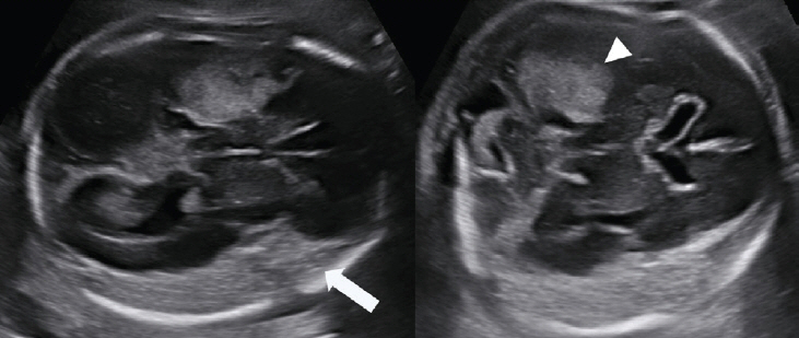
Fig. 1. Image showing subdural hemorrhage. Patient was referred to our hospital due to a history of previous intrauterine fetal death (IUFD). First visit at 26 weeks showed IUFD, ventriculomegaly, left subdural space hemorrhage (arrow), right temporoparietal area hemorrhage (arrowhead), fetal ascites, pleural effusion, scalp edema, and intracardiac hematoma. After hysterotomy, cord next generation sequencing and parental gene screening was performed, but significant result were not found.
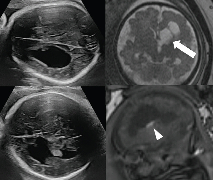
Fig. 2. Image demonstrating progressive ventriculomegaly. Ultrasound image (left) showing left ventriculomegaly with a hyperechogenic blood clot in the lateral ventricle. Magnetic resonance imaging (right) showing the same results, indicating ventriculomegaly (arrow) and a hyperechogenic blood clot (arrowhead).
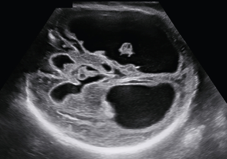
Fig. 3. Image showing increased periventricular echogenicity. Increased periventricular echogenicity accompanied by severe bilateral ventriculomegaly (both sides >30 mm).
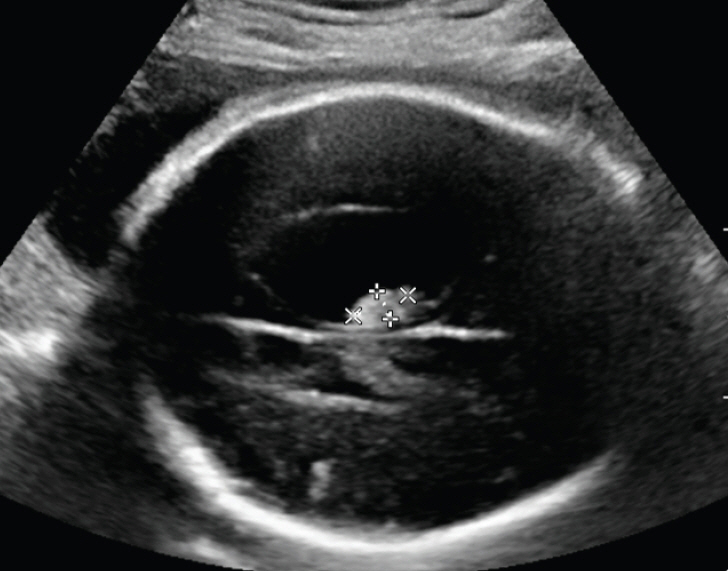
Fig. 4. Image demonstrating echogenic intraventricular clot. Ultrasound image showing an echogenic space-occupying lesion. A blood clot (1.2×0.6 cm) was noted in the ventricle with unilateral ventriculomegaly. Postnatal magnetic resonance imaging confirmed the presence of an old intraventricular hemorrhage.

Fig. 5. Image showing an irregular shaped choroid plexus. The patient showed an irregular choroid plexus (arrowhead) with severe bilateral ventriculomegaly and increased periventricular echogenicity.
Reference
-
References
1. Kutuk MS, Yikilmaz A, Ozgun MT, Dolanbay M, Canpolat M, Uludag S, et al. Prenatal diagnosis and postnatal outcome of fetal intracranial hemorrhage. Childs Nerv Syst. 2014; 30:411–8.
Article2. Vergani P, Strobelt N, Locatelli A, Paterlini G, Tagliabue P, Parravicini E, et al. Clinical significance of fetal intracranial hemorrhage. Am J Obstet Gynecol. 1996; 175:536–43.3. Putbrese B, Kennedy A. Findings and differential diagnosis of fetal intracranial haemorrhage and fetal ischaemic brain injury: what is the role of fetal MRI? Br J Radiol. 2017; 90:20160253.
Article4. Sherer DM, Anyaegbunam A, Onyeije C. Antepartum fetal intracranial hemorrhage, predisposing factors and prenatal sonography: a review. Am J Perinatol. 1998; 15:431–41.
Article5. Monteagudo A, Kuller JA, Craigo S, Fox NS, Norton ME, Post A, et al. SMFM fetal anomalies consult series #3: intracranial anomalies. Am J Obstet Gynecol. 2020; 223:B2–50.
Article6. Sanapo L, Whitehead MT, Bulas DI, Ahmadzia HK, Pesacreta L, Chang T, et al. Fetal intracranial hemorrhage: role of fetal MRI. Prenat Diagn. 2017; 37:827–36.
Article7. Monteagudo A. Intracranial hemorrhage. Am J Obstet Gynecol. 2020; 223:B34–7.8. Ghi T, Simonazzi G, Perolo A, Savelli L, Sandri F, Bernardi B, et al. Outcome of antenatally diagnosed intracranial hemorrhage: case series and review of the literature. Ultrasound Obstet Gynecol. 2003; 22:121–30.
Article9. Sileo FG, Zöllner J, D’Antonio F, Islam S, Papageorghiou AT, Khalil A. Perinatal and long-term outcome of fetal intracranial hemorrhage: systematic review and meta-analysis. Ultrasound Obstet Gynecol. 2022; 59:585–95.
Article10. Gedik Özköse Z, Oğlak SC, Bestel A, Behram M, Süzen Çaypınar S, Ölmez F, et al. Fetal intracranial hemorrhage: prenatal sonographic diagnosis criteria and postnatal outcomes. J Turk Ger Gynecol Assoc. 2022; 23:268–74.
Article11. Adiego B, Martínez-Ten P, Bermejo C, Estévez M, Recio Rodriguez M, Illescas T. Fetal intracranial hemorrhage. Prenatal diagnosis and postnatal outcomes. J Matern Fetal Neonatal Med. 2019; 32:21–30.
Article12. Ahn KH, Lee KS. Artificial intelligence in obstetrics. Obstet Gynecol Sci. 2022; 65:113–24.13. Malinger G, Paladini D, Haratz KK, Monteagudo A, Pilu GL, Timor-Tritsch IE. ISUOG practice guidelines (updated): sonographic examination of the fetal central nervous system. Part 1: performance of screening examination and indications for targeted neurosonography. Ultrasound Obstet Gynecol. 2020; 56:476–84.14. Abdelkader MA, Ramadan W, Gabr AA, Kamel A, Abdelrahman RW. Fetal intracranial hemorrhage: sonographic criteria and merits of prenatal diagnosis. J Matern Fetal Neonatal Med. 2017; 30:2250–6.
Article15. Papile LA, Burstein J, Burstein R, Koffler H. Incidence and evolution of subependymal and intraventricular hemorrhage: a study of infants with birth weights less than 1,500 gm. J Pediatr. 1978; 92:529–34.
Article16. Dunbar MJ, Woodward K, Leijser LM, Kirton A. Antenatal diagnosis of fetal intraventricular hemorrhage: systematic review and meta-analysis. Dev Med Child Neurol. 2021; 63:144–55.
Article17. Qi W, Luo JY, Li ZL, Zhang QJ, Liu ZD, Liao QP, et al. Clinical analysis of eight cases of fetal intracranial hemorrhage in pregnancy. J Matern Fetal Neonatal Med. 2021; 34:2609–15.18. Epstein KN, Kline-Fath BM, Zhang B, Venkatesan C, Habli M, Dowd D, et al. Prenatal evaluation of intracranial hemorrhage on fetal MRI: a retrospective review. AJNR Am J Neuroradiol. 2021; 42:2222–8.
Article19. Van Mieghem T, Van Schoubroeck D, Depiere M, Debeer A, Hanssens M. Fetal cerebral hemorrhage caused by vitamin K deficiency after complicated bariatric surgery. Obstet Gynecol. 2008; 112:434–6.
Article20. Minami H, Furuhashi M, Minami K, Miyazaki K, Yoshida K, Ishikawa K. Fetal intraventricular bleeding possibly due to maternal vitamin K deficiency. Fetal Diagn Ther. 2009; 24:357–60.
Article21. Shigemi D, Nakanishi K, Miyazaki M, Shibata Y, Suzuki S. A case of maternal vitamin K deficiency associated with hyperemesis gravidarum: its potential impact on fetal blood coagulability. J Nippon Med Sch. 2015; 82:54–8.
Article22. Coste T, Vincent-Delorme C, Stichelbout M, Devisme L, Gelot A, Deryabin I, et al. COL4A1/COL4A2 and inherited platelet disorder gene variants in fetuses showing intracranial hemorrhage. Prenat Diagn. 2022; 42:601–10.23. Kutuk MS, Balta B, Kodera H, Matsumoto N, Saitsu H, Doganay S, et al. Is there relation between COL4A1/A2 mutations and antenatally detected fetal intraventricular hemorrhage? Childs Nerv Syst. 2014; 30:419–24.
Article24. Zdravic D, Yougbare I, Vadasz B, Li C, Marshall AH, Chen P, et al. Fetal and neonatal alloimmune thrombocytopenia. Semin Fetal Neonatal Med. 2016; 21:19–27.
Article25. Gupta V, Schlatterer SD, Bulas DI, du Plessis AJ, Mulkey SB. Pregnancy and child outcomes following fetal intracranial hemorrhage. Pediatr Neurol. 2023; 140:68–75.
Article26. Hayashi M, Poretti A, Gorra M, Farzin A, Graham EM, Huisman TA, et al. Prenatal cerebellar hemorrhage: fetal and postnatal neuroimaging findings and postnatal outcome. Pediatr Neurol. 2015; 52:529–34.
Article27. ENSO Working Group. Role of prenatal magnetic resonance imaging in fetuses with isolated mild or moderate ventriculomegaly in the era of neurosonography: international multicenter study. Ultrasound Obstet Gynecol. 2020; 56:340–7.28. Parodi A, Govaert P, Horsch S, Bravo MC, Ramenghi LA. Cranial ultrasound findings in preterm germinal matrix haemorrhage, sequelae and outcome. Pediatr Res. 2020; 87:13–24.
Article29. Jocelyn LJ, Casiro OG. Neurodevelopmental outcome of term infants with intraventricular hemorrhage. Am J Dis Child. 1992; 146:194–7.
Article30. Winkelhorst D, Kamphuis MM, Steggerda SJ, Rijken M, Oepkes D, Lopriore E, et al. Perinatal outcome and long-term neurodevelopment after intracranial haemorrhage due to fetal and neonatal alloimmune thrombocytopenia. Fetal Diagn Ther. 2019; 45:184–91.
Article31. Eldad K, Ya’ara G, Simon L, Omer BY. The association between fetal intracranial hemorrhages detected on MRI and neurodevelopment. Eur J Radiol. 2024; 173:111380.
Article32. Elchalal U, Yagel S, Gomori JM, Porat S, Beni-Adani L, Yanai N, et al. Fetal intracranial hemorrhage (fetal stroke): does grade matter? Ultrasound Obstet Gynecol. 2005; 26:233–43.
- Full Text Links
-
- Actions
-
Cited
- CITED
-
- Close
- Share
-
- Similar articles
-
- A case of fetal intracranial hemorrhage diagnosed by antenatal ultrasonographic examination
- The Usefulness of Fetal MRI for Prenatal Diagnosis
- Fetal Ultrasonography for Prenatal Detection of Tuberous Sclerosis Associated with Cardiac Rhabdomyoma
- Prenatal diagnosis of fetal adrenal hemorrhage and endocrinologic evaluation
- Fetal urologic abnormalities detected by prenatal ultrasonography
