Fetal Ultrasonography for Prenatal Detection of Tuberous Sclerosis Associated with Cardiac Rhabdomyoma
- Affiliations
-
- 1Department of Obstetrics and Gynecology, School of Medicine, Catholic University of Daegu, Daegu, Korea. duchess7@hanmail.net
- 2Department of Pediatrics, School of Medicine, Catholic University of Daegu, Daegu, Korea.
- KMID: 2468546
- DOI: http://doi.org/10.14734/PN.2019.30.4.240
Abstract
- Cardiac rhabdomyoma is common cardiac mass found during the fetal period. Cardiac rhabdomyoma and tuberous sclerosis have significant associations. Tuberous sclerosis in newborns can cause disability in nearly all organs. Prenatal diagnosis of fetal tuberous sclerosis enables early evaluation and management of the affected infant. In Daegu Catholic University Hospital, a total of three cases of fetal intracranial tuberous sclerosis were diagnosed among five cases of fetal cardiac rhabdomyoma. The diagnosis in all three cases was confirmed by postnatal brain magnetic resonance imaging. Intracranial lesions appeared as multiple small, round and relatively hyperechoic masses on prenatal ultrasonography. Prenatal ultrasonography using transabdominal and transvaginal probes in various angles is helpful. As tuberous sclerosis is not observed in a single instance, regular follow-up examinations are necessary. Fetuses with cardiac rhabdomyoma require detailed prenatal evaluations for tuberous sclerosis, especially in the brain. For this, prenatal ultrasonography is a very useful technique.
MeSH Terms
Figure
-
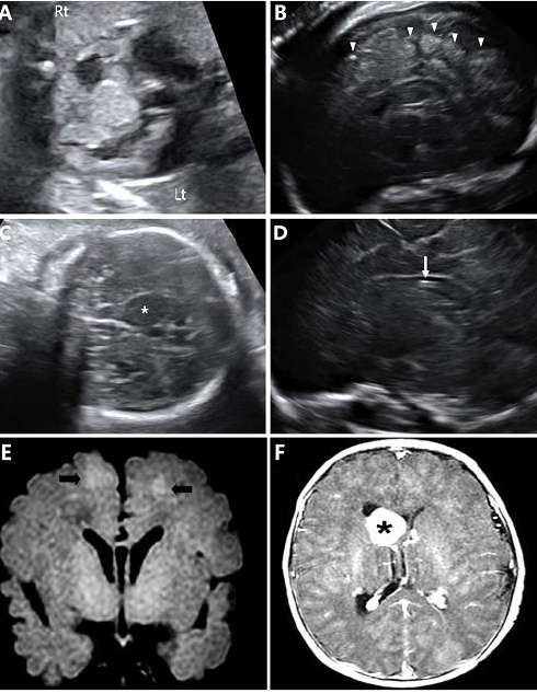
Fig. 1 (A) Fetal echocardiography showed multiple cardiac rhabdomyomas in the right ventricle, the left ventricle and the interventricular septum. (B) The para-sagittal view of the fetal brain showed multiple cortical tubers at the cerebral cortex as hyperechoic dots and round masses (arrowheads). (C) The coronal view of the fetal brain showed a single subependymal giant cell astrocytoma (SEGA) as a hypoechoic round mass adjacent to the foramen of Monro (asterisk). (D) The sagittal view of the fetal brain showed a subependymal nodule as an echogenic spot along the ependymal lining of the lateral ventricle (arrow). (E) Postnatal brain magnetic resonance imaging (MRI) of case no. 1. Hyperintensity of cortical and subcortical tubers in T1-weighted images (black arrows). (F) Postnatal brain MRI of case no. 4. SEGA within the lateral ventricle near the foramen of Monro (asterisk).
Reference
-
1. Yuan SM. Fetal primary cardiac tumors during perinatal period. Pediatr Neonatol. 2017; 58:205–210.
Article2. Dragoumi P, O'Callaghan F, Zafeiriou DI. Diagnosis of tuberous sclerosis complex in the fetus. Eur J Paediatr Neurol. 2018; 22:1027–1034.
Article3. Northrup H, Krueger DA. International Tuberous Sclerosis Complex Consensus Group. Tuberous sclerosis complex diagnostic criteria update: recommendations of the 2012 international tuberous sclerosis complex consensus conference. Pediatr Neurol. 2013; 49:243–254.4. Saxena A, Sampson JR. Epilepsy in tuberous sclerosis: phenotypes, mechanisms, and treatments. Semin Neurol. 2015; 35:269–276.
Article5. Kapoor A, Girard L, Lattouf JB, Pei Y, Rendon R, Card P, et al. Evolving strategies in the treatment of tuberous sclerosis complex-associated angiomyolipomas (TSC-AML). Urology. 2016; 89:19–26.
Article6. Wang CC, Wang CY, Lai YJ, Chang TY, Su HY. Prenatal diagnosis of tuberous sclerosis complex using fetal ultrasonography and magnetic resonance imaging and genetic testing. Taiwan J Obstet Gynecol. 2018; 57:163–165.
Article7. Ekmekci E, Ozkan BO, Yildiz MS, Kocakaya B. Prenatal diagnosis of fetal cardiac rhabdomyoma associated with tuberous sclerosis: a case report. Case Rep Womens Health. 2018; 19:e00070.
Article8. Tworetzky W, McElhinney DB, Margossian R, Moon-Grady AJ, Sallee D, Goldmuntz E, et al. Association between cardiac tumors and tuberous sclerosis in the fetus and neonate. Am J Cardiol. 2003; 92:487–489.
Article9. Saada J, Hadj Rabia S, Fermont L, Le Bidois J, Bernardes LS, Martinovic J, et al. Prenatal diagnosis of cardiac rhabdomyomas: incidence of associated cerebral lesions of tuberous sclerosis complex. Ultrasound Obstet Gynecol. 2009; 34:155–159.
Article
- Full Text Links
-
- Actions
-
Cited
- CITED
-
- Close
- Share
-
- Similar articles
-
- A Case of Fetal Hydrops combined with Intracardiac Rhabdomyoma and Tuberous Sclerosis
- A Case of Fetal Cardiac Tumor Diagnosed by Ultrasonography
- Two Cases of Fetal Cardiac Tumor Diagnosed by Ultrasonography
- Sirolimus therapy for fetal cardiac rhabdomyoma in a pregnant woman with tuberous sclerosis
- A Case of Infantile Spasms in Tuberous Sclerosis with Fetal Cardiac Tumors
