Catheter detection by transthoracic echocardiography during placement of peripherally inserted central catheters: a real-time method for eliminating misplacement
- Affiliations
-
- 1Department of Thoracic and Cardiovascular Surgery, Chungnam National University Hospital, Chungnam National University School of Medicine, Daejeon, Korea
- 2Division of Pulmonology, Department of Internal Medicine, Chungnam National University Hospital, Chungnam National University School of Medicine, Daejeon, Korea
- KMID: 2557243
- DOI: http://doi.org/10.4266/acc.2024.00150
Abstract
- Background
Although guidelines and protocols are available for central venous access, existing methods lack specificity and sensitivity, especially when placing peripherally inserted central catheters (PICCs). We evaluated the feasibility of catheter detection in the right atrial cavity using transthoracic echocardiography (TTE) during PICC placement. Methods: This single-center, retrospective study included consecutive patients who underwent PICC placement between January 2022 and March 2023. TTE was performed to detect the arrival of the catheter in the right atrial cavity. Catheter misplacement was defined as an aberrant catheter position on chest x-ray (CXR). The primary endpoint was predicting catheter misplacement based on catheter detection in the right atrial cavity. The secondary endpoint was optimizing catheter placement and examining catheter-associated complications. Results: Of the 110 patients identified, 10 were excluded because of poor echogenicity and vein access failure. The remaining 100 patients underwent PICC placement with TTE. The catheter was visualized in the right atrial cavity in 90 patients. CXR exams revealed catheter misplacement in seven cases. Eight patients with catheter misplacement underwent the same procedure in the other arm. In two patients, PICC placement failed due to anatomical reasons. Catheter misplacement was detected using TTE with sensitivity, specificity, positive predictive value, and negative predictive value of 97% confidence interval (CI; 91.31%–99.36%), 90% CI (55.50%–99.75%), 99%, and 75%, respectively. Conclusions: TTE is a reliable tool for detecting catheter misplacement and optimizing catheter tip positioning during PICC placement.
Figure
-
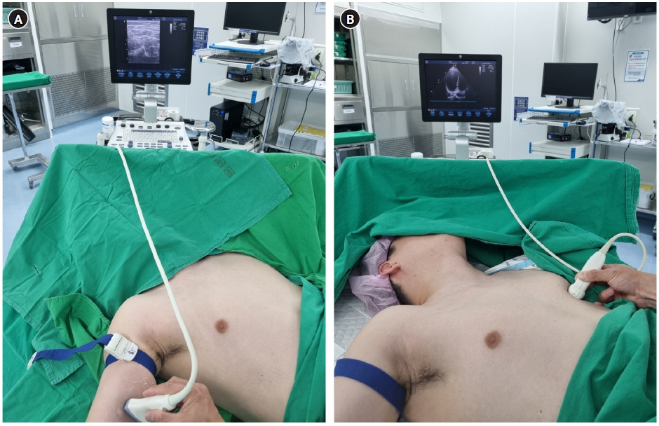
Figure 1. Before sterile draping: (A) rapid assessment of the peripheral vein by linear probe (12L) and (B) the best point for echocardiography in the apical four-chamber or subcostal view clearly shows the right atrial cavity by the echocardiographic probe (3Sc-RS) (GE Vivid S5 Ultrasound System).
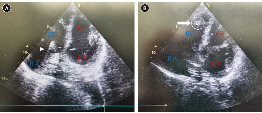
Figure 2. Transthoracic echocardiography during peripherally inserted central catheter, apical four-chamber view: (A) hyperechogenic line (arrowhead represents the catheter heading from the RA to the RV) and (B) hyperechogenic artifact (arrow represents the catheter tip at the RV). LV: left ventricle; RV: right ventricle; RA: right atrium; LA: left atrium.
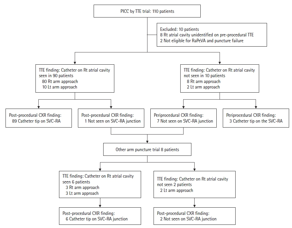
Figure 3. Study protocol used to analyze the diagnostic test. PICC: peripherally inserted central catheter; TTE: transthoracic echocardiography; RaPeVa: rapid peripheral vein assessment; Rt: right; Lt: left; CXR: chest x-ray; SVC–RA: superior vena cava–right atrium.
Reference
-
1. Montanarella MJ, Agarwal A, Moon B. Peripherally inserted central catheter (PICC) line placement. StatPearls Publishing;2024. [cited 2024 Mar 1]. Available from: https://pubmed.ncbi.nlm.nih.gov/34424637/.2. Royce B. Use of C-arm fluoroscopy by nurses for placement of PICC lines. J Assoc Vasc Access. 2009; 14:138–41.
Article3. Storm ES, Miller DL, Hoover LJ, Georgia JD, Bivens T. Radiation doses from venous access procedures. Radiology. 2006; 238:1044–50.
Article4. Andreucci M, Solomon R, Tasanarong A. Side effects of radiographic contrast media: pathogenesis, risk factors, and prevention. Biomed Res Int. 2014; 2014:741018.
Article5. Bedel J, Vallée F, Mari A, Riu B, Planquette B, Geeraerts T, et al. Guidewire localization by transthoracic echocardiography during central venous catheter insertion: a periprocedural method to evaluate catheter placement. Intensive Care Med. 2013; 39:1932–7.
Article6. Díaz-Gómez JL, Mayo PH, Koenig SJ. Point-of-care ultrasonography. N Engl J Med. 2021; 385:1593–602.
Article7. Santos FK, Flumignan RL, Areias LL, Sarpe AK, Amaral FC, Ávila RB, et al. Peripherally inserted central catheter versus central venous catheter for intravenous access: a protocol for systematic review and meta-analysis. Medicine (Baltimore). 2020; 99:e20352.8. Johansson E, Hammarskjöld F, Lundberg D, Arnlind MH. Advantages and disadvantages of peripherally inserted central venous catheters (PICC) compared to other central venous lines: a systematic review of the literature. Acta Oncol. 2013; 52:886–92.
Article9. Greca A, Iacobone E, Elisei D, Biasucci DG, D’Andrea V, Barone G, et al. ECHOTIP: a structured protocol for ultrasound-based tip navigation and tip location during placement of central venous access devices in adult patients. J Vasc Access. 2023; 24:535–44.
Article10. Brescia F, Pittiruti M, Spencer TR, Dawson RB. The SIP protocol update: Eight strategies, incorporating Rapid Peripheral Vein Assessment (RaPeVA), to minimize complications associated with peripherally inserted central catheter insertion. J Vasc Access. 2024; 25:5–13.
Article11. Emoli A, Cappuccio S, Marche B, Musarò A, Scoppettuolo G, Pittiruti M, et al. The ISP (Safe Insertion of PICCs) protocol: a bundle of 8 recommendations to minimize the complications related to the peripherally inserted central venous catheters (PICC). Assist Inferm Ric. 2014; 33:82–9.12. Tomaszewski KJ, Ferko N, Hollmann SS, Eng SC, Richard HM, Rowe L, et al. Time and resources of peripherally inserted central catheter insertion procedures: a comparison between blind insertion/chest X-ray and a real time tip navigation and confirmation system. Clinicoecon Outcomes Res. 2017; 9:115–25.
Article13. Gullo G, Colin A, Frossard P, Jouannic AM, Knebel JF, Qanadli SD. Appropriateness of replacing fluoroscopic guidance with ECG-electromagnetic guidance for PICC insertion: a randomized controlled trial. AJR Am J Roentgenol. 2021; 216:981–8.
Article14. Kwon S, Son SM, Lee SH, Kim JH, Kim H, Kim JY, et al. Outcomes of bedside peripherally inserted central catheter placement: a retrospective study at a single institution. Acute Crit Care. 2020; 35:31–7.
Article15. Ploton G, Pistorius MA, Raimbeau A, Denis Le Seve J, Bergère G, Ngohou C, et al. A STROBE cohort study of 755 deep and superficial upper-extremity vein thrombosis. Medicine (Baltimore). 2020; 99:e18996.
Article16. Moran J, Colbert CY, Song J, Mathews J, Arroliga AC, Varghees S, et al. Screening for novel risk factors related to peripherally inserted central catheter-associated complications. J Hosp Med. 2014; 9:481–9.
Article17. Sharifkazemi M, Rezaian G, Hosseininejad E, Arjangzadeh A. Three simple but interesting transthoracic echocardiographic road maps for proximal superior vena cava visualisation in healthy young adults. Int J Cardiol Heart Vasc. 2022; 39:101004.
Article
- Full Text Links
-
- Actions
-
Cited
- CITED
-
- Close
- Share
-
- Similar articles
-
- Misplacement of Central Venous Catheter Tip
- Insertion and Management of Central Venous Catheters
- Removal of a Peripherally Inserted Central Catheter Remnant using Cardiac Catheterization in Preterm Infant
- Safety and Efficacy of Peripherally Inserted Central Catheter Placement by Surgical Intensivist–Led Vascular Access Team
- Early-onset Pericardial Effusion after Peripherally Inserted Central Venous Catheterization in a Preterm Infant
