Acute Crit Care.
2024 May;39(2):214-225. 10.4266/acc.2023.01571.
Microbial infections in burn patients
- Affiliations
-
- 1Postgraduate and Research Department of Biotechnology, St. Xavier’s College (Autonomous), West Bengal, India
- 2Department of Microbiology, Sarsuna College (under Calcutta University), West Bengal, India
- KMID: 2557237
- DOI: http://doi.org/10.4266/acc.2023.01571
Abstract
- Polymicrobial infections are the leading causes of complications incurred from injuries that burn patients develop. Such patients admitted to the hospital have a high risk of developing hospital-acquired infections, with longer patient stays leading to increased chances of acquiring such drug-resistant infections. Acinetobacter baumannii, Klebsiella pneumoniae, Pseudomonas aeruginosa, and Proteus mirabilis are the most common multidrug-resistant (MDR) Gram-negative bacteria identified in burn wound infections (BWIs). BWIs caused by viruses, like Herpes Simplex and Varicella Zoster, and fungi-like Candida spp. appear to occur occasionally. However, the preponderance of infection by opportunistic pathogens is very high in burn patients. Variations in the causative agents of BWIs are due to differences in geographic location and infection control measures. Overall, burn injuries are characterized by elevated serum cytokine levels, systemic immune response, and immunosuppression. Hence, early detection and treatment can accelerate the wound-healing process and reduce the risk of further infections at the site of injury. A multidisciplinary collaboration between burn surgeons and infectious disease specialists is also needed to properly monitor antibiotic resistance in BWI pathogens, help check the super-spread of MDR pathogens, and improve treatment outcomes as a result.
Figure
-
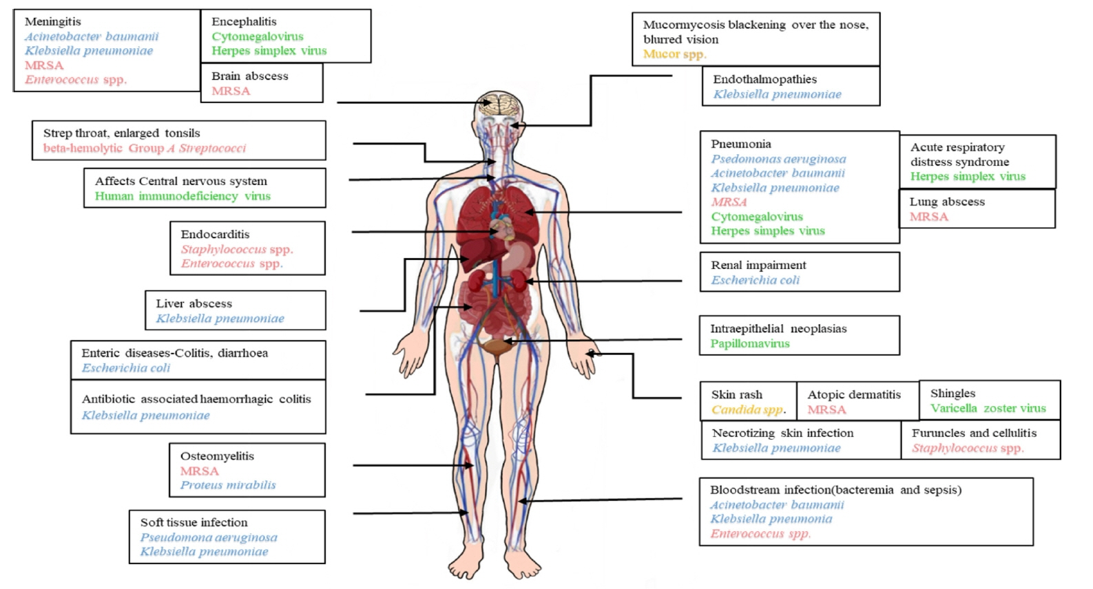
Figure 1. Burn wound infection microbes and their effect on a burn patient. MRSA: methicillin-resistant Staphylococcus aureus; spp.: species.
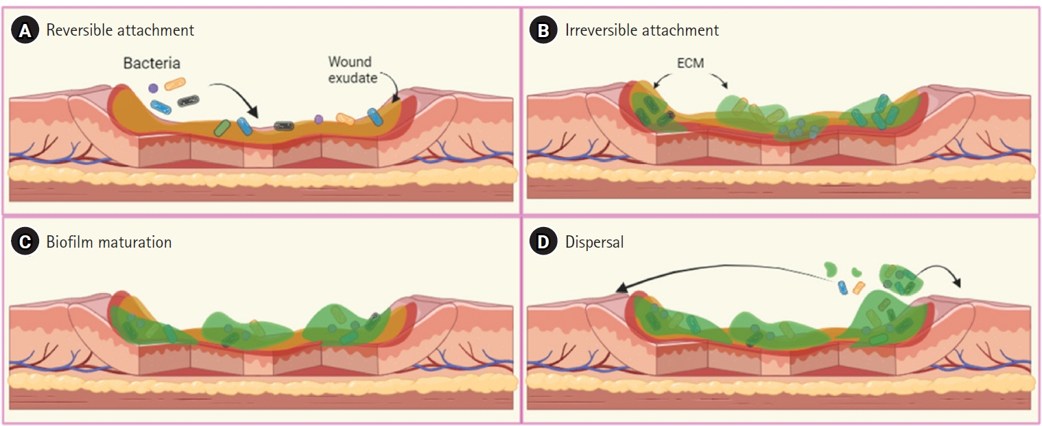
Figure 2. (A) Burn wounds typically contain burn wound exudates, which facilitate the initial inoculation and reversible attachment by bacterial pathogens. (B) Bacteria begin to produce extracellular matrix (ECM) and form micro-colonies during the process of irreversible attachment. (C) During the maturation stage, the biofilm grows in size and structural complexity. (D) The mature biofilm enters the dispersal stage, releasing bacterial cells from the ECM, which can then colonize new sites within the wound. Adapted from Maslova et al. NPJ Biofilms Microbiomes 2021;7:73 [3].
Reference
-
1. Mohapatra S, Gupta A, Agrawal K. Bacteriological profiles in burn patients within first twenty-four hours of injury. Int J Med Microbiol Trop Dis. 2016; 2:71–4.
Article2. Chen YY, Wu PF, Chen CS, Chen IH, Huang WT, Wang FD. Trends in microbial profile of burn patients following an event of dust explosion at a tertiary medical center. BMC Infect Dis. 2020; 20:193.
Article3. Maslova E, Eisaiankhongi L, Sjöberg F, McCarthy RR. Burns and biofilms: priority pathogens and in vivo models. NPJ Biofilms Microbiomes. 2021; 7:73.
Article4. López-Jácome LE, Chávez-Heres T, Becerra-Lobato N, García-Hernández ML, Vanegas-Rodríguez ES, Colin-Castro CA, et al. Microbiology and infection profile of electric burned patients in a referral burn hospital in Mexico City. J Burn Care Res. 2020; 41:390–7.
Article5. Forjuoh SN. Burns in low- and middle-income countries: a review of available literature on descriptive epidemiology, risk factors, treatment, and prevention. Burns. 2006; 32:529–37.
Article6. Peck MD. Epidemiology of burns throughout the world. Part I: distribution and risk factors. Burns. 2011; 37:1087–100.
Article7. Ho WS, Ying SY. An epidemiological study of 1063 hospitalized burn patients in a tertiary burns centre in Hong Kong. Burns. 2001; 27:119–23.
Article8. Opriessnig E, Luze H, Smolle C, Draschl A, Zrim R, Giretzlehner M, et al. Epidemiology of burn injury and the ideal dressing in global burn care: regional differences explored. Burns. 2023; 49:1–14.
Article9. Peck MD. Epidemiology of burns throughout the World. Part II: intentional burns in adults. Burns. 2012; 38:630–7.
Article10. Kara YA. Hot topics in burn injuries. Chapter 2: burn etiology and pathogenesis. Intechope;2018.11. Elsous A, Ouda M, Mohsen S, Al-Shaikh M, Mokayad S, Abo-Shaban N, et al. Epidemiology and outcomes of hospitalized burn patients in Gaza strip: a descriptive study. Ethiop J Health Sci. 2016; 26:9–16.
Article12. Bhardwaj S, Bhatia S, Singh S, Franco F Jr. Growing emergence of drug-resistant Pseudomonas aeruginosa and attenuation of its virulence using quorum sensing inhibitors: a critical review. Iran J Basic Med Sci. 2021; 24:699–719.13. Jeschke MG, van Baar ME, Choudhry MA, Chung KK, Gibran NS, Logsetty S. Burn injury. Nat Rev Dis Primers. 2020; 6:11.
Article14. Norbury W, Herndon DN, Tanksley J, Jeschke MG, Finnerty CC. Infection in burns. Surg Infect (Larchmt). 2016; 17:250–5.
Article15. El Hamzaoui N, Barguigua A, Larouz S, Maouloua M. Epidemiology of burn wound bacterial infections at a Meknes hospital, Morocco. New Microbes New Infect. 2020; 38:100764.
Article16. Thabet L, Messadi Aa, Mbarek M, Turki A, Meddeb B, Ben Redjeb S. Surveillance of multidrug resistant bacteria in a Tunisian hospital. Tunis Med. 2008; 86:992–5.17. Thabet L, Turki A, Ben Redjeb S, Messadi A. Bacteriological profile and antibiotic resistance of bacteria isolates in a burn department. Tunis Med. 2008; 86:1051–4.
Article18. von Baum H, Ober JF, Wendt C, Wenzel RP, Edmond MB. Antibiotic-resistant bloodstream infections in hospitalized patients: specific risk factors in a high-risk population? Infection. 2005; 33:320–6.
Article19. Wilson GR, French GW, Sully L. Loss of split thickness skin grafts due to non-group A beta-haemolytic streptococci. Ann R Coll Surg Engl. 1988; 70:217–9.20. Williams FN, Herndon DN, Hawkins HK, Lee JO, Cox RA, Kulp GA, et al. The leading causes of death after burn injury in a single pediatric burn center. Crit Care. 2009; 13:R183.
Article21. McManus AT, Mason AD, McManus WF, Pruitt BA. Twenty-five year review of Pseudomonas aeruginosa bacteremia in a burn center. Eur J Clin Microbiol. 1985; 4:219–23.22. Walton MA, Villarreal C, Herndon DN, Heggers JP. The use of aztreonam as an alternate therapy for multi-resistant Pseudomonas aeruginosa. Burns. 1997; 23:225–7.
Article23. Corbella X, Montero A, Pujol M, Domínguez MA, Ayats J, Argerich MJ, et al. Emergence and rapid spread of carbapenem resistance during a large and sustained hospital outbreak of multiresistant Acinetobacter baumannii. J Clin Microbiol. 2000; 38:4086–95.
Article24. Thomas RE, Thomas BC. Reducing biofilm infections in burn patients' wounds and biofilms on surfaces in hospitals, medical facilities and medical equipment to improve burn care: a systematic review. Int J Environ Res Public Health. 2021; 18:13195.
Article25. Khan BA, Yeh AJ, Cheung GY, Otto M. Investigational therapies targeting quorum-sensing for the treatment of Staphylococcus aureus infections. Expert Opin Investig Drugs. 2015; 24:689–704.
Article26. Ramakrishnan M, Putli Bai S, Babu M. Study on biofilm formation in burn wound infection in a pediatric hospital in Chennai, India. Ann Burns Fire Disasters. 2016; 29:276–80.27. Avni T, Levcovich A, Ad-El DD, Leibovici L, Paul M. Prophylactic antibiotics for burns patients: systematic review and meta-analysis. BMJ. 2010; 340:c241.
Article28. Falagas ME, Koletsi PK, Bliziotis IA. The diversity of definitions of multidrug-resistant (MDR) and pandrug-resistant (PDR) Acinetobacter baumannii and Pseudomonas aeruginosa. J Med Microbiol. 2006; 55(Pt 12):1619–29.
Article29. Gupta M, Naik AK, Singh SK. Bacteriological profile and antimicrobial resistance patterns of burn wound infections in a tertiary care hospital. Heliyon. 2019; 5:e02956.
Article30. BioMérieux. Burn patients often have higher rates of multidrug-resistant infections—but there are ways to help [Internet]. BioMérieux;2023. [cited 2024 May 1]. Available from: https://www.biomerieux.com/nl/en/blog/antimicrobial-resistance-stewardship/Burn-Patients-Multidrug-Resistant-Infections.html.31. Lachiewicz AM, Hauck CG, Weber DJ, Cairns BA, van Duin D. Bacterial infections after burn injuries: impact of multidrug resistance. Clin Infect Dis. 2017; 65:2130–6.
Article32. Wanis M, Walker SA, Daneman N, Elligsen M, Palmay L, Simor A, et al. Impact of hospital length of stay on the distribution of Gram negative bacteria and likelihood of isolating a resistant organism in a Canadian burn center. Burns. 2016; 42:104–11.
Article33. Sheridan R, Weber J, Chang P. Multi-drug resistant Gram-negative bacteria colonization and infection in burned children: lessons learned from a 20-year experience. Burns Open. 2018; 2:43–6.
Article34. Rosanova MT, Stamboulian D, Lede R. Risk factors for mortality in burn children. Braz J Infect Dis. 2014; 18:144–9.
Article35. Taneja N, Emmanuel R, Chari PS, Sharma M. A prospective study of hospital-acquired infections in burn patients at a tertiary care referral centre in North India. Burns. 2004; 30:665–9.
Article36. Kabanangi F, Joachim A, Nkuwi EJ, Manyahi J, Moyo S, Majigo M. High level of multidrug-resistant gram-negative pathogens causing burn wound infections in hospitalized children in Dar es Salaam, Tanzania. Int J Microbiol. 2021; 2021:6644185.
Article37. Magiorakos AP, Srinivasan A, Carey RB, Carmeli Y, Falagas ME, Giske CG, et al. Multidrug-resistant, extensively drug-resistant and pandrug-resistant bacteria: an international expert proposal for interim standard definitions for acquired resistance. Clin Microbiol Infect. 2012; 18:268–81.
Article38. Gupta N, Haque A, Lattif AA, Narayan RP, Mukhopadhyay G, Prasad R. Epidemiology and molecular typing of Candida isolates from burn patients. Mycopathologia. 2004; 158:397–405.
Article39. Horvath EE, Murray CK, Vaughan GM, Chung KK, Hospenthal DR, Wade CE, et al. Fungal wound infection (not colonization) is independently associated with mortality in burn patients. Ann Surg. 2007; 245:978–85.
Article40. Mousa HA. Fungal infection of burn wounds in patients with open and occlusive treatment methods. East Mediterr Health J. 1999; 5:333–6.
Article41. Ahmad S, Khan Z, Mustafa AS, Khan ZU. Epidemiology of Candida colonization in an intensive care unit of a teaching hospital in Kuwait. Med Mycol. 2003; 41:487–93.
Article42. Murray CK, Loo FL, Hospenthal DR, Cancio LC, Jones JA, Kim SH, et al. Incidence of systemic fungal infection and related mortality following severe burns. Burns. 2008; 34:1108–12.
Article43. Becker WK, Cioffi WG, McManus AT, Kim SH, McManus WF, Mason AD, et al. Fungal burn wound infection: a 10-year experience. Arch Surg. 1991; 126:44–8.44. Ballard J, Edelman L, Saffle J, Sheridan R, Kagan R, Bracco D, et al. Positive fungal cultures in burn patients: a multicenter review. J Burn Care Res. 2008; 29:213–21.
Article45. Greenhalgh DG, Saffle JR, Holmes JH, Gamelli RL, Palmieri TL, Horton JW, et al. American Burn Association consensus conference to define sepsis and infection in burns. J Burn Care Res. 2007; 28:776–90.
Article46. Mathew BP, Nath M. Recent approaches to antifungal therapy for invasive mycoses. ChemMedChem. 2009; 4:310–23.
Article47. Dries DJ. Management of burn injuries: recent developments in resuscitation, infection control and outcomes research. Scand J Trauma Resusc Emerg Med. 2009; 17:14.
Article48. Struck MF. Infection control in burn patients: are fungal infections underestimated? Scand J Trauma Resusc Emerg Med. 2009; 17:51–6.
Article49. de Macedo JL, Santos JB. Bacterial and fungal colonization of burn wounds. Mem Inst Oswaldo Cruz. 2005; 100:535–9.
Article50. Church D, Elsayed S, Reid O, Winston B, Lindsay R. Burn wound infections. Clin Microbiol Rev. 2006; 19:403–34.
Article51. Capoor MR, Sarabahi S, Tiwari VK, Narayanan RP. Fungal infections in burns: diagnosis and management. Indian J Plast Surg. 2010; 43(Suppl):S37–42.
Article52. National Committee for Clinical Laboratory Standards. Reference method for broth dilution antifungal susceptibility testing of yeasts; approved standard. Second edition. NCCLS document M27-A2 [ISBN 1-56238-469-4] [Internet]. National Committee for Clinical Laboratory Standards;2002. [cited 2024 May 1]. Available from: https://webstore.ansi.org/preview-pages/CLSI/preview_M27-A2.pdf.53. Mousa HA. Aerobic, anaerobic and fungal burn wound infections. J Hosp Infect. 1997; 37:317–23.
Article54. Baj J, Korona-Głowniak I, Buszewicz G, Forma A, Sitarz M, Teresiński G. Viral infections in burn patients: a state-of-the-art review. Viruses. 2020; 12:1315.
Article55. Xu H, Su C, Pearson A, Mody CH, Zheng C. Herpes simplex virus 1 UL24 abrogates the DNA sensing signal pathway by inhibiting NF-κB activation. J Virol. 2017; 91:e00025–17.
Article56. Wood JJ, O'Mahony JB, Rodrick ML, Eaton R, Demling RH, Mannick JA. Abnormalities of antibody production after thermal injury: an association with reduced interleukin 2 production. Arch Surg. 1986; 121:108–15.
Article57. Haik J, Weissman O, Stavrou D, Ben-noon HI, Liran A, Tessone A, et al. Is prophylactic acyclovir treatment warranted for prevention of herpes simplex virus infections in facial burns? A review of the literature. J Burn Care Res. 2011; 32:358–62.
Article58. Rennekampff HO, Hamprecht K. Cytomegalovirus infection in burns: a review. J Med Microbiol. 2006; 55:483–7.
Article59. Schwacha MG. Macrophages and post-burn immune dysfunction. Burns. 2003; 29:1–14.
Article60. Bordes J, Maslin J, Prunet B, d'Aranda E, Lacroix G, Goutorbe P, et al. Cytomegalovirus infection in severe burn patients monitoring by real-time polymerase chain reaction: a prospective study. Burns. 2011; 37:434–9.
Article61. Wurzer P, Guillory A, Parvizi D, Clayton RP, Branski LK, Kamolz LP, et al. Human herpes viruses in burn patients: a systematic review. Burns. 2017; 43:25–33.
Article62. Sheridan RL, Weber JM, Pasternak MM, Mulligan JM, Tompkins RG. A 15-year experience with varicella infections in a pediatric burn unit. Burns. 1999; 25:353–6.
Article63. Midilli K, Erkiliç A, Kuşkucu M, Analay H, Erkiliç S, Benzonana N, et al. Nosocomial outbreak of disseminated orf infection in a burn unit, Gaziantep, Turkey, October to December 2012. Euro Surveill. 2013; 18:20425.
Article64. Bergqvist C, Kurban M, Abbas O. Orf virus infection. Rev Med Virol. 2017; 27.
Article65. Al-Qattan MM. Orf infection of the hand. J Hand Surg Am. 2011; 36:1855–8.
Article66. Edge JM, Van der Merwe AE, Pieper CH, Bouic P. Clinical outcome of HIV positive patients with moderate to severe burns. Burns. 2001; 27:111–4.
Article67. Allgöwer M, Schoenenberger GA, Sparkes BG. Burning the largest immune organ. Burns. 1995; 21 Suppl 1:S7–47.
Article68. Camilleri IG, Milner RH. Human papilloma virus proliferation in a healing burn. Burns. 1996; 22:162–3.
Article69. Espinoza LF, Friedstat J, Faoro N, Chang PH, McMullen KA, Simko LC, et al. Geographic variation in outcomes after burn injury: a burn model system national database study. Ann Plast Surg. 2020; 84:644–50.70. Gupta VK, Paul S, Dutta C. Geography, ethnicity or subsistence-specific variations in human microbiome composition and diversity. Front Microbiol. 2017; 8:1162.
Article71. Ozumba UC, Jiburum BC. Bacteriology of burn wounds in Enugu, Nigeria. Burns. 2000; 26:178–80.
Article72. Laura P, José A, Nikki A, Khaled A, Barret J, Jeffery C, et al. Impact of COVID-19 on global burn care. Burns. 2022; 48:1301–10.
Article73. Sierawska O, Małkowska P, Taskin C, Hrynkiewicz R, Mertowska P, Grywalska E, et al. Innate immune system response to burn damage-focus on cytokine alteration. Int J Mol Sci. 2022; 23:716.
Article74. Vinish M, Cui W, Stafford E, Bae L, Hawkins H, Cox R, et al. Dendritic cells modulate burn wound healing by enhancing early proliferation. Wound Repair Regen. 2016; 24:6–13.
Article75. Rani M, Schwacha MG. Aging and the pathogenic response to burn. Aging Dis. 2012; 3:171–80.76. Grogan JB. Altered neutrophil phagocytic function in burn patients. J Trauma. 1976; 16:734–8.
Article77. Rich RR. Clinical immunology principles and practices. 2001. Mosby.78. Altman LC, Furukawa CT, Klebanoff SJ. Depressed mononuclear leukocyte chemotaxis in thermally injured patients. J Immunol. 1977; 119:199–205.
Article79. Moins-Teisserenc H, Cordeiro DJ, Audigier V, Ressaire Q, Benyamina M, Lambert J, et al. Severe altered immune status after burn injury is associated with bacterial infection and septic shock. Front Immunol. 2021; 12:586195.
Article80. Coban YK. Infection control in severely burned patients. World J Crit Care Med. 2012; 1:94–101.
Article81. Boyce JM, White RL, Causey WA, Lockwood WR. Burn units as a source of methicillin-resistant Staphylococcus aureus infections. JAMA. 1983; 249:2803–7.
Article82. Wolvos T. Wound instillation: the next step in negative pressure wound therapy. Lessons learned from initial experiences. Ostomy Wound Manage. 2004; 50:56–66.83. Wilson JR, Mills JG, Prather ID, Dimitrijevich SD. A toxicity index of skin and wound cleansers used on in vitro fibroblasts and keratinocytes. Adv Skin Wound Care. 2005; 18:373–8.
Article84. Coban YK, Erkiliç A, Analay H. Our 18-month experience at a new burn center in Gaziantep, Turkey. Ulus Travma Acil Cerrahi Derg. 2010; 16:353–6.
- Full Text Links
-
- Actions
-
Cited
- CITED
-
- Close
- Share
-
- Similar articles
-
- Herpes Zoster Manifestation in the Treatment of a Facial Scald Burn: A Case Report
- Roles of the Burn Clinical Nurse Specialist (BCNS) in Burn Center
- The Role of Medical Social Worker for Burn Patients and Family
- Central Venous Catheter-related Infection in Major Burn Patients: Comparison of Subclavian Vein and Femoral Vein
- Antimicrobial Central Venous Catheter Dose Not Reduce Catheter-related Bacteremia in Major Burn Injured Patients
