Korean J Physiol Pharmacol.
2024 Jul;28(4):335-344. 10.4196/kjpp.2024.28.4.335.
The NADPH oxidase inhibitor diphenyleneiodonium suppresses Ca2+ signaling and contraction in rat cardiac myocytes
- Affiliations
-
- 1College of Pharmacy, Chungnam National University, Daejeon 34134, Korea
- 2Nexel Co. Ltd., Seoul 07802, Korea
- KMID: 2557206
- DOI: http://doi.org/10.4196/kjpp.2024.28.4.335
Abstract
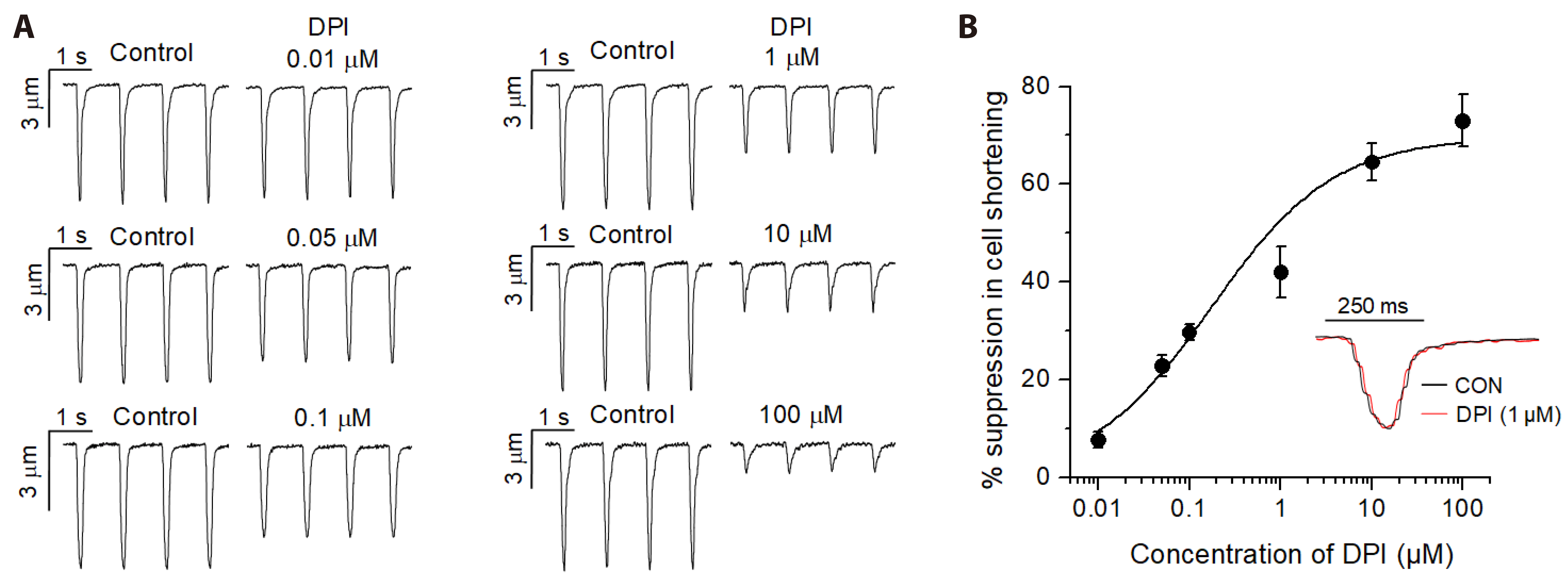
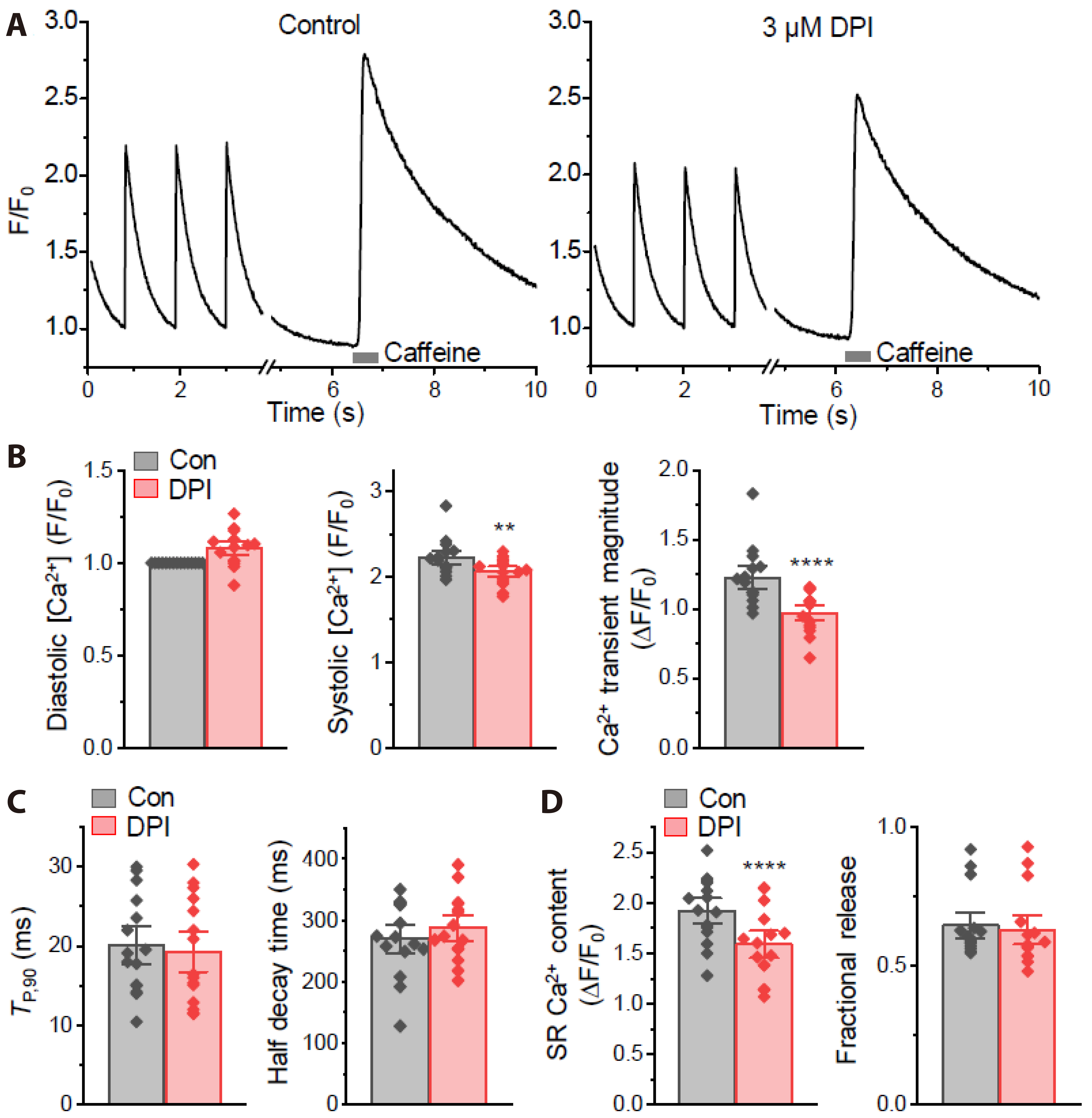
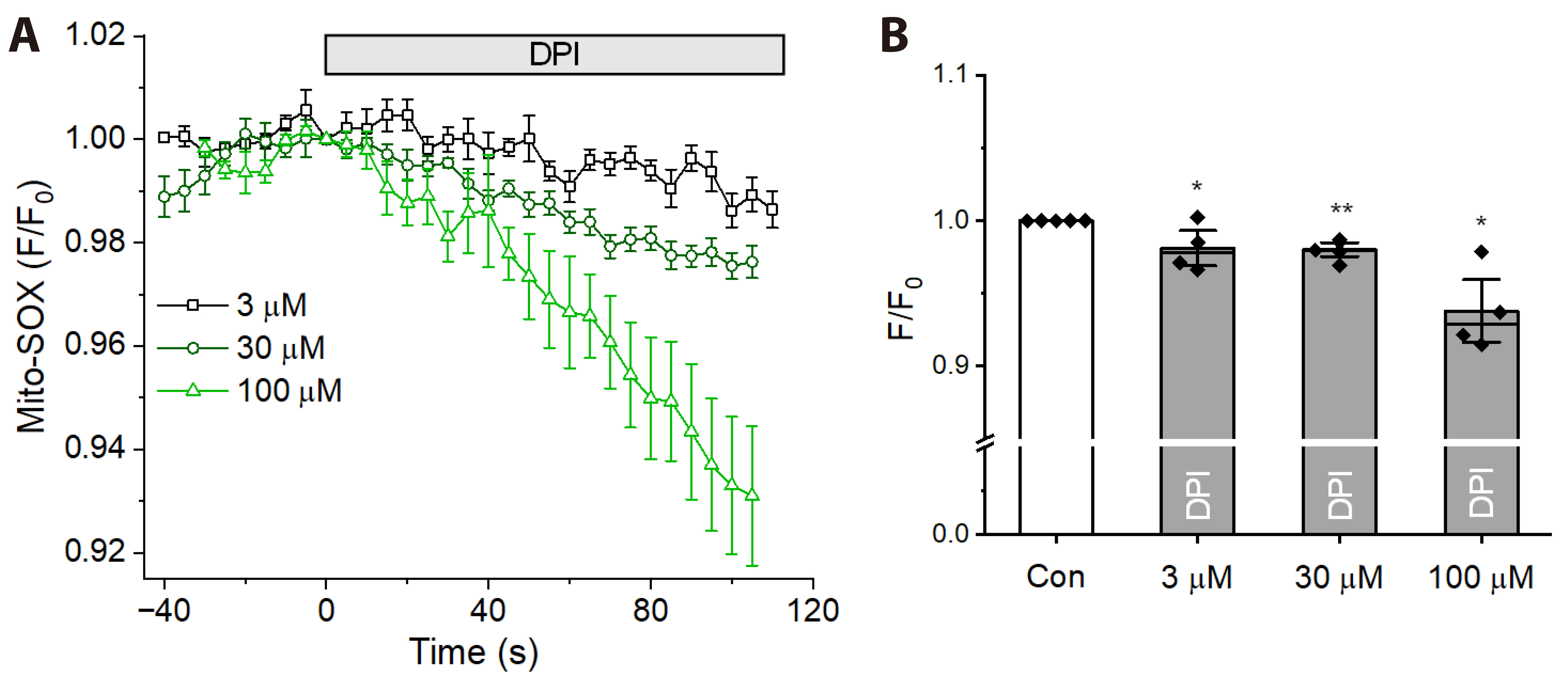
- Diphenyleneiodonium (DPI) has been widely used as an inhibitor of NADPH oxidase (Nox) to discover its function in cardiac myocytes under various stimuli. However, the effects of DPI itself on Ca2+ signaling and contraction in cardiac myocytes under control conditions have not been understood. We investigated the effects of DPI on contraction and Ca2+ signaling and their underlying mechanisms using video edge detection, confocal imaging, and whole-cell patch clamp technique in isolated rat cardiac myocytes. Application of DPI suppressed cell shortenings in a concentration-dependent manner (IC50 of ≅0.17 µM) with a maximal inhibition of ~70% at ~100 µM. DPI decreased the magnitude of Ca2+ transient and sarcoplasmic reticulum Ca2+ content by 20%–30% at 3 µM that is usually used to remove the Nox activity, with no effect on fractional release. There was no significant change in the half-decay time of Ca2+ transients by DPI. The L-type Ca2+ current (ICa) was decreased concentration-dependently by DPI (IC50 of ≅40.3 µM) with ≅13.1%-inhibition at 3 µM. The frequency of Ca2+ sparks was reduced by 3 µM DPI (by ~25%), which was resistant to a brief removal of external Ca2+ and Na+. Mitochondrial superoxide level was reduced by DPI at 3–100 µM. Our data suggest that DPI may suppress L-type Ca2+ channel and RyR, thereby attenuating Ca2+-induced Ca2+ release and contractility in cardiac myocytes, and that such DPI effects may be related to mitochondrial metabolic suppression.
Figure
Reference
-
1. Fabiato A. 1983; Calcium-induced release of calcium from the cardiac sarcoplasmic reticulum. Am J Physiol. 245:C1–C14. DOI: 10.1152/ajpcell.1983.245.1.C1. PMID: 6346892.2. Beuckelmann DJ, Wier WG. 1988; Mechanism of release of calcium from sarcoplasmic reticulum of guinea-pig cardiac cells. J Physiol. 405:233–255. DOI: 10.1113/jphysiol.1988.sp017331. PMID: 2475607. PMCID: PMC1190974.3. Näbauer M, Callewaert G, Cleemann L, Morad M. 1989; Regulation of calcium release is gated by calcium current, not gating charge, in cardiac myocytes. Science. 244:800–803. DOI: 10.1126/science.2543067. PMID: 2543067.4. Niggli E, Lederer WJ. 1990; Voltage-independent calcium release in heart muscle. Science. 250:565–568. DOI: 10.1126/science.2173135. PMID: 2173135.5. Cheng H, Lederer WJ, Cannell MB. 1993; Calcium sparks: elementary events underlying excitation-contraction coupling in heart muscle. Science. 262:740–744. DOI: 10.1126/science.8235594. PMID: 8235594.6. Cannell MB, Cheng H, Lederer WJ. 1994; Spatial non-uniformities in [Ca2+]i during excitation-contraction coupling in cardiac myocytes. Biophys J. 67:1942–1956. DOI: 10.1016/S0006-3495(94)80677-0. PMID: 7858131.7. Wier WG, Egan TM, López-López JR, Balke CW. 1994; Local control of excitation-contraction coupling in rat heart cells. J Physiol. 474:463–471. DOI: 10.1113/jphysiol.1994.sp020037. PMID: 8014907. PMCID: PMC1160337.8. Shacklock PS, Wier WG, Balke CW. 1995; Local Ca2+ transients (Ca2+ sparks) originate at transverse tubules in rat heart cells. J Physiol. 487:601–608. DOI: 10.1113/jphysiol.1995.sp020903. PMID: 8544124. PMCID: PMC1156648.9. Parker I, Zang WJ, Wier WG. 1996; Ca2+ sparks involving multiple Ca2+ release sites along Z-lines in rat heart cells. J Physiol. 497:31–38. DOI: 10.1113/jphysiol.1996.sp021747. PMID: 8951709. PMCID: PMC1160910.10. Zima AV, Blatter LA. 2006; Redox regulation of cardiac calcium channels and transporters. Cardiovasc Res. 71:310–321. DOI: 10.1016/j.cardiores.2006.02.019. PMID: 16581043.11. Yan Y, Liu J, Wei C, Li K, Xie W, Wang Y, Cheng H. 2008; Bidirectional regulation of Ca2+ sparks by mitochondria-derived reactive oxygen species in cardiac myocytes. Cardiovasc Res. 77:432–441. DOI: 10.1093/cvr/cvm047. PMID: 18006452.12. Terentyev D, Györke I, Belevych AE, Terentyeva R, idhar A Sr, Nishijima Y, de Blanco EC, Khanna S, Sen CK, Cardounel AJ, Carnes CA, Györke S. 2008; Redox modification of ryanodine receptors contributes to sarcoplasmic reticulum Ca2+ leak in chronic heart failure. Circ Res. 103:1466–1472. DOI: 10.1161/CIRCRESAHA.108.184457. PMID: 19008475. PMCID: PMC3274754.13. Cooper LL, Li W, Lu Y, Centracchio J, Terentyeva R, Koren G, Terentyev D. 2013; Redox modification of ryanodine receptors by mitochondria-derived reactive oxygen species contributes to aberrant Ca2+ handling in ageing rabbit hearts. J Physiol. 591:5895–5911. DOI: 10.1113/jphysiol.2013.260521. PMID: 24042501. PMCID: PMC3872760.14. Bedard K, Krause KH. 2007; The NOX family of ROS-generating NADPH oxidases: physiology and pathophysiology. Physiol Rev. 87:245–313. DOI: 10.1152/physrev.00044.2005. PMID: 17237347.15. Akki A, Zhang M, Murdoch C, Brewer A, Shah AM. 2009; NADPH oxidase signaling and cardiac myocyte function. J Mol Cell Cardiol. 47:15–22. DOI: 10.1016/j.yjmcc.2009.04.004. PMID: 19374908.16. Prosser BL, Ward CW, Lederer WJ. 2011; X-ROS signaling: rapid mechano-chemo transduction in heart. Science. 333:1440–1445. DOI: 10.1126/science.1202768. PMID: 21903813.17. Jian Z, Han H, Zhang T, Puglisi J, Izu LT, Shaw JA, Onofiok E, Erickson JR, Chen YJ, Horvath B, Shimkunas R, Xiao W, Li Y, Pan T, Chan J, Banyasz T, Tardiff JC, Chiamvimonvat N, Bers DM, Lam KS, et al. 2014; Mechanochemotransduction during cardiomyocyte contraction is mediated by localized nitric oxide signaling. Sci Signal. 7:ra27. DOI: 10.1126/scisignal.2005046. PMID: 24643800. PMCID: PMC4103414.18. Kim JC, Wang J, Son MJ, Woo SH. 2017; Shear stress enhances Ca2+ sparks through Nox2-dependent mitochondrial reactive oxygen species generation in rat ventricular myocytes. Biochim Biophys Acta Mol Cell Res. 1864:1121–1131. DOI: 10.1016/j.bbamcr.2017.02.009. PMID: 28213332.19. Looi YH, Grieve DJ, Siva A, Walker SJ, Anilkumar N, Cave AC, Marber M, Monaghan MJ, Shah AM. 2008; Involvement of Nox2 NADPH oxidase in adverse cardiac remodeling after myocardial infarction. Hypertension. 51:319–325. DOI: 10.1161/HYPERTENSIONAHA.107.101980. PMID: 18180403.20. Cross AR, Jones OT. 1986; The effect of the inhibitor diphenylene iodonium on the superoxide-generating system of neutrophils. Specific labelling of a component polypeptide of the oxidase. Biochem J. 237:111–116. DOI: 10.1042/bj2370111. PMID: 3800872. PMCID: PMC1146954.21. Zhang M, Perino A, Ghigo A, Hirsch E, Shah AM. 2013; NADPH oxidases in heart failure: poachers or gamekeepers? Antioxid Redox Signal. 18:1024–1041. DOI: 10.1089/ars.2012.4550. PMID: 22747566. PMCID: PMC3567780.22. Bloxham DP. 1979; The relationship of diphenyleneiodonium-induced hypoglycaemia to the specific covalent modification of NADH-ubiquinone oxidoreductase. Biochem Soc Trans. 7:103–106. DOI: 10.1042/bst0070103. PMID: 437247.23. Cooper JM, Petty RK, Hayes DJ, Morgan-Hughes JA, Clark JB. 1988; Chronic administration of the oral hypoglycaemic agent diphenyleneiodonium to rats. An animal model of impaired oxidative phosphorylation (mitochondrial myopathy). Biochem Pharmacol. 37:687–694. DOI: 10.1016/0006-2952(88)90143-8. PMID: 3342100.24. Majander A, Finel M, Wikström M. 1994; Diphenyleneiodonium inhibits reduction of iron-sulfur clusters in the mitochondrial NADH-ubiquinone oxidoreductase (Complex I). J Biol Chem. 269:21037–21042. DOI: 10.1016/S0021-9258(17)31926-9. PMID: 8063722.25. Paradies G, Petrosillo G, Pistolese M, Di Venosa N, Federici A, Ruggiero FM. 2004; Decrease in mitochondrial complex I activity in ischemic/reperfused rat heart: involvement of reactive oxygen species and cardiolipin. Circ Res. 94:53–59. DOI: 10.1161/01.RES.0000109416.56608.64. PMID: 14656928.26. Tazzeo T, Worek F. Janssen Lj. 2009; The NADPH oxidase inhibitor diphenyleneiodonium is also a potent inhibitor of cholinesterases and the internal Ca(2+) pump. Br J Pharmacol. 158:790–796. DOI: 10.1111/j.1476-5381.2009.00394.x. PMID: 19788497. PMCID: PMC2765598.27. Stuehr DJ, Fasehun OA, Kwon NS, Gross SS, Gonzalez JA, Levi R, Nathan CF. 1991; Inhibition of macrophage and endothelial cell nitric oxide synthase by diphenyleneiodonium and its analogs. FASEB J. 5:98–103. DOI: 10.1096/fasebj.5.1.1703974. PMID: 1703974.28. Dodd-o JM, Zheng G, Silverman HS, Lakatta EG, Ziegelstein RC. 1997; Endothelium-independent relaxation of aortic rings by the nitric oxide synthase inhibitor diphenyleneiodonium. Br J Pharmacol. 120:857–864. DOI: 10.1038/sj.bjp.0701014. PMID: 9138692. PMCID: PMC1564554.29. Sanders SA, Eisenthal R, Harrison R. 1997; NADH oxidase activity of human xanthine oxidoreductase--generation of superoxide anion. Eur J Biochem. 245:541–548. DOI: 10.1111/j.1432-1033.1997.00541.x. PMID: 9182988.30. Wang J, Trinh TN, Vu ATV, Kim JC, Hoang ATN, Ohk CJ, Zhang YH, Nguyen CM, Woo SH. 2022; Chrysosplenol-C increases contraction by augmentation of sarcoplasmic reticulum Ca2+ loading and release via protein kinase C in rat ventricular myocytes. Mol Pharmacol. 101:13–23. DOI: 10.1124/molpharm.121.000365. PMID: 34764211.31. Lee S, Kim JC, Li Y, Son MJ, Woo SH. 2008; Fluid pressure modulates L-type Ca2+ channel via enhancement of Ca2+-induced Ca2+ release in rat ventricular myocytes. Am J Physiol Cell Physiol. 294:C966–C976. DOI: 10.1152/ajpcell.00381.2007. PMID: 18272819.32. Byrne JA, Grieve DJ, Bendall JK, Li JM, Gove C, Lambeth JD, Cave AC, Shah AM. 2003; Contrasting roles of NADPH oxidase isoforms in pressure-overload versus angiotensin II-induced cardiac hypertrophy. Circ Res. 93:802–805. DOI: 10.1161/01.RES.0000099504.30207.F5. PMID: 14551238.33. Li Y, Trush MA. 1998; Diphenyleneiodonium, an NAD(P)H oxidase inhibitor, also potently inhibits mitochondrial reactive oxygen species production. Biochem Biophys Res Commun. 253:295–299. DOI: 10.1006/bbrc.1998.9729. PMID: 9878531.34. Hool LC, Di Maria CA, Viola HM, Arthur PG. 2005; Role of NAD(P)H oxidase in the regulation of cardiac L-type Ca2+ channel function during acute hypoxia. Cardiovasc Res. 67:624–635. DOI: 10.1016/j.cardiores.2005.04.025. PMID: 15913584.35. Trimm JL, Salama G, Abramson JJ. 1986; Sulfhydryl oxidation induces rapid calcium release from sarcoplasmic reticulum vesicles. J Biol Chem. 261:16092–16098. DOI: 10.1016/S0021-9258(18)66682-7. PMID: 3782109.36. Boraso A, Williams AJ. 1994; Modification of the gating of the cardiac sarcoplasmic reticulum Ca(2+)-release channel by H2O2 and dithiothreitol. Am J Physiol. 267:H1010–H1016. DOI: 10.1152/ajpheart.1994.267.3.H1010. PMID: 8092267.37. Marengo JJ, Hidalgo C, Bull R. 1998; Sulfhydryl oxidation modifies the calcium dependence of ryanodine-sensitive calcium channels of excitable cells. Biophys J. 74:1263–1277. DOI: 10.1016/S0006-3495(98)77840-3. PMID: 9512024.38. Bers DM. 2002; Cardiac excitation-contraction coupling. Nature. 415:198–205. DOI: 10.1038/415198a. PMID: 11805843.39. Zima AV, Copello JA, Blatter LA. 2004; Effects of cytosolic NADH/NAD(+) levels on sarcoplasmic reticulum Ca(2+) release in permeabilized rat ventricular myocytes. J Physiol. 555:727–741. DOI: 10.1113/jphysiol.2003.055848. PMID: 14724208. PMCID: PMC1664876.40. Györke S, Lukyanenko V, Györke I. 1997; Dual effects of tetracaine on spontaneous calcium release in rat ventricular myocytes. J Physiol. 500:297–309. DOI: 10.1113/jphysiol.1997.sp022021. PMID: 9147318. PMCID: PMC1159384.41. Satoh H, Blatter LA, Bers DM. 1997; Effects of [Ca2+]i, SR Ca2+ load, and rest on Ca2+ spark frequency in ventricular myocytes. Am J Physiol. 272:H657–H668. DOI: 10.1152/ajpheart.1997.272.2.H657. PMID: 9124422.42. Vendrov AE, Xiao H, Lozhkin A, Hayami T, Hu G, Brody MJ, Sadoshima J, Zhang YY, Runge MS, Madamanchi NR. 2023; Cardiomyocyte NOX4 regulates resident macrophage-mediated inflammation and diastolic dysfunction in stress cardiomyopathy. Redox Biol. 67:102937. DOI: 10.1016/j.redox.2023.102937. PMID: 37871532. PMCID: PMC10598408.43. Rana M, Setia M, Suvas PK, Chakraborty A, Suvas S. 2022; Diphenyleneiodonium treatment inhibits the development of severe herpes stromal keratitis lesions. J Virol. 96:e0101422. DOI: 10.1128/jvi.01014-22. PMID: 35946937. PMCID: PMC9472634.44. Weir EK, Wyatt CN, Reeve HL, Huang J, Archer SL, Peers C. 1994; Diphenyleneiodonium inhibits both potassium and calcium currents in isolated pulmonary artery smooth muscle cells. J Appl Physiol. 76:2611–2615. DOI: 10.1152/jappl.1994.76.6.2611. PMID: 7928890.45. Wyatt CN, Weir EK, Peers C. 1994; Diphenylene iodonium blocks K+ and Ca2+ currents in type I cells isolated from the neonatal rat carotid body. Neurosci Lett. 172:63–66. DOI: 10.1016/0304-3940(94)90663-7. PMID: 8084538.46. Sánchez JA, García MC, Sharma VK, Young KC, Matlib MA, Sheu SS. 2001; Mitochondria regulate inactivation of L-type Ca2+ channels in rat heart. J Physiol. 536.2:387–396. DOI: 10.1111/j.1469-7793.2001.0387c.xd. PMID: 11600674. PMCID: PMC2278878.47. Irisawa H, Sato R. 1986; Intra- and extracellular actions of proton on the calcium current of isolated guinea pig ventricular cells. Circ Res. 59:48–355. DOI: 10.1161/01.RES.59.3.348. PMID: 2429781.48. Chantawansri C, Huynh N, Yamanaka J, Garfinkel A, Lamp ST, Inoue M, Bridge JH, Goldhaber JI. 2008; Effect of metabolic inhibition on couplon behavior in rabbit ventricular myocytes. Biophys J. 94:1656–1666. DOI: 10.1529/biophysj.107.114892. PMID: 18024504. PMCID: PMC2242765.49. Haouzi P, Sonobe T, Judenherc-Haouzi A. 2016; Developing effective countermeasures against acute hydrogen sulfide intoxication: challenges and limitations. Ann N Y Acad Sci. 1374:29–40. DOI: 10.1111/nyas.13015. PMID: 26945701. PMCID: PMC4940262.50. Cheung JY, Wang J, Zhang XQ, Song J, Davidyock JM, Prado FJ, Shanmughapriya S, Worth AM, Madesh M, Judenherc-Haouzi A, Haouzi P. 2018; Methylene blue counteracts H2S-induced cardiac ion channel dysfunction and ATP reduction. Cardiovasc Toxicol. 18:407–419. DOI: 10.1007/s12012-018-9451-5. PMID: 29603116. PMCID: PMC6126975.
- Full Text Links
- Actions
-
Cited
- CITED
-
- Close
- Share
- Similar articles
-
- Efficacy of Diphenyleneiodonium Chloride (DPIC) Against Diverse Plant Pathogens
- NADPH oxidase mediated oxidative stress in hepatic fibrogenesis
- Diphenyleneiodonium Inhibits Apoptotic Cell Death of Gastric Epithelial Cells Infected with Helicobacter pylori in a Korean Isolate
- YJI-7 Suppresses ROS Production and Expression of Inflammatory Mediators via Modulation of p38MAPK and JNK Signaling in RAW 264.7 Macrophages
- Involvement of Vascular NAD(P)H Oxidase-derived Superoxide in Cerebral Vasospasm after Subarachnoid Hemorrhage in Rats


