J Cerebrovasc Endovasc Neurosurg.
2024 Jun;26(2):223-226. 10.7461/jcen.2024.E2023.06.002.
Percutaneous femoral access: Stuck guide wire, decannulation difficulty due to unravelling and knotting
- Affiliations
-
- 1Department of Neurosurgery, GB Pant Institute of Post Graduate Medical Education and Research, New Delhi, India
- KMID: 2556986
- DOI: http://doi.org/10.7461/jcen.2024.E2023.06.002
Abstract
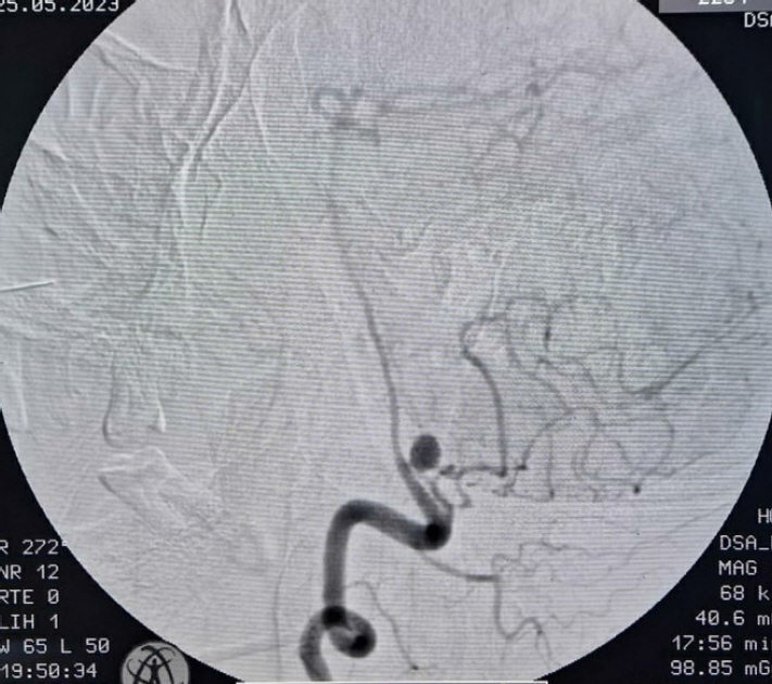
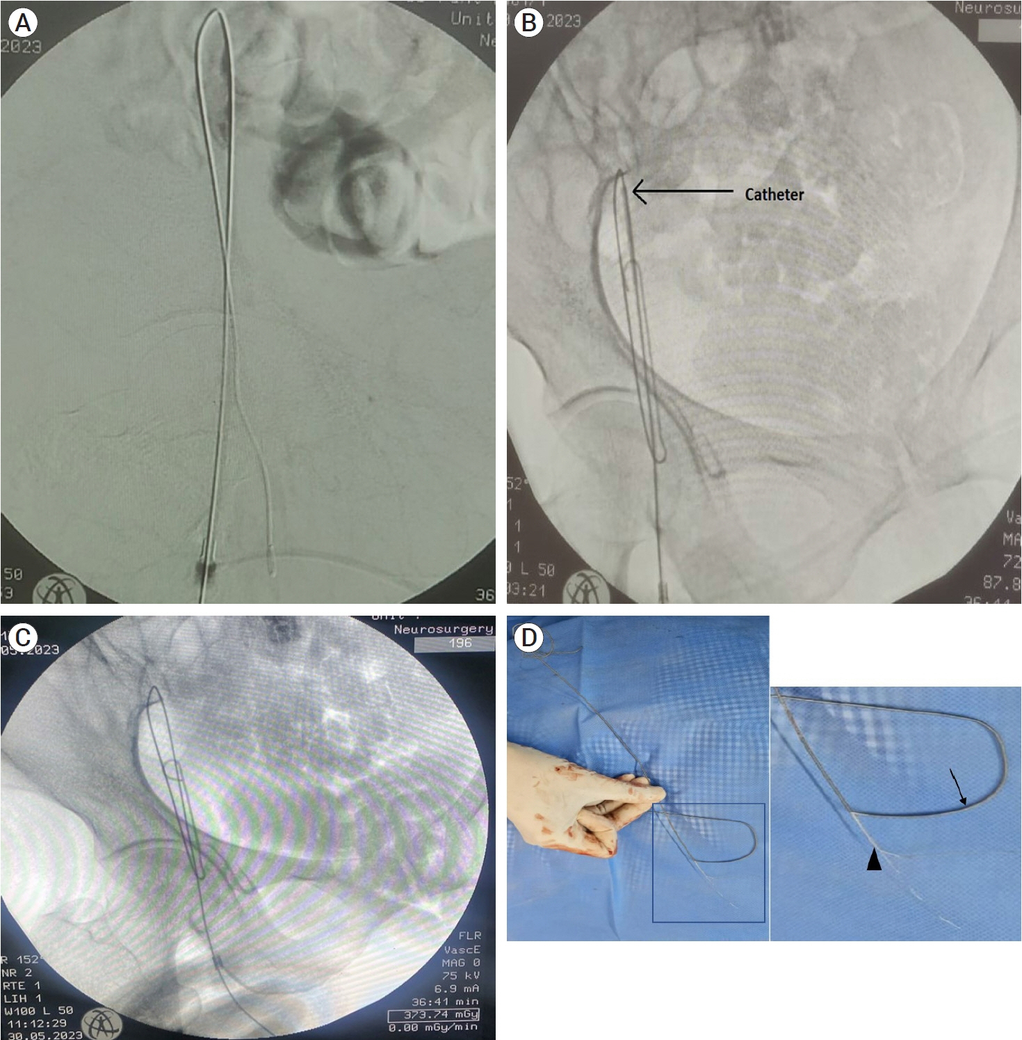
- Percutaneous techniques for femoral arterial access are increasingly being performed due to advances in endovascular cerebral procedures, as they provide a less morbid and minimally invasive approach than open procedures. Common complications associated with this peripheral puncture include hematoma, bleeding, pseudoaneurysm, arteriovenous fistula, retroperitoneal bleeding, inadvertent venous puncture, dissection, etc. The retrograde femoral access is currently the most frequently used arterial access as it is technically straightforward, allows for the use of larger size sheaths and catheters, allows repeated attempts, etc. Although being technically less challenging, grave complications can occur due to hardware failure. Here, we present a case of unruptured posterior inferior cerebellar artery (PICA) aneurysm, who underwent uneventful diagnostic cerebral digital substraction angiography (DSA) via right femoral artery route on first attempt, but on second attempt for therapeutic intervention, landed up with stuck guide wire and faced decannulation difficulty due to unravelling of guide wire and multiple knot formation, which was finally removed after multiple attempts at pulling and improvised manoeuvres. Such cannulation and decannulation difficulties have been reported multiple times for central venous access, but extremely rarely for femoral routes, making this case a rarity and worth reporting.
Keyword
Figure
Reference
-
1. Transradial vs. Transfemoral approach in cardiac catheterization: A literature review. Cureus. 2017; Jun. 9(6):e1309.2. Barik AK, Gupta P, Andleeb R, Chaturvedi J. Retrieval of self-knotted and unraveled guide wire in the femoral artery: Focus on point-of-care ultrasound (POCUS). Curr Med Res Pr. 2019; 9(6):249–50.
Article3. Jalwal GK, Rajagopalan V, Bindra A, Rath GP, Goyal K, Kumar A, et al. Percutaneous retrieval of malpositioned, kinked and unraveled guide wire under fluoroscopic guidance during central venous cannulation. J Anaesthesiol Clin Pharmacol. 2014; Apr. 30(2):267–9.
Article4. Khan KZ, Graham D, Ermenyi A, Pillay WR. Case report: Managing a knotted Seldinger wire in the subclavian vein during central venous cannulation. Can J Anaesth. 2007; May. 54(5):375–9.
Article5. Ndrepepa G, Berger PB, Mehilli J, Seyfarth M, Neumann FJ, Schömig A, et al. Periprocedural bleeding and 1 year outcome after percutaneous coronary intervention: Appropriateness of including bleeding as a component of a quadruple end point. J Am Coll Cardiol. 2008; Feb. 51(7):690–7.
- Full Text Links
- Actions
-
Cited
- CITED
-
- Close
- Share
- Similar articles
-
- Accidental Insertion of Entire Guide - Wire in the Inferior Vena Cava During Central Venous Catheterization
- Percutaneous Interlocking Intramedullary Nailing of Femoral Shaft Fracture with Retrograde Guide Wire Insertion Technique
- J-guide Wire Knotting during the Central Venous Catheterization: A case report
- Guide wire fracture during percutaneous coronary intervention
- Removal of a Broken Intramedullary Nail with a Narrow Hollow Using a Bulb-tipped Guide Wire and Kirschner Wire: A Case Report


