J Cerebrovasc Endovasc Neurosurg.
2024 Jun;26(2):187-195. 10.7461/jcen.2023.E2023.02.001.
Symptomatic perianeursymal cyst development 20 years after endovascular treatment of a ruptured giant aneurysm: Case report and updated review
- Affiliations
-
- 1Department of Neurosurgery, Massachusetts General Hospital, Harvard Medical School, Boston, MA, USA
- KMID: 2556981
- DOI: http://doi.org/10.7461/jcen.2023.E2023.02.001
Abstract
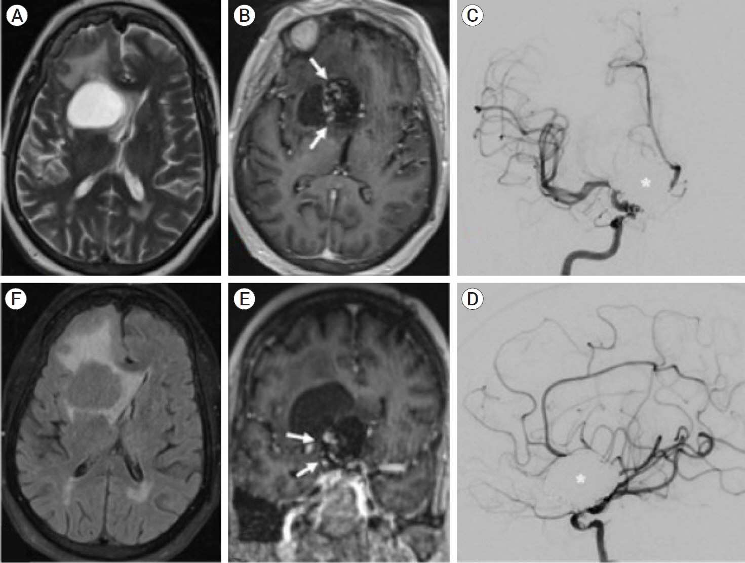
- Perianeurysmal cysts are a rare and poorly understood finding in patients both with treated and untreated aneurysms. While the prior literature suggests that a minority of perianeurysmal cysts develop 1-4 years following endovascular aneurysm treatment, this updated review demonstrates that nearly half of perianeurysmal cysts were diagnosed following aneurysm coiling, with the other half diagnosed concurrently with an associated aneurysm prior to treatment. 64% of perianeurysmal cysts were surgically decompressed, with a 39% rate of recurrence requiring re-operation. We report a case of a 71-year-old woman who presented with vertigo and nausea and was found to have a 3.4 cm perianeurysmal cyst 20 years after initial endovascular coiling of a ruptured giant ophthalmic aneurysm. The cyst was treated with endoscopic fenestration followed by open fenestration upon recurrence. The case represents the longest latency from initial aneurysm treatment to cyst diagnosis reported in the literature and indicates that the diagnosis of perianeurysmal cyst should remain on the differential even decades after treatment. Based on a case discussion and updated literature review, this report highlights proposed etiologies of development and management strategies for a challenging lesion.
Keyword
Figure
Reference
-
1. Barber SM, Al-Zubidi N, Diaz OM, Zhang YJ, Lee AG. Delayed hydrocephalus and perianeurysmal cyst formation after stent-assisted coil embolization of a large, unruptured basilar apex aneurysm. Asia Pac J Ophthalmol (Phila). 2014; Nov-Dec. 3(6):354–60.
Article2. Benvenuti L, Gagliardi R, Scazzeri F, Gaglianone S. Parenchymal perianeurysmal cyst in the brain: Case report. Neurosurgery. 2006; Apr. 58(4):e788.
Article3. Birua GJS, Tyagi G, Beniwal M, Srinivas D, Rao S. Large parenchymal perianeurysmal cyst: A case report. J Neurosci Rural Pract. 2021; Sep. 12(4):800–3.
Article4. Friedman JA, McIver JI, Collignon FP, Nichols DA, Piepgras DG. Development of a pontine cyst after endovascular coil occlusion of a basilar artery trunk aneurysm: Case report. Neurosurgery. 2003; Mar. 52(3):694–9.
Article5. Galdamez MM, Lorente PS, Rico AP, Pérez-Higueras A. Intracranial perianeurysmal cyst: Still a dilemma. Neuroradiol J. 2011; Oct. 24(5):743–8.
Article6. Grandhi R, Miller RA, Zwagerman NT, Lunsford LD, Horowitz M. Perianeursymal cyst development after endovascular treatment of a ruptured giant aneurysm. J Neuroimaging. 2014; Sep-Oct. 24(5):515–7.
Article7. Hirota N, Ueno J, Naitoh H, Sugiyama K, Karasawa H, Kin H, et al. Giant aneurysm associated with a large cyst. Case illustration. J Neurosurg. 1999; Jul. 91(1):160.8. Jayakumar N, Ughratdar I, White E. Obstructive hydrocephalus secondary to a perianeurysmal cyst: A case report. Br J Neurosurg. 2022; Oct. 36(5):658–60.
Article9. Kobayashi H, Enomoto Y, Yamada T, Egashira Y, Nakayama N, Ohe N, et al. Perianeurysmal cyst formation in the brainstem after coil embolization: Illustrative case. J Neurosurg Case Lessons. 2022; May. 3(22):CASE21690.
Article10. König M, Bakke SJ, Scheie D, Sorteberg W, Meling TR. Reactive expansive intracerebral process as a complication of endovascular coil treatment of an unruptured intracranial aneurysm: Case report. Neurosurgery. 2011; May. 68(5):e1468–74.
Article11. Kulwin CG, Gandhi RH, Patel NB, Payner TD. Symptomatic perianeurysmal parenchymal cyst: Case illustration. J Neurosurg. 2015; Aug. 123(2):470–1.
Article12. Liang ES, Efendy M, Winter C, Coulthard A. Intracranial perianeurysmal cysts: Case series and review of the literature. J Neurointerv Surg. 2021; Aug. 14(8):837–41.
Article13. Marcoux J, Roy D, Bojanowski MW. Acquired arachnoid cyst after a coil-ruptured aneurysm. Case illustration. J Neurosurg. 2002; Sep. 97(3):722.14. Norris SA, Derdeyn CP, Perlmutter JS. Levodopa‐responsive hemiparkinsonism secondary to cystic expansion from a coiled cerebral aneurysm. J Neuroimaging. 2015; Mar-Apr. 25(2):316–8.
Article15. Pedro KM, Sih IMY. Perianeurysmal parenchymal cyst of the pons: A case report and review of literature. Interdiscip Neurosurg. 2020; 21:100731.
Article16. Sato N, Sze G, Awad IA, Putman CM, Shibazaki T, Endo K. Parenchymal perianeurysmal cystic changes in the brain: Report of five cases. Radiology. 2000; Apr. 215(1):229–33.
Article17. Shetler KE, Parikh NS, Sekar K, Williams OA. Clinical reasoning: An unusual case of auditory hallucinations in a middle-aged man. Neurology. 2020; May. 94(20):e2180–6.
Article18. Takai K, Nishihara T, Nemoto S, Ueki K, Miyauchi H, Mishima K, et al. Multilocular cystic lesion associated with a giant aneurysm: Case illustration. J Neurosurg. 2001; Dec. 95(6):1081.19. Zammit A, Tudose A, Khan N, Renowden S, Teo M. Perianeurysmal parenchymal cysts – Case series and literature review. Brain Spine. 2022; Jul. 2:100920.
Article
- Full Text Links
- Actions
-
Cited
- CITED
-
- Close
- Share
- Similar articles
-
- Ruptured Intracranial Dermoid Cyst Associated with Rupture of Cerebral Aneurysm
- Progressive Visual Loss after Endovascular Coiling Treatment of a Large Paraclinoid Aneurysm
- Rupture of Giant Posterior Communicating Artery Aneurysm During Carotid Angiography: A Case Report
- Giant vertebral artery aneurysms presenting acutely with WFNS grade five subarachnoid haemorrhage, report of 4 cases treated with endovascular or surgical proximal parent artery occlusion achieving good functional outcome
- A Case of Ruptured Giant Intracranial Aneurysm During Carotid Angiography


