Two consecutive ruptured intracranial aneurysm in patient with multiple intracranial aneurysms
- Affiliations
-
- 1Department of Neurosurgery, Kangwon National University Hospital, Kangwon National University College of Medicine, Chuncheon, Korea
- 2Department of Neurosurgery, Wonju Severance Christian Hospital, Wonju College of Medicine, Yonsei University, Wonju, Korea
- KMID: 2556979
- DOI: http://doi.org/10.7461/jcen.2023.E2023.08.010
Abstract
- When aneurysmal subarachnoid hemorrhage due to multiple aneurysms is suspected, identifying the rupture site is essential to determine the exact surgical site, but it may not be easy. Even if embolization is adequately performed, complications may remain. Typical complications include rebleeding and hydrocephalus in the early phase and delayed cerebral ischemia in the delayed phase. Herein, we describe a case of rupture of an intracranial aneurysm after performing embolization for a different ruptured intracranial aneurysm in a patient with multiple intracranial aneurysms. Patients with multiple intracranial aneurysms need to be considered for closer observation than those with a single ruptured intracranial aneurysm, even if the patient’s prognosis is good.
Keyword
Figure
-
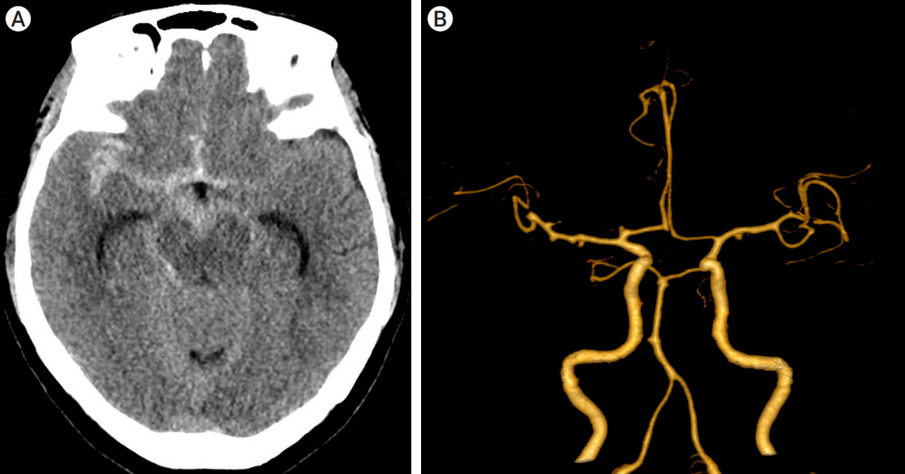
Fig. 1. Initial radiographic findings. (A) CT scan showing subarachnoid hemorrhage in the basal cistern. (B) CT angiography showing an aneurysm arising from the proximal A1 segment. CT, computed tomography
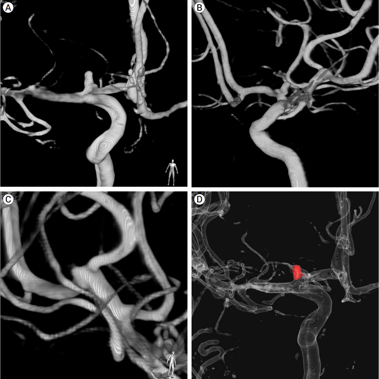
Fig. 2. DSA before and after endovascular treatment. (A) Anteroposterior view of the digital subtraction angiography showing an aneurysm arising from the proximal A1 segment. It is irregular shape of aneurysm with height of 3.66 mm and transverse diameter is 1.68 mm, with a neck size of 3.04 mm. (B) Oblique view of the digital subtraction angiography showing an aneurysm arising from the proximal A1 segment. (C) Anterolateral working view of the digital subtraction angiography suggests the presence an aneurysm from right distal M1 segment. (D) Postoperative DSA. Red colored area indicates the embolization materials (Coil). DSA, digital subtraction angiography
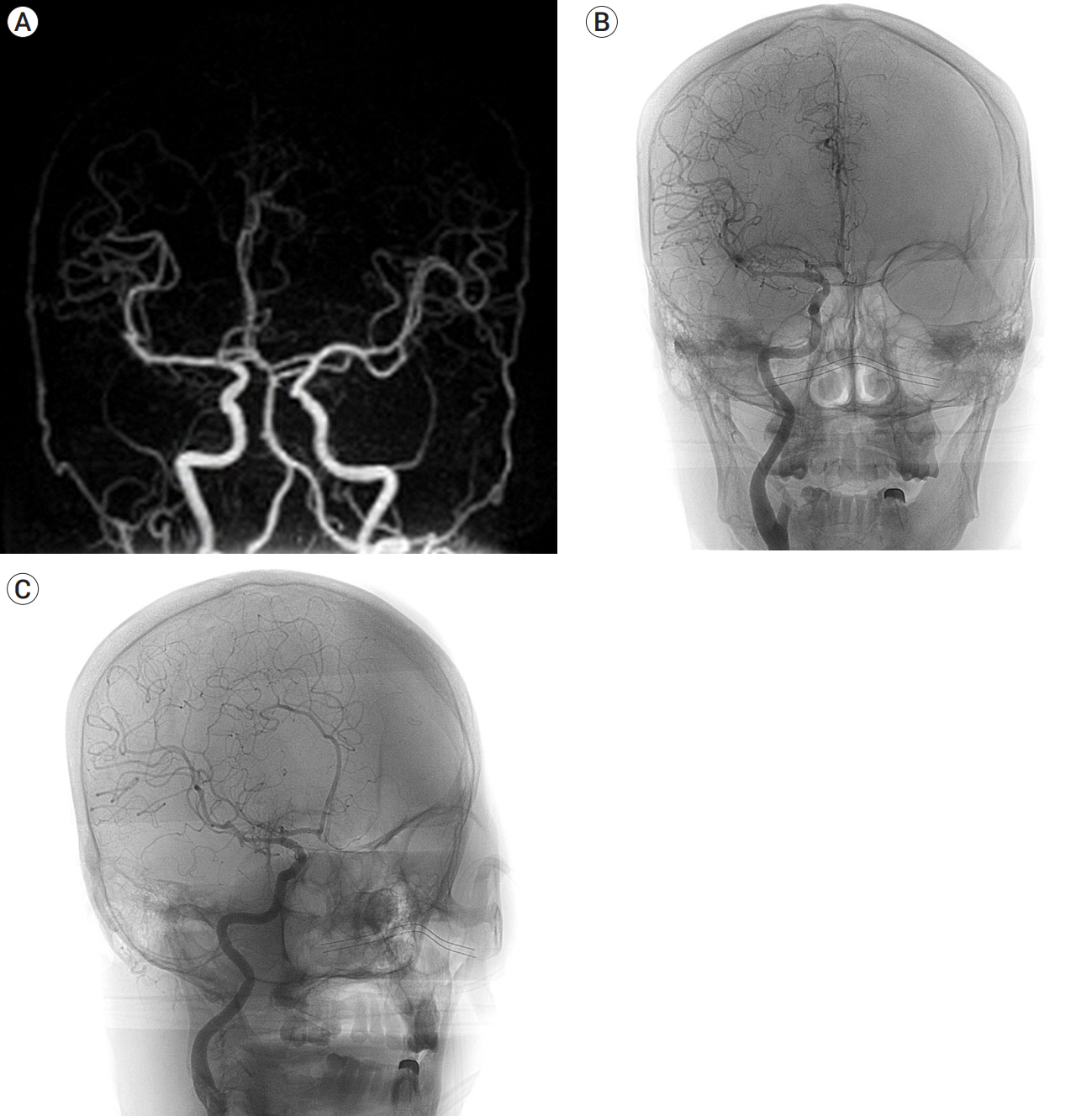
Fig. 3. (A) MRA on the third day after endovascular treatment. It showed that there was no significant change in aneurysm in both aneurysms. (B and C) DSA on the 7th day after endovascular treatment. It showed that there was no significant change in both aneurysms. Anteroposterior view (B) and working view (C). DSA, digital subtraction angiography
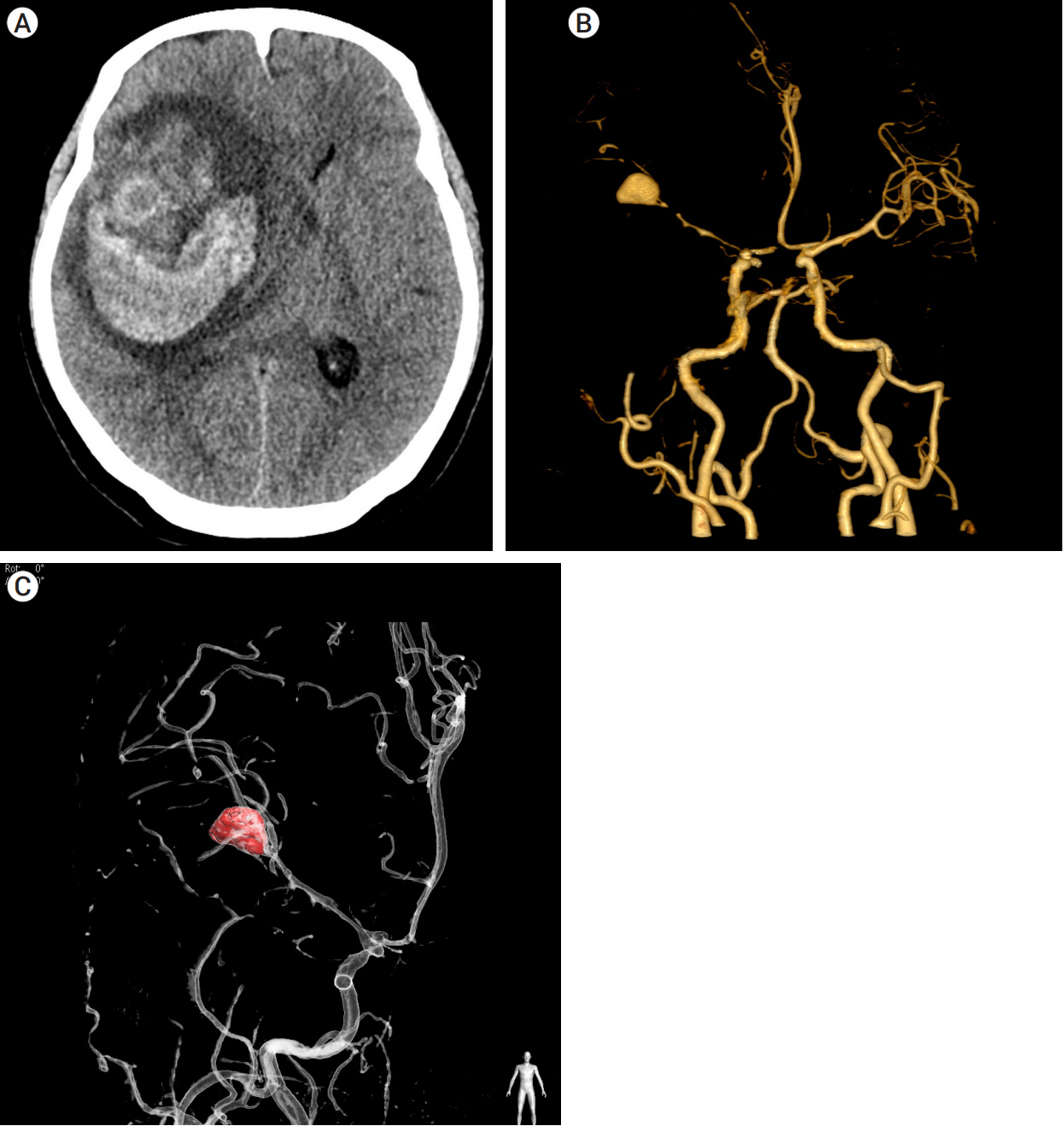
Fig. 4. CT and DSA on the 23th day after endovascular treatment, immediately after sudden mental change. (A) CT scan showing intracerebral hemorrhage in the right frontal lobe. (B) CT angiography showing a newly noted 14 mm aneurysm in the right distal M1 portion with Irregular narrowing of the right MCA. (C) Postoperative DSA. Red colored area indicates the embolization materials (Coil). CT, computed tomography; DSA, digital subtraction angiography
Reference
-
1. Chung DY, Abdalkader M, Nguyen TN. Aneurysmal subarachnoid hemorrhage. Neurol Clin. 2021; May. 39(2):419–42.
Article2. Nehls DG, Flom RA, Carter LP, Spetzler RF. Multiple intracranial aneurysms: Determining the site of rupture. J Neurosurg. 1985; Sep. 63(3):342–8.
Article3. Neifert SN, Chapman EK, Martini ML, Shuman WH, Schupper AJ, Oermann EK, et al. Aneurysmal subarachnoid hemorrhage: The last decade. Transl Stroke Res. 2021; Jun. 12(3):428–46.
Article4. Orning JL, Shakur SF, Alaraj A, Behbahani M, Charbel FT, Aletich VA, et al. Accuracy in identifying the source of subarachnoid hemorrhage in the setting of multiple intracranial aneurysms. Neurosurgery. 2018; Jul. 83(1):62–8.
Article5. Osgood ML. Aneurysmal subarachnoid hemorrhage: Review of the pathophysiology and management strategies. Curr Neurol Neurosci Rep. 2021; Jul. 21(9):50.
Article6. Sato H, Kamide T, Kikkawa Y, Kimura T, Kuribara S, Yanagawa T, et al. Clinical characteristics of ruptured intracranial aneurysm in patients with multiple intracranial aneurysms. World Neurosurg. 2021; May. 149:e935–41.
Article7. Suarez JI. Diagnosis and management of subarachnoid hemorrhage. Continuum (Minneap Minn). 2015; Oct. (5 Neurocritical Care):21:1263–87.
Article
- Full Text Links
-
- Actions
-
Cited
- CITED
-
- Close
- Share
-
- Similar articles
-
- Surgical Treatment of Unruptured Cerebral Aneurysms
- Surgical Intervention for Cases of Ruptured Intracranial Aneurysms Accompanying Large Intracranial Hematomas
- Current Update on the Randomized Controlled Trials of Intracranial Aneurysms
- Comprehension of Two Modalities: Endovascular Coiling and Microsurgical Clipping in Treatment of Intracranial Aneurysms
- Two Bilateral Symmetrical Cases of 4 Multiple Intracranial Aneurysms
