J Korean Neurosurg Soc.
2024 Jul;67(4):477-482. 10.3340/jkns.2023.0158.
Real-Time Visualization of Thrombus during Suction Thrombectomy : Contrast-in-Stasis Technique
- Affiliations
-
- 1Department of Neurosurgery, Daegu Fatima Hospital, Daegu, Korea
- KMID: 2556745
- DOI: http://doi.org/10.3340/jkns.2023.0158
Abstract
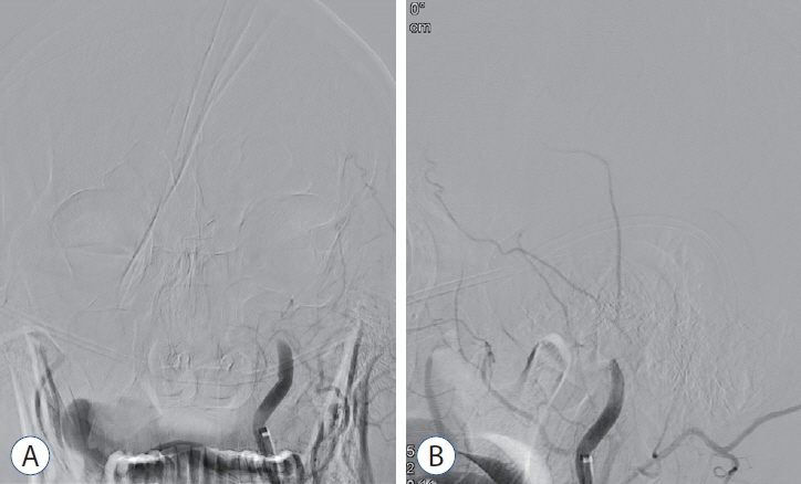
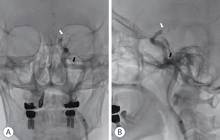
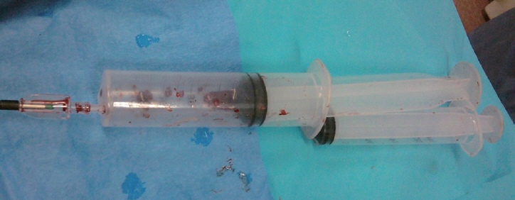
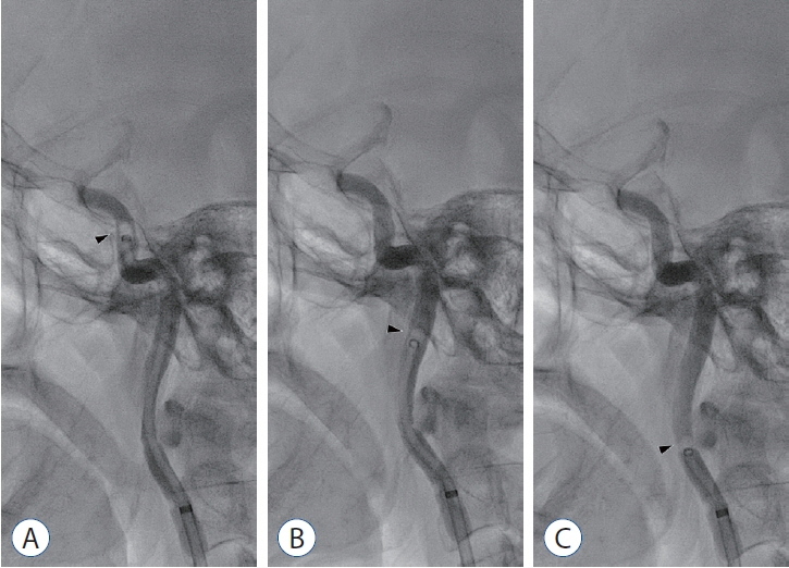
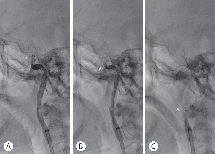
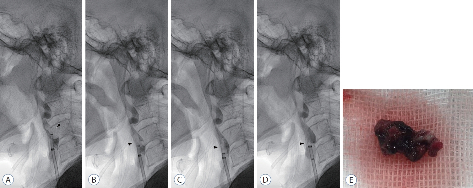
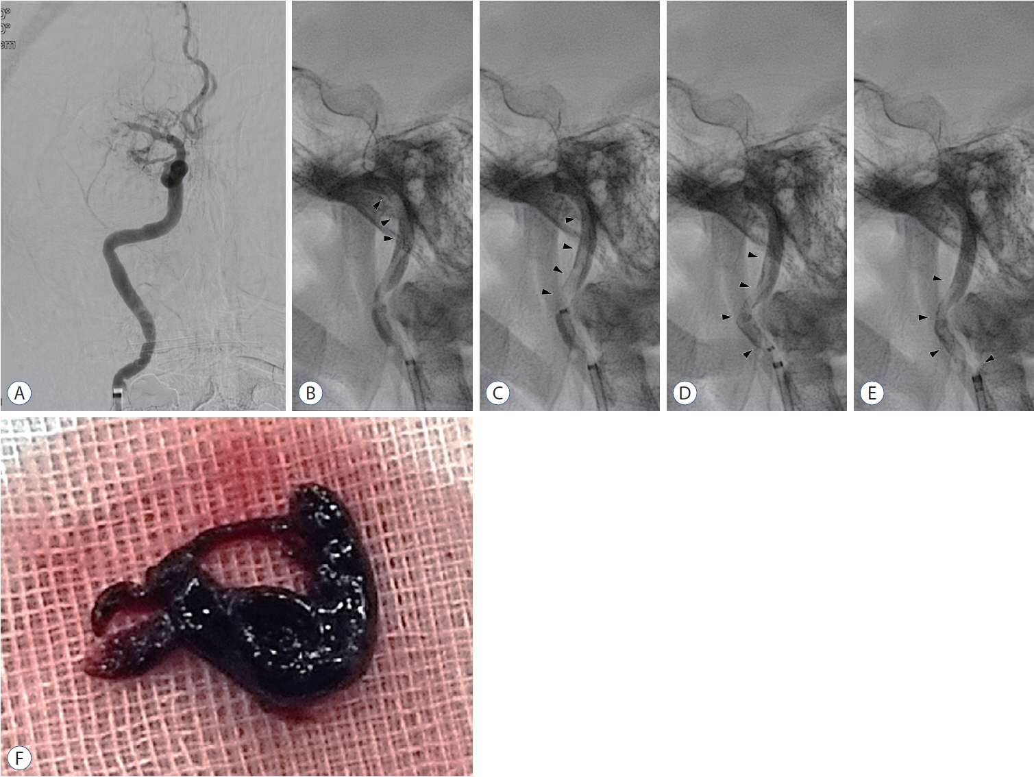
- This report introduces a simple method to visualize the captured thrombus in real-time during suction thrombectomy using “contrast-in-stasis technique”. It enables visualization of the thrombus captured by a suction catheter as it is being retrieved through the tortuous course of the carotid artery eventually into the guiding catheter. It also offers visual identification of important findings such as fragmentation of thrombus into pieces or loss of thrombus during retrieval, and, therefore, helps clinicians to make further critical decisions during the procedure.
Keyword
Figure
Reference
-
References
1. Park H. A suction thrombectomy technique: a rapid and effective method for intra-arterial thrombolysis. J Cerebrovasc Endovasc Neurosurg. 17:13–19. 2015.
Article2. Powers WJ, Rabinstein AA, Ackerson T, Adeoye OM, Bambakidis NC, Becker K, et al. Guidelines for the early management of patients with acute ischemic stroke: 2019 update to the 2018 guidelines for the early management of acute ischemic stroke: a guideline for healthcare professionals from the American Heart Association/American Stroke Association. Stroke. 50:e344–e418. 2019.
Article3. Roh HK, Ju MW, Byoun HS, Park B, Park KH, Lim J. Forced suction thrombectomy in patients with acute ischemic stroke using the SOFIA Plus device. J Cerebrovasc Endovasc Neurosurg. 24:241–248. 2022.
Article4. Suh CH, Lee YJ. Recent trend for endovascular treatment in patients with acute ischemic stroke: balloon guide catheter. Neurointervention. 14:142–143. 2019.
Article
- Full Text Links
- Actions
-
Cited
- CITED
-
- Close
- Share
- Similar articles
-
- Suction thrombectomy of distal medium vessel occlusion using microcatheter during mechanical thrombectomy for acute ischemic stroke: A case series
- Pseudoaneurysm Formation after Repetitive Suction Thrombectomy Using a Penumbra Suction Catheter
- Forced Arterial Suction Thrombectomy Using Distal Access Catheter in Acute Ischemic Stroke
- A Suction Thrombectomy Technique: A Rapid and Effective Method for Intra-Arterial Thrombolysis
- Directly Retrieved Entire Clot from Forced Suction Thrombectomy Using the Penumbra System in Acute Middle Cerebral Artery Occlusion

