Garre’s osteomyelitis of the mandible managed by nonsurgical reendodontic treatment
- Affiliations
-
- 1Department of Conservative Dentistry, School of Dentistry, Kyung Hee University, Seoul, Korea
- KMID: 2556542
- DOI: http://doi.org/10.5395/rde.2024.49.e13
Abstract
- Chronic osteomyelitis with proliferative periostitis, known as Garre’s osteomyelitis, is a type of osteomyelitis characterized by a distinctive gross thickening of the periosteum of bones. Peripheral reactive bone formation can be caused by mild irritation or infection. Garre’s osteomyelitis is usually diagnosed in children and young adults, and the mandible is more affected than the maxilla. The following is a case report of a 12-year-old female patient with Garre’s osteomyelitis of the mandible due to an infection of a root canal-treated tooth. Without surgical intervention, the patient’s symptoms were relieved through nonsurgical root canal re-treatment with long-term calcium hydroxide placement. A cone-beam computed tomography image obtained 6 months after treatment completion displayed complete healing of the periapical lesion and resolution of the peripheral reactive buccal bone. Due to the clinical features of Garre's osteomyelitis, which is characterized by thickening of the periosteum, it can be mistaken for other diseases such as fibrous dysplasia. It is important to correctly diagnose Garre's osteomyelitis based on its distinctive clinical features to avoid unnecessary surgical intervention, and it can lead to minimally invasive treatment options.
Figure
-
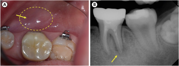
Figure 1 Preoperative examination. (A) A clinical photograph displaying the normal coral-pink color of the buccal vestibule of tooth #36 and the absence of a sinus tract. (B) A periapical radiograph displaying apical radiolucency associated with #36.
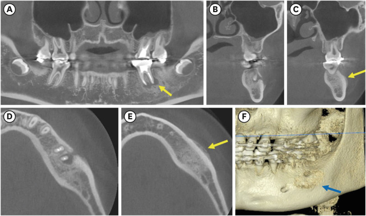
Figure 2 Cone beam computed tomography images of tooth #36. (A) A sagittal image displaying periapical radiolucency of tooth #36. (B, C) The coronal view exhibits unique proliferative buccal cortical bone formation (arrow). (D, E) The axial view displays a typical “onion skin-like appearance over the cortical bone (arrow). (F) Three-dimensional reconstruction image presenting buccal bone changes which indicate the neo-hard tissue formation (arrow).
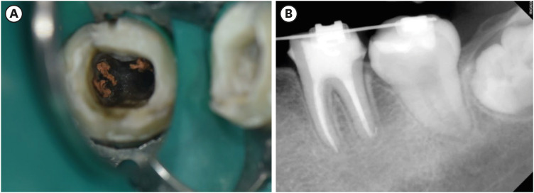
Figure 3 Intraoperative photograph and follow-up periapical radiograph. (A) After removal of composite resin core, contaminated gutta-percha cones were observed. (B) Six-month follow-up after nonsurgical root canal retreatment revealed the resolution of periapical radiolucency. Tooth #36 is asymptomatic and functional.
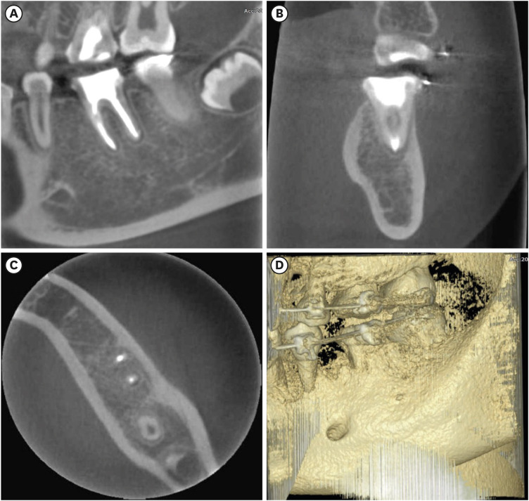
Figure 4 Cone beam computed tomography images of tooth #36 after 6-month follow-up. (A) A sagittal image displaying the complete resolution of periapical radiolucency of tooth #36. (B, C) Coronal and axial views display the absence of buccal cortical bone formation. (D) Three-dimensional reconstruction image presents normal buccal bone anatomy of the mandible.
Reference
-
1. Garré C. Über besondere formen und folgezustande der akuten infekfionsen osteomyelitis. Beitr Z Klin Chir. 1893; 10:241–298.2. Lichty G, Langlais RP, Aufdemorte T. Garré’s osteomyelitis. Literature review and case report. Oral Surg Oral Med Oral Pathol. 1980; 50:309–313. PMID: 6935581.3. Seok H, Kim SG, Song JY. Proliferative periostitis of the mandibular ramus and condyle: a case report. J Korean Assoc Oral Maxillofac Surg. 2015; 41:198–202. PMID: 26339579.4. Suma R, Vinay C, Shashikanth MC, Subba Reddy VV. Garre’s sclerosing osteomyelitis. J Indian Soc Pedod Prev Dent. 2007; 25(Supplment):S30–S33. PMID: 17921638.5. Eisenbud L, Miller J, Roberts IL. Garré’s proliferative periostitis occurring simultaneously in four quadrants of the jaws. Oral Surg Oral Med Oral Pathol. 1981; 51:172–178. PMID: 6937840.6. Batcheldor GD Jr, Giansanti JS, Hibbard ED, Waldron CA. Garré’s osteomyelitis of the jaws: a review and report of two cases. J Am Dent Assoc. 1973; 87:892–897. PMID: 4516757.7. Kannan SK, Sandhya G, Selvarani R. Periostitis ossificans (Garrè’s osteomyelitis) radiographic study of two cases. Int J Paediatr Dent. 2006; 16:59–64. PMID: 16364095.8. Dym H, Zeidan J. Microbiology of acute and chronic osteomyelitis and antibiotic treatment. Dent Clin North Am. 2017; 61:271–282. PMID: 28317566.9. Jalali P, Riccobono J, Augsburger RA, Tahmasbi-Arashlow M. Radiographic patterns of periosteal bone reactions associated with endodontic lesions. Restor Dent Endod. 2023; 48:e23. PMID: 37675448.10. McWalter GM, Schaberg SJ. Garre’s osteomyelitis of the mandible resolved by endodontic treatment. J Am Dent Assoc. 1984; 108:193–195. PMID: 6584491.11. Zand V, Lotfi M, Vosoughhosseini S. Proliferative periostitis: a case report. J Endod. 2008; 34:481–483. PMID: 18358903.12. Aeran H, Tuli A, Pokhiriyal A, Chaudhary A. Management of Garré’s sclerosing osteomyelitis by endodontic therapy: a case report. Int J Oral Health Dent. 2018; 4:55–57.13. Kim D, Kim E. Antimicrobial effect of calcium hydroxide as an intracanal medicament in root canal treatment: a literature review - part I. In vitro studies. Restor Dent Endod. 2014; 39:241–252. PMID: 25383341.14. Kim D, Kim E. Antimicrobial effect of calcium hydroxide as an intracanal medicament in root canal treatment: a literature review - part II. In vivo studies. Restor Dent Endod. 2015; 40:97–103. PMID: 25984470.15. Ibrahim AM, Zakhary SY, Amin SA. Calcium hydroxide intracanal medication effects on pain and flare-up: a systematic review and meta-analysis. Restor Dent Endod. 2020; 45:e26. PMID: 32839707.16. Petrikowski CG, Pharoah MJ, Lee L, Grace MG. Radiographic differentiation of osteogenic sarcoma, osteomyelitis, and fibrous dysplasia of the jaws. Oral Surg Oral Med Oral Pathol Oral Radiol Endod. 1995; 80:744–750. PMID: 8680984.
- Full Text Links
-
- Actions
-
Cited
- CITED
-
- Close
- Share
-
- Similar articles
-
- A CASE REPORT OF GARRE
- A Case of Tuberculous Osteomyelitis of the Mandible
- Pathologic Fractures of the Mandible
- Treatment of osteomyelitis in the rear area of the lingula of the mandible using sagittal split ramus osteotomy: a case report
- Reconstruction of a pathologic fracture following osteomyelitis of the mandible using a fibula osteocutaneous flap
