Electrically Evoked Auditory Brainstem Response Using Extracochlear Stimulation at Different Cochlear Sites: A Comparison With Intracochlear Stimulation
- Affiliations
-
- 1Department of Otorhinolaryngology-Head and Neck Surgery, Gil Medical Center, Gachon University College of Medicine, Incheon, Korea
- 2Department of Otorhinolaryngology-Head and Neck Surgery, Seoul National University Hospital, Seoul National University College of Medicine, Seoul, Korea
- KMID: 2556000
- DOI: http://doi.org/10.21053/ceo.2023.00034
Abstract
Objectives
. The distribution and extent of excitable spiral ganglion neurons (SGNs) have been investigated using the electrically evoked auditory brainstem response (EABR) during preoperative and perioperative periods. In this study, we investigated the EABR with extracochlear stimulation (eEABR) as a preoperative test to estimate these factors.
Methods
. Sixteen male Sprague-Dawley rats were used in this study. Experiments were conducted in nine rats with normal hearing and seven rats that were partially deafened with ouabain treatment. Each experiment involved the following steps: extracochlear stimulating electrode placement at three different sites along the axis of the cochlea and eEABR recordings; cochleostomy and four-channel intracochlear array implantation, followed by EABR recordings with various electrode pair combinations; and after electrophysiological measurements, harvest of the cochleae for histopathological evaluation. The slope characteristics of the amplitude growth function measured from eEABR and EABR, frequency-specific auditory thresholds, and the density of SGNs were compared.
Results
. Similar trends were observed in slope changes on different sites of stimulation with both types of stimulation in normal-hearing animals—specifically, a monotonically increasing slope with increasing distance between bipolar pairs. In addition, eEABR slopes showed significant correlations with EABR slopes when the expected cochlear regions of stimulation were similar in normal-hearing animals. In partially deaf animals, the auditory thresholds at several frequencies had a significant correlation with the eEABR slopes of each extracochlear electrode at the apical, middle, and basal cochlear positions. This indicated that increasing the regions of cochlear stimulation had a differential impact on eEABR slopes, depending on the neural conditions.
Conclusion
. Our results indicated that eEABR slopes showed significant spatial correlations with the functionality of the auditory nerve. Therefore, eEABR tests at various cochlear positions might be used for estimating the extent of excitable SGNs in cochlear implant candidates prior to implantation.
Keyword
Figure
-
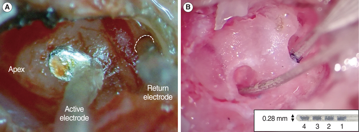
Fig. 1. Placement of the bipolar electrodes for electrical stimulation. (A) The extracochlear implant consists of a raddle-shaped active electrode on the cochlear surface and a ball-type return electrode in the round window niche (dotted line). (B) For intracochlear stimulation, two electrode arrays were inserted into the basal and middle turn via cochleostomy. The inset image shows the intracochlear electrode array inserted into the scala, which has four contacts. The contacts are numbered from 1 (most basal) to 4 (most apical).
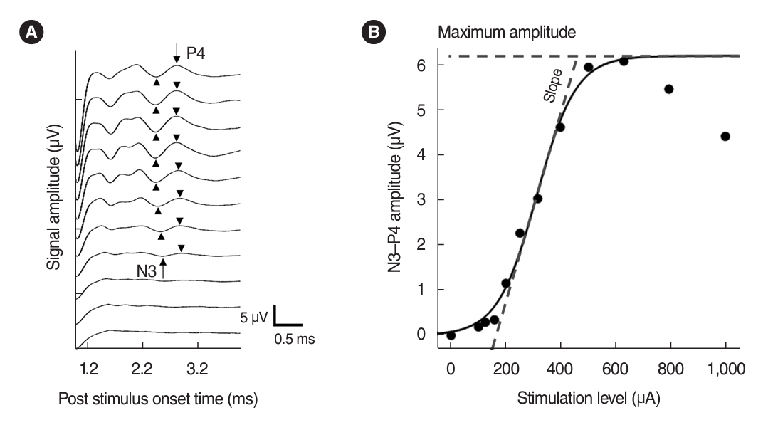
Fig. 2. Example of electrically evoked auditory brainstem responses for the various stimulation levels (μA) recorded in a normal-hearing animal. (A) Arrow and arrowheads indicate the minima and maxima detected as N3 and P4 components. (B) The amplitude growth function for the N3-P4 peak-to-peak amplitudes is plotted for the same example data. The dashed lines indicate the slope characteristics and the maximum amplitude computed from the Boltzmann sigmoidal equation.
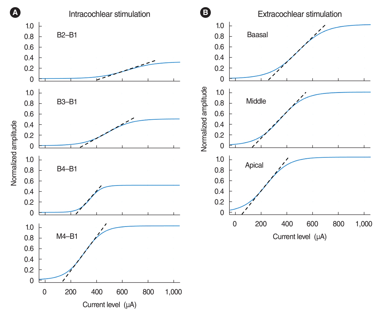
Fig. 3. The normalized amplitude growth functions in response to intracochlear (A) and extracochlear stimulation (B) from a single normalhearing animal. The slope of the growth function is marked in the dashed line. The bipolar electrode pair and extracochlear electrode positions are shown in the legend of each panel.
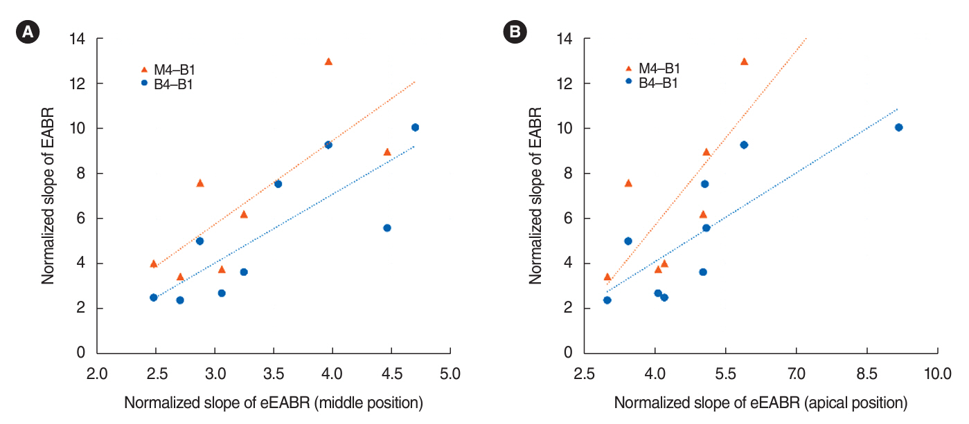
Fig. 4. Correlation of normalized slope (normalized amplitude/mA) of the electrically evoked auditory brainstem response (EABR) and EABR with extracochlear stimulation (eEABR), recorded in the normal-hearing group. Depicted are individual data (points) and linear regression lines of EABR to eEABR slopes. The EABR slopes were obtained with the two most widely spaced intracochlear electrodes. Electrode pairs are indicated by different symbols, as the legend (color online) notes. For eEABR slopes, the two different positions were analyzed separately (A: middle position, B: apical position). Pearson correlation coefficients (r) and P-values for each comparison are shown in Table 1.
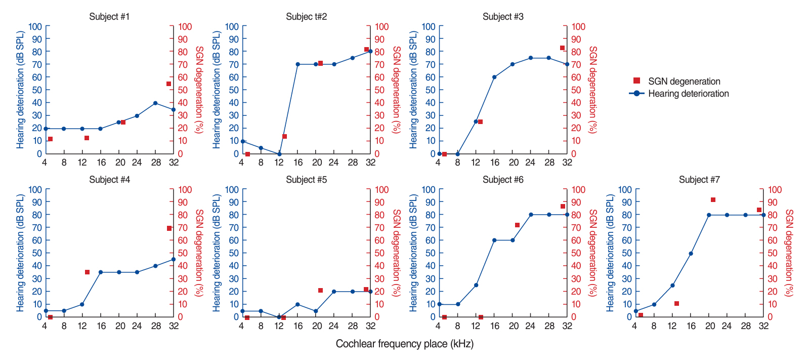
Fig. 5. Tone-pip auditory brainstem response (ABR) audiograms and percent spiral ganglion neuron (SGN) loss as a function of cochlear frequency place (kHz) related to each cochlear region for individual partially deafened animals. Hearing degeneration (dB SPL, ●) is on the primary left axis, and the corresponding degree of SGN loss (%, ■) is plotted on the right axis.

Fig. 6. Scatter plots showing the relation between electrically evoked auditory brainstem response with extracochlear stimulation (eEABR) slope (normalized amplitude/mA) and auditory brainstem response (ABR) thresholds (dB SPL) at 20 kHz for the seven animals in the partially deaf group. Electrode regions are indicated by different symbols, as noted in the legend (color online). Pearson correlation coefficients (r) and P-values for each comparison are shown in Table 2.
Reference
-
1. Brown CJ, Abbas PJ, Gantz B. Electrically evoked whole-nerve action potentials: data from human cochlear implant users. J Acoust Soc Am. 1990; Sep. 88(3):1385–91.
Article2. Brown CJ, Abbas PJ, Gantz BJ. Preliminary experience with neural response telemetry in the nucleus CI24M cochlear implant. Am J Otol. 1998; May. 19(3):320–7.3. Starr A, Brackmann DE. Brain stem potentials evoked by electrical stimulation of the cochlea in human subjects. Ann Otol Rhinol Laryngol. 1979; Jul-Aug. 88(4 Pt 1):550–6.
Article4. Chouard CH, Meyer B, Donadieu F. Auditory brainstem potentials in man evoked by electrical stimulation of the round window. Acta Otolaryngol. 1979; Mar-Apr. 87(3-4):287–93.
Article5. van den Honert C, Stypulkowski PH. Characterization of the electrically evoked auditory brainstem response (ABR) in cats and humans. Hear Res. 1986; 21(2):109–26.
Article6. Nikolopoulos TP, Mason SM, Gibbin KP, O’Donoghue GM. The prognostic value of promontory electric auditory brain stem response in pediatric cochlear implantation. Ear Hear. 2000; Jun. 21(3):236–41.
Article7. Kileny PR, Zwolan TA. Pre-perioperative, transtympanic electrically evoked auditory brainstem response in children. Int J Audiol. 2004; Dec. 43 Suppl 1:S16–21.8. Lassaletta L, Polak M, Huesers J, Diaz-Gomez M, Calvino M, VarelaNieto I, et al. Usefulness of electrical auditory brainstem responses to assess the functionality of the cochlear nerve using an intracochlear test electrode. Otol Neurotol. 2017; Dec. 38(10):e413–20.
Article9. Fernandez NM, Vernetta CP, Garrido LC, Gomez MD, Perez CM. Electrically evoked auditory brainstem response over round window by bipolar stimulation. J Int Adv Otol. 2018; Dec. 14(3):370–4.
Article10. Pau H, Gibson WP, Sanli H. Trans-tympanic electric auditory brainstem response (TT-EABR): the importance of the positioning of the stimulating electrode. Cochlear Implants Int. 2006; Dec. 7(4):183–7.
Article11. Smith L, Simmons FB. Estimating eighth nerve survival by electrical stimulation. Ann Otol Rhinol Laryngol. 1983; Jan-Feb. 92(1 Pt 1):19–23.
Article12. Hall RD. Estimation of surviving spiral ganglion cells in the deaf rat using the electrically evoked auditory brainstem response. Hear Res. 1990; Nov. 49(1-3):155–68.
Article13. Miller CA, Abbas PJ, Robinson BK. The use of long-duration current pulses to assess nerve survival. Hear Res. 1994; Jul. 78(1):11–26.
Article14. Miller CA, Abbas PJ, Brown CJ. Electrically evoked auditory brainstem response to stimulation of different sites in the cochlea. Hear Res. 1993; Apr. 66(2):130–42.
Article15. Shepherd RK, Hatsushika S, Clark GM. Electrical stimulation of the auditory nerve: the effect of electrode position on neural excitation. Hear Res. 1993; Mar. 66(1):108–20.
Article16. Abbas PJ, Brown CJ, Shallop JK, Firszt JB, Hughes ML, Hong SH, et al. Summary of results using the nucleus CI24M implant to record the electrically evoked compound action potential. Ear Hear. 1999; Feb. 20(1):45–59.
Article17. Snyder RL, Middlebrooks JC, Bonham BH. Cochlear implant electrode configuration effects on activation threshold and tonotopic selectivity. Hear Res. 2008; Jan. 235(1-2):23–38.
Article18. Ramekers D, Versnel H, Strahl SB, Smeets EM, Klis SF, Grolman W. Auditory-nerve responses to varied inter-phase gap and phase duration of the electric pulse stimulus as predictors for neuronal degeneration. J Assoc Res Otolaryngol. 2014; Apr. 15(2):187–202.
Article19. Muller M. Frequency representation in the rat cochlea. Hear Res. 1991; Feb. 51(2):247–54.
Article20. Kim JR, Abbas PJ, Brown CJ, Etler CP, O’Brien S, Kim LS. The relationship between electrically evoked compound action potential and speech perception: a study in cochlear implant users with short electrode array. Otol Neurotol. 2010; Sep. 31(7):1041–8.21. Nehme A, El Zir E, Moukarzel N, Haidar H, Vanpoucke F, Arnold L. Measures of the electrically evoked compound action potential threshold and slope in HiRes 90K(TM) users. Cochlear Implants Int. 2014; Jan. 15(1):53–60.22. Dziemba OC, Aristeidou A, Brill S. Slope of electrically evoked compound action potential amplitude growth function is site-dependent. Cochlear Implants Int. 2021; May. 22(3):136–47.
Article23. Kuo SC, Gibson WP. The role of the promontory stimulation test in cochlear implantation. Cochlear Implants Int. 2002; Mar. 3(1):19–28.
Article24. Lenarz T, Hoth S. Comparison of different methods of preoperative electrical testing in cochlear implant patients. In : In : Banfai P, editor. International cochlear implant symposium; Bermann;1987. p. 97–100.25. Smoorenburg GF, Van Olphen AF. Pre-operative electrostimulation of the auditory nerve and postoperative results with the house/3m cochlear implant. In : International cochlear implant symposium; Test Promontoria;1987. Düren (west Germany).26. Kileny PR, Zwolan TA, Zimmerman-Phillips S, Kemink JL. A comparison of round-window and transtympanic promontory electric stimulation in cochlear implant candidates. Ear Hear. 1992; Oct. 13(5):294–9.
Article27. Cousillas H, Patuzzi RB, Johnstone BM. Post-stimulatory effects of direct current stimulation of the cochlea on auditory nerve activity. Hear Res. 1988; Oct. 36(1):21–39.
Article28. Laurikainen E, Kanninen P, Aho H, Saukko P. The anatomy of the human promontory for laser Doppler flowmetry. Eur Arch Otorhinolaryngol. 1997; 254(6):264–8.
Article
- Full Text Links
-
- Actions
-
Cited
- CITED
-
- Close
- Share
-
- Similar articles
-
- Utility of Electrically Evoked Potentials in Cochlear Implant Users
- A Study of Electrical Middle Latency Response in a Cat: Effect of Stimulation Rate and Pulse Duration
- Auditory brainstem response evoked by electrical stimulation (II): estimation of surviving spiral ganglion cells
- Comparison of Brainstem Auditory Evoked Response in Normal Infants, Male and Female, Right and Left Ear
- Computer Generated Three-Dimensional Reconstruction of the Auditory Pathway Structures of Brainstem
