Solitary Skull Langerhans Cell Histiocytosis Presenting With a Pus Draining Fistula: An Unusual Presentation and Review of Literature
- Affiliations
-
- 1Liaquat National Hospital, Karachi, Pakistan
- 2Section of Neurosurgery, Department of Surgery, Aga Khan University Hospital, Karachi, Pakistan
- 3Department of Pathology and Laboratory Medicine, Aga Khan University Hospital, Karachi, Pakistan
- KMID: 2555869
- DOI: http://doi.org/10.14791/btrt.2023.0043
Abstract
- Langerhans cell histiocytosis (LCH) is a rare condition in adults, especially when it is limited to a single area of the skull, known as solitary calvarial involvement. In this case report, we present a unique instance of LCH affecting the parietal bone with a pus-draining fistula. This is a rare and unusual presentation at this location, which has been scarcely reported in medical literature. A 30-year-old woman with no prior comorbidity presented with complaints of headache that persisted for a year. She also had swelling on her scalp and a yellowish discharge for 3 weeks, but no neurological problems were observed. Radiology revealed thinning of the calvaria, with ragged margins along the inner table, multiple focal erosions, and involvement of overlying soft tissue and bony sequestrum. The patient underwent biparietal craniotomy and excision of the lesion. The histopathology report showed LCH. After 8 months of follow-up, there was no recurrence. The management of solitary calvarial involvement by LCH with masquerading presentation as a scalp infection can be achieved through complete excision of the lesions, resulting in a favorable outcome.
Keyword
Figure
-
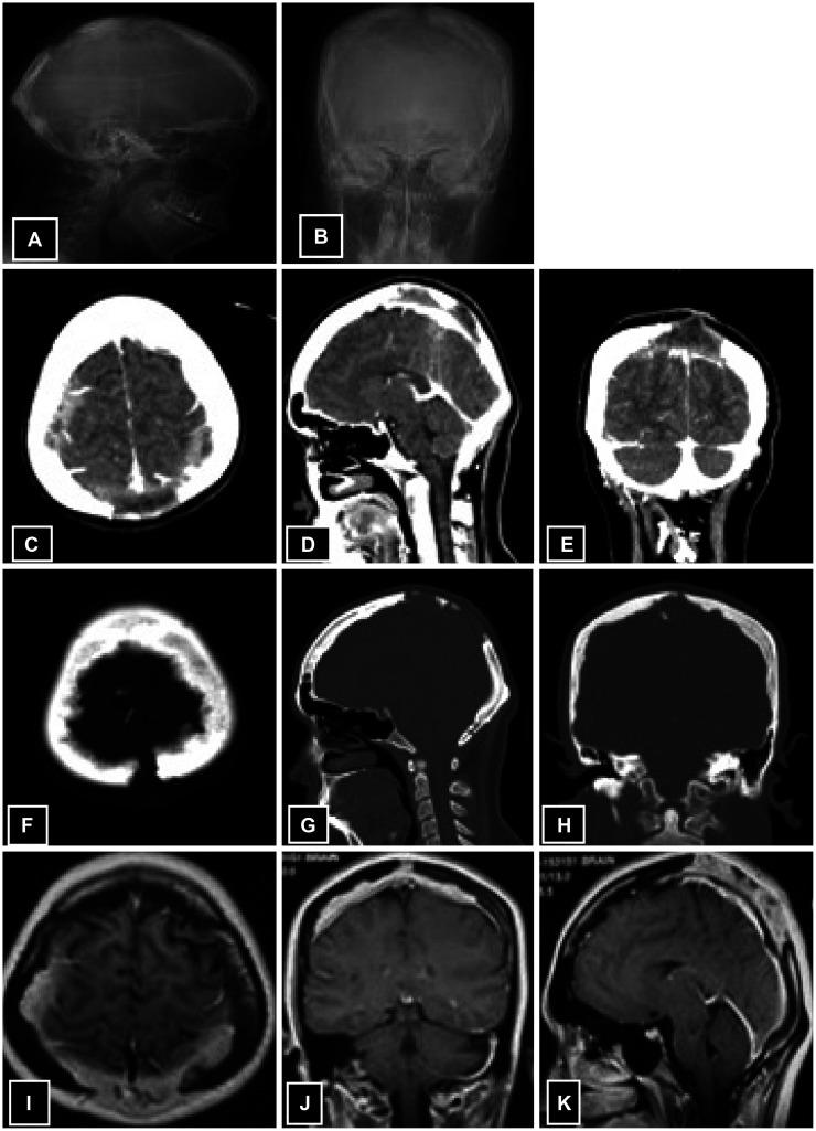
Fig. 1 Preoperative images of the patient. A and B: X-rays showing solitary punched-out midline lytic lesion extending from vertex to lambda with beveled margins without sclerosis. C-E: CT scan with contrast; axial (C), sagittal (D), and coronal (E) views showing extra-axial lesion involving bilateral parietal bones with subcutaneous tissue involvement of scalp. F-H: CT scan with post-contrast enhancement representing bone window showing erosion in parietal bones bilaterally. I-K: MRI with contrast; axial (I), coronal (J), and sagittal (K) views showing ill-defined heterogeneously enhancing lesion at vertex with enhancing extradural collection.
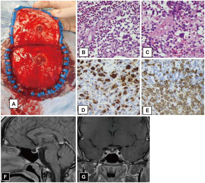
Fig. 2 Intraoperative image (A) showing a nodular lesion arising from parietal bone with galeal involvement. B and C: Microscopic description: fibro collagenous tissue composed of sheets of Langerhan cells, having moderate to abundant eosinophilic cytoplasm with elongated, pale nuclei having longitudinal grooves, inconspicuous nucleoli, fine chromatin, and thin nuclear membranes. D and E: CD68 (D) and CD1a (E) reactivity. F and G: MRI sagittal view (F) and coronal view (G) showing thickened pituitary stalk.
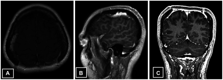
Fig. 3 Postoperative MRI. Axial (A), sagittal (B), and coronal (C) view showing residual dural thickening and enhancement.
Reference
-
1. Badia RR, Wong D, Ghandour R, Chertack N, Meng X, Hutchinson R, et al. Validation of testicular germ cell tumor staging in nationwide cancer registries. Urol Oncol. 2021; 39:838.e1–838.e6.2. Tatli M, Guzel A, Guzel E. Solitary eosinophilic granuloma of the parietal bone in an adult patient. Neurosciences (Riyadh). 2007; 12:160–162. PMID: 21857602.3. El-Arab KK, Luedke AI, Julian BT, Ferrauiola J, Miller FR, Wang HT. Langerhans cell histiocytosis in an adult: a discussion of epidemiology and treatment options. J Craniofac Surg. 2020; 31:e70–e73. PMID: 31634312.4. Kaul R, Gupta N, Gupta S, Gupta M. Eosinophilic granuloma of skull bone. J Cytol. 2009; 26:156–157. PMID: 21938183.5. Kitsoulis PV, Paraskevas G, Vrettakos A, Marini A. A case of eosinophilic granuloma of the skull in an adult man: a case report. Cases J. 2009; 2:9144. PMID: 20062661.6. Samara A, Nepute J, Lu HC, Perrin RJ, Eldaya RW. Calvarial Langerhans cell histiocytosis in an adult: typical imaging findings in an atypical age group. Radiol Case Rep. 2019; 14:1478–1482. PMID: 31641396.7. Gannaban RAP, Calimag PP Jr, Zapanta ZS. Langerhans cell histiocystosis presenting as a scalp abscess: a case report. Philipp J Surg Spec. 2011; 66:74–79.8. Mosiewicz A, Rola R, Jarosz B, Trojanowska A, Trojanowski T. Langerhans cell histiocytosis of the parietal bone with epidural and extracranial expansion–case report and a review of the literature. Neurol Neurochir Pol. 2010; 44:196–203. PMID: 20496290.9. Girschikofsky M, Arico M, Castillo D, Chu A, Doberauer C, Fichter J, et al. Management of adult patients with Langerhans cell histiocytosis: recommendations from an expert panel on behalf of Euro-Histio-Net. Orphanet J Rare Dis. 2013; 8:72. PMID: 23672541.10. Sugiyama H, Tsutsumi S, Hashizume A, Kuroda K, Sugiyama N, Ueno H, et al. Calvarial Langerhans cell histiocytosis in an adult presenting rapid growth. Surg Neurol Int. 2022; 13:347. PMID: 36128163.11. Sunder G, Mukesh P, Deepika P. Intradural Langerhans’ cell histiocytosis invading frontal bone of skull: a case report with a brief review. Ann Clin Med Case Rep. 2020; 5:1–3.12. Zaveri J, La Q, Yarmish G, Neuman J. More than just Langerhans cell histiocytosis: a radiologic review of histiocytic disorders. Radiographics. 2014; 34:2008–2024. PMID: 25384298.13. Gadner H, Grois N, Pötschger U, Minkov M, Aricò M, Braier J, et al. Improved outcome in multisystem Langerhans cell histiocytosis is associated with therapy intensification. Blood. 2008; 111:2556–2562. PMID: 18089850.14. Lee SK, Jung TY, Jung S, Han DK, Lee JK, Baek HJ. Solitary Langerhans cell histocytosis of skull and spine in pediatric and adult patients. Childs Nerv Syst. 2014; 30:271–275. PMID: 23780406.15. Hashimoto K, Nishimura S, Sakata N, Inoue M, Sawada A, Akagi M. Treatment outcomes of Langerhans cell histiocytosis: a retrospective study. Medicina (Kaunas). 2021; 57:356. PMID: 33917120.16. Cantu MA, Lupo PJ, Bilgi M, Hicks MJ, Allen CE, McClain KL. Optimal therapy for adults with Langerhans cell histiocytosis bone lesions. PLoS One. 2012; 7:e43257. PMID: 22916233.
- Full Text Links
-
- Actions
-
Cited
- CITED
-
- Close
- Share
-
- Similar articles
-
- Langerhans Cell Histiocytosis Presenting as a Solitary Nodule
- Eosinophilic Granuloma Presenting as an Epidural Hematoma and Cyst
- Adult Scapular Langerhans Cell Histiocytosis Mistaken for Acute Osteomyelitis
- Two Cases of Langerhans' Cell Histiocytosis: Report of Two Cases
- Langerhans cell histiocytosis of the mandible: two case reports and literature review
