J Rhinol.
2024 Mar;31(1):52-56. 10.18787/jr.2023.00076.
Two Cases of Cerebrospinal Fluid Rhinorrhea Repair Surgery Using TachoComb
- Affiliations
-
- 1Department of Otolaryngology-Head and Neck Surgery, Bucheon Saint Mary’s Hospital, College of Medicine, The Catholic University of Korea, Seoul, Republic of Korea
- KMID: 2554092
- DOI: http://doi.org/10.18787/jr.2023.00076
Abstract
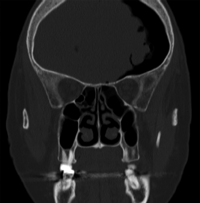
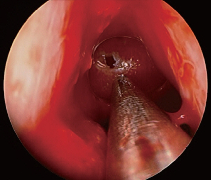
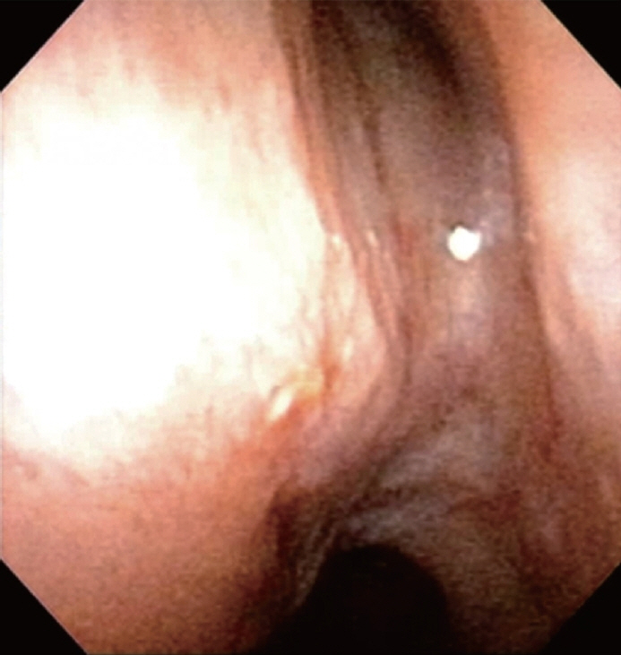
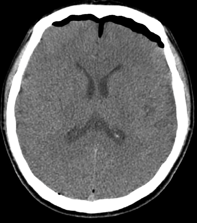
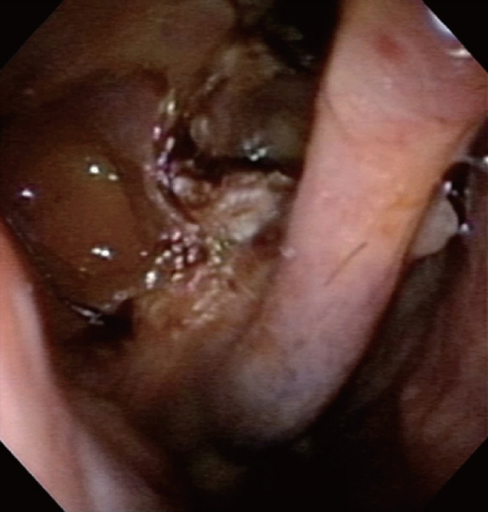
- Cerebrospinal fluid (CSF) rhinorrhea is a rare condition characterized by the leakage of CSF through the nose. The diagnosis is established through comprehensive history taking, brain imaging, and nasal endoscopy. Surgical intervention is considered a secondary option for CSF leakage when conservative treatments, including behavioral therapy, pharmacotherapy, or lumbar puncture, fail to elicit a response. In recent years, endoscopic intranasal surgery has been favored over craniotomy for such surgical treatment. When repairing CSF leakage defects via endoscopic intranasal surgery, autologous fat and muscle flaps are commonly employed. However, these grafts may lead to complications, including donor site infection, edema, and wound dehiscence. Therefore, in this article, we would like to introduce two cases of CSF rhinorrhea repair surgery using TachoComb. While previous studies have employed TachoComb as a supplementary material for the repair of CSF leak defects, in the cases we describe, the primary reconstruction of the defect area was achieved using TachoComb, supported by free grafts such as septal bone or turbinate mucosal flap, which were smaller than the size of the CSF leakage defects.
Keyword
Figure
Reference
-
References
1. Abuabara A. Cerebrospinal fluid rhinorrhoea: diagnosis and management. Med Oral Patol Oral Cir Bucal. 2007; 12(5):E397–400.2. Yilmazlar S, Arslan E, Kocaeli H, Dogan S, Aksoy K, Korfali E, et al. Cerebrospinal fluid leakage complicating skull base fractures: analysis of 81 cases. Neurosurg Rev. 2006; 29(1):64–71.
Article3. Brodie HA, Thompson TC. Management of complications from 820 temporal bone fractures. Am J Otol. 1997; 18(2):188–97.4. Tosun F, Gonul E, Yetiser S, Gerek M. Analysis of different surgical approaches for the treatment of cerebrospinal fluid rhinorrhea. Minim Invasive Neurosurg. 2005; 48(6):355–60.
Article5. Stankiewicz JA. Cerebrospinal f luid fistula and endoscopic sinus surgery. Laryngoscope. 1991; 101(3):250–6.6. Zanation AM, Carrau RL, Snyderman CH, Germanwala AV, Gardner PA, Prevedello DM, et al. Nasoseptal flap reconstruction of high f low intraoperative cerebral spinal f luid leaks during endoscopic skull base surgery. Am J Rhinol Allergy. 2009; 23(5):518–21.7. Hadad G, Bassagasteguy L, Carrau RL, Mataza JC, Kassam A, Snyderman CH, et al. A novel reconstructive technique after endoscopic expanded endonasal approaches: vascular pedicle nasoseptal flap. Laryngoscope. 2006; 116(10):1882–6.
Article8. Jones ME, Reino T, Gnoy A, Guillory S, Wackym P, Lawson W. Identification of intranasal cerebrospinal fluid leaks by topical application with fluorescein dye. Am J Rhinol. 2000; 14(2):93–6.
Article9. Kassam AB, Prevedello DM, Carrau RL, Snyderman CH, Thomas A, Gardner P, et al. Endoscopic endonasal skull base surgery: analysis of complications in the authors’ initial 800 patients. J Neurosurg. 2011; 114(6):1544–68.
Article10. Harvey RJ, Parmar P, Sacks R, Zanation AM. Endoscopic skull base reconstruction of large dural defects: a systematic review of published evidence. Laryngoscope. 2012; 122(2):452–9.
Article11. Pinheiro-Neto CD, Ramos HF, Peris-Celda M, Fernandez-Miranda JC, Gardner PA, Snyderman CH, et al. Study of the nasoseptal flap for endoscopic anterior cranial base reconstruction. Laryngoscope. 2011; 121(12):2514–20.
Article
- Full Text Links
- Actions
-
Cited
- CITED
-
- Close
- Share
- Similar articles
-
- A Case of Acute Hydrocephalus Occurring after Operative Treatment on Nontraumatic Cerebrospinal Fluid Rhinorrhea
- Surgical Repair of Cerebrospinal Fluid Rhinorrhea with Mucoperichondrial Free Graft
- Endoscopic Repair of Spontaneous Cerebrospinal Fluid Rhinorrhea with a Nasoseptal Flap
- Management of Cerebrospinal Fluid Rhinorrhea
- Traumatic Cerebrospinal Fluid Rhinorrhea: Successful Closure under the Surgical Microscope

