J Dent Rehabil Appl Sci.
2024 Feb;40(1):31-38. 10.14368/jdras.2024.40.1.31.
Root canal therapy of anterior teeth with dens invaginatus
- Affiliations
-
- 1Department of Conservative Dentistry, School of Dentistry, Dental Science Research Institute, Chonnam National University, Gwangju, Republic of Korea
- KMID: 2553629
- DOI: http://doi.org/10.14368/jdras.2024.40.1.31
Abstract
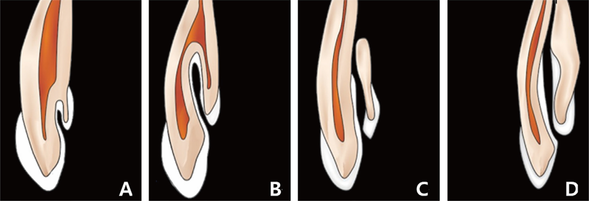
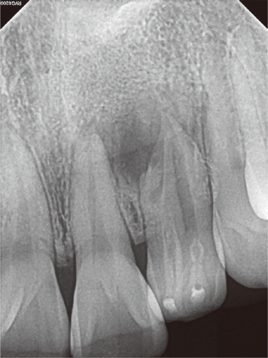
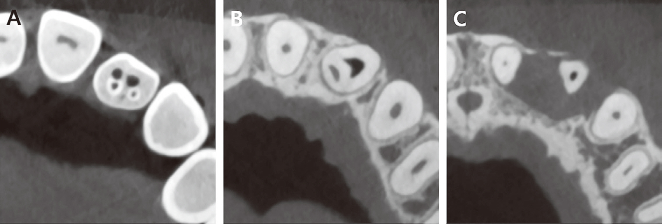
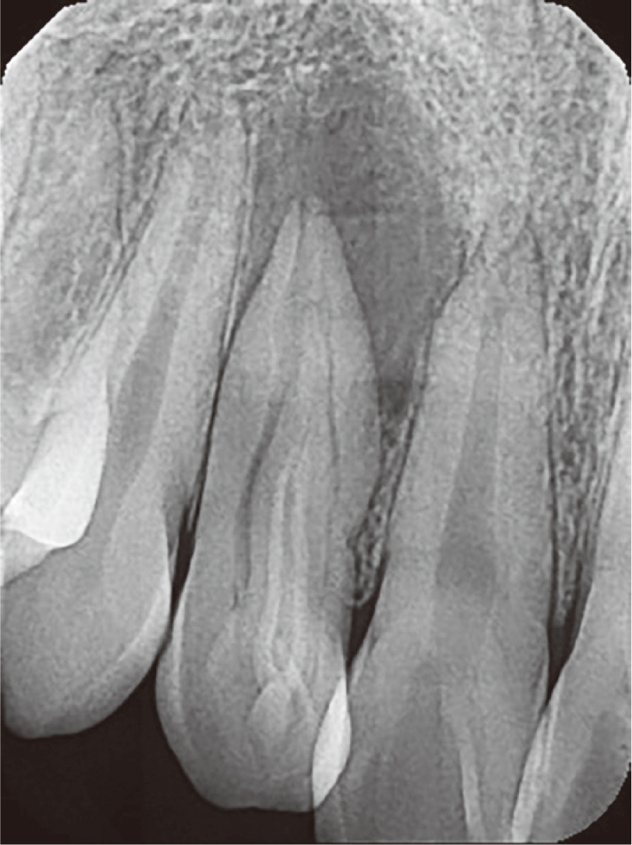
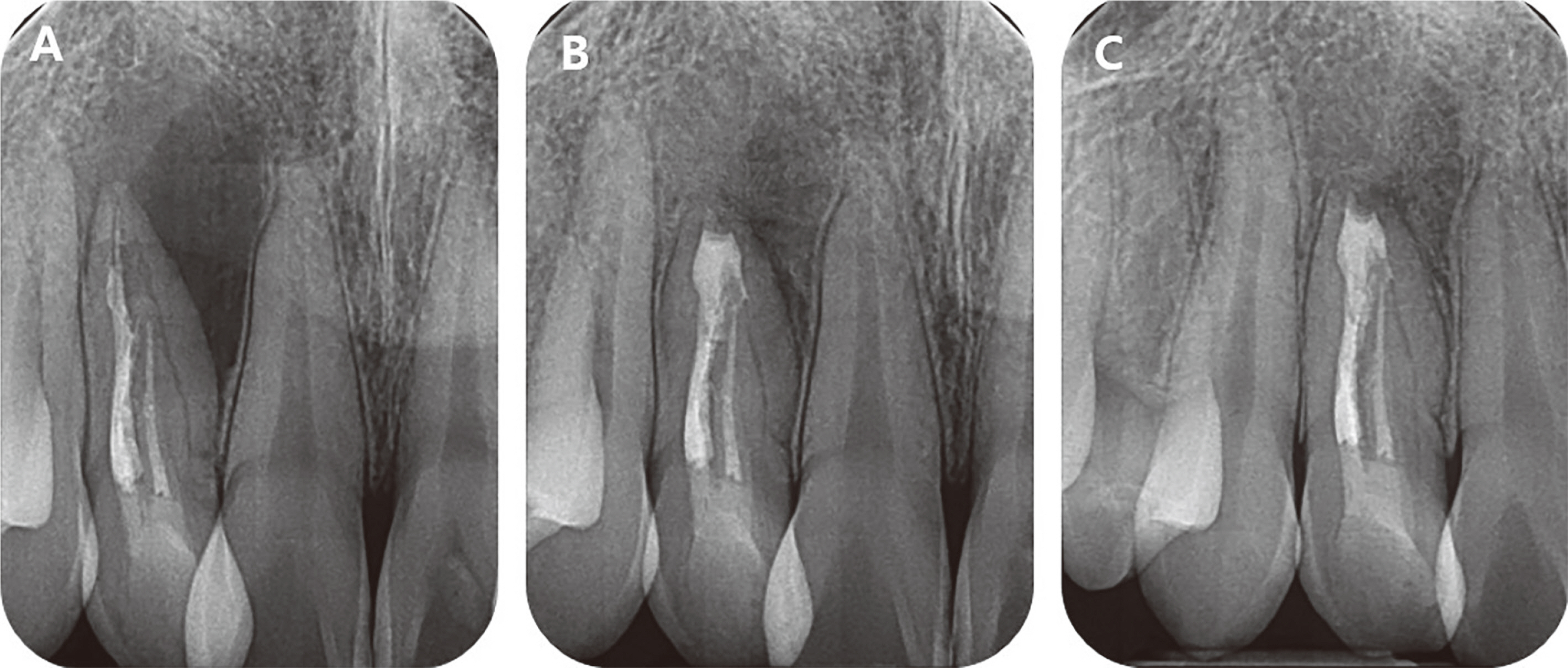
- Dens in dente is a developmental anomaly resulting from infolding of the enamel organ into dental papilla prior to calcification of dental tissue. The pulpal tissue of the tooth can be vulnerable for bacterial invasion through direct exposure to the oral cavity or through defective enamel and dentin of the infolding part, thereby increasing the possibility of pulpal necrosis and subsequent apical periodontitis. Treatment planning of teeth with dens invaginatus may be difficult due to the complex root canal morphology. Therefore, thorough knowledge of anatomical variations of dens invaginatus is of great importance for proper treatment planning. The focus of this case report is on Oehler’s type II and III dens invaginatus. The infolding of type III dens invaginatus extends beyond the crown and CEJ. Bacterial invasion through the infolding can easily cause inflammation of the pulpal and periradicular tissue. This case report presents endodontic treatment of type II and III dens invaginatus with the aid of CBCT.
Keyword
Figure
Reference
-
References
1. Gallacher A, Ali R, Bhakta S. 2016; Dens invaginatus: diagnosis and management strategies. Br Dent J. 221:383–7. DOI: 10.1038/sj.bdj.2016.724. PMID: 27713460.
Article2. Hülsmann M. 1997; Dens Invaginatus: aetiology, classification, prevalence, diagnosis, and treatment considerations. Int Endod J. 30:79–90. DOI: 10.1111/j.1365-2591.1997.tb00679.x. PMID: 10332241.
Article3. Gonçalves A, Gonçalves M, Oliveira DP, Gonçalves N. 2002; Dens invaginatus type III: report of a case and 10-year radiographic follow-up. Int Endod J. 35:873–9. DOI: 10.1046/j.1365-2591.2002.00575.x. PMID: 12406383.
Article4. Oehlers FA. 1957; Dens invaginatus (dilated composite odontome). I. Variations of the invagination process and associated anterior crown forms. Oral Surg Oral Med Oral Pathol. 10:1204–18. DOI: 10.1016/0030-4220(57)90077-4. PMID: 13477660.5. Fregnani ER, Spinola LFB, Sônego JRO, Bueno CES, De Martin AS. 2008; Complex endodontic treatment of an immature type III dens invaginatus. A case report. Int Endod J. 41:913–9. DOI: 10.1111/j.1365-2591.2008.01414.x. PMID: 18699792.
Article6. Hata G, Toda T. 1987; Treatment of dens invaginatus by endodontic therapy, apicocurettage, and retrofilling. J Endod. 13:469–72. DOI: 10.1016/S0099-2399(87)80067-5. PMID: 3482106.7. Jung M. 2004; Endodontic treatment of dens invaginatus type III with three root canals and open apical foramen. Int Endod J. 37:205–13. DOI: 10.1111/j.0143-2885.2004.00778.x. PMID: 15032229.
Article8. Mupparapu M, Singer SR. 2004; A rare presentation of dens invaginatus in a mandibular lateral incisor occurring concurrently with bilateral maxillary dens invaginatus: case report and review of literature. Aust Dent J. 49:90–3. DOI: 10.1111/j.1834-7819.2004.tb00056.x. PMID: 15293820.9. Kharangate N, Figueiredo NR, Fernandes M, Lambor R. 2015; Bilateral dens invaginatus in the mandibular premolars - Diagnosis and treatment. Contemp Clin Dent. 6:428–31. DOI: 10.4103/0976-237X.161911. PMID: 26321850. PMCID: PMC4550002.
Article10. Skoner JR, Wallace JA. 1994; Dens invaginatus: another use for the ultrasonic. J Endod. 20:138–40. DOI: 10.1016/S0099-2399(06)80060-9. PMID: 7996087.
Article11. Roizenblit RN, Soares FO, Lopes RT, Dos Santos BC, Gusman H. 2020; Root canal filling quality of mandibular molars with EndoSequence BC and AH Plus sealers: A micro-CT study. Aust Endod J. 46:82–7. DOI: 10.1111/aej.12373. PMID: 31556201.12. Kim JH, Cho SY, Choi Y, Kim DH, Shin SJ, Jung IY. 2022; Clinical Efficacy of Sealer-based Obturation Using Calcium Silicate Sealers: A Randomized Clinical Trial. J Endod. 48:144–51. DOI: 10.1016/j.joen.2021.11.011. PMID: 34856212.
Article13. Zhou HM, Shen Y, Zheng W, Li L, Zheng YF, Haapasalo M. 2013; Physical properties of 5 root canal sealers. J Endod. 39:1281–6. DOI: 10.1016/j.joen.2013.06.012. PMID: 24041392.
Article14. Yu YH, Kushnir L, Kohli M, Karabucak B. 2021; Comparing the incidence of postoperative pain after root canal filling with warm vertical obturation with resin-based sealer and sealer-based obturation with calcium silicate-based sealer: a prospective clinical trial. Clin Oral Investig. 25:5033–42. DOI: 10.1007/s00784-021-03814-x. PMID: 33555456.
Article
- Full Text Links
- Actions
-
Cited
- CITED
-
- Close
- Share
- Similar articles
-
- Endodontic management of a maxillary lateral incisor with dens invaginatus and external root irregularity using cone-beam computed tomography
- A case report of multiple bilateral dens invaginatus in maxillary anteriors
- Successful nonsurgical treatment of type II dens invaginatus with 5 root canals using a self-adjusting file: a case report
- Guided endodontics: a case report of maxillary lateral incisors with multiple dens invaginatus
- Generalized Short Root Anomaly with Various Dental Anomalies : A Case Report with a 5 - Year Follow - up

