Korean Circ J.
2024 Feb;54(2):110-112. 10.4070/kcj.2023.0314.
Identification of Coronary Healed Plaque Using a Combined HighDefinition 35–65 MHz Intravascular Ultrasound and Near-Infrared Spectroscopy
- Affiliations
-
- 1Cardiovascular Center, Korea University Ansan Hospital, Ansan, Korea
- KMID: 2552230
- DOI: http://doi.org/10.4070/kcj.2023.0314
Figure
-
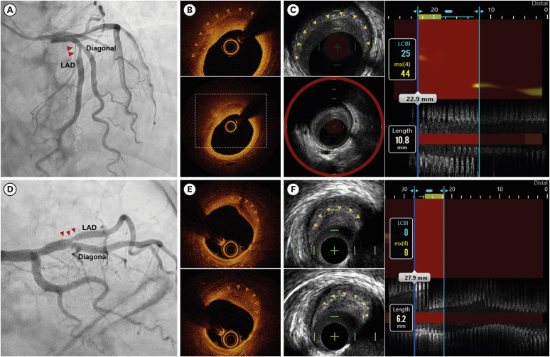
Figure 1 Coronary angiographies (A, D) showing focal bifurcation lesions. OCT identified atheromas with a characteristic layered appearance, confirming the presence of healed plaques (B, E). High-definition 35–65 MHz IVUS revealed crescent-shaped, hypoechoic layers clearly demarcating underlying hyperechoic plaques (C, F). These layered plaques exhibited low lipids on NIRS.IVUS = intravascular ultrasound; NIRS = near-infrared spectroscopy; OCT = optical coherence tomography.
Reference
-
1. Burke AP, Kolodgie FD, Farb A, et al. Healed plaque ruptures and sudden coronary death: evidence that subclinical rupture has a role in plaque progression. Circulation. 2001; 103:934–940. PMID: 11181466.2. Vergallo R, Crea F. Atherosclerotic plaque healing. N Engl J Med. 2020; 383:846–857. PMID: 32846063.3. Shimokado A, Matsuo Y, Kubo T, et al. In vivo optical coherence tomography imaging and histopathology of healed coronary plaques. Atherosclerosis. 2018; 275:35–42. PMID: 29859471.4. Ma T, Yu M, Li J, et al. Multi-frequency intravascular ultrasound (IVUS) imaging. IEEE Trans Ultrason Ferroelectr Freq Control. 2015; 62:97–107. PMID: 25585394.5. García-García HM, Finizio M, del Val D, Rivero F, Waksman R, Alfonso F. High-definition intravascular ultrasound: current clinical uses. Int J Cardiovasc Imaging. 2022; 38:1213–1220.
- Full Text Links
-
- Actions
-
Cited
- CITED
-
- Close
- Share
-
- Similar articles
-
- An Overview of Near-Infrared Spectroscopy-Intravascular Ultrasound and Its Applications in Coronary Artery Disease
- Practical Application of Coronary Imaging Devices in Cardiovascular Intervention
- Advances in Intravascular Imaging: New Insights into the Vulnerable Plaque from Imaging Studies
- A Case of Coronary Pseudostenosis, Diagnosed by Intravascular Ultrasound
- Multimodal intravascular photoacoustic and ultrasound imaging
