Accuracy of posteroanterior cephalogram landmarks and measurements identification using a cascaded convolutional neural network algorithm: A multicenter study
- Han SH
 1
1 - Lim J2
- Kim JS2
- Cho JH3
- Hong M4
- Kim M5
- Kim SJ6
- Kim YJ7
- Kim Y8
- Lim SH9
- Sung SJ7
- Kang KH10
- Baek SH11
- Choi SK10
- Kim N2
- Affiliations
-
- 1Department of Orthodontics, Seoul St. Mary's Hospital, College of Medicine, The Catholic University of Korea, Seoul, Korea
- 2Department of Convergence Medicine, Asan Medical Institute of Convergence Science and Technology, Asan Medical Center, University of Ulsan College of Medicine, Seoul, Korea
- 3Department of Orthodontics, School of Dentistry, Chonnam National University, Gwangju, Korea
- 4Department of Orthodontics, School of Dentistry, Kyungpook National University, Daegu, Korea
- 5Department of Orthodontics, College of Medicine, Ewha Womans University, Seoul, Korea
- 6Department of Orthodontics, Kyung Hee University School of Dentistry, Seoul, Korea
- 7Department of Orthodontics, Asan Medical Center, University of Ulsan College of Medicine, Seoul, Korea
- 8Department of Orthodontics, Institute of Oral Health Science, Ajou University School of Medicine, Suwon, Korea
- 9Department of Orthodontics, College of Dentistry, Chosun University, Gwangju, Korea
- 10Department of Orthodontics, School of Dentistry, Wonkwang University, Iksan, Korea
- 11Department of Orthodontics, School of Dentistry, Dental Research Institute, Seoul National University, Seoul, Korea
- KMID: 2551474
- DOI: http://doi.org/10.4041/kjod23.075
Abstract
Objective
To quantify the effects of midline-related landmark identification on midline deviation measurements in posteroanterior (PA) cephalograms using a cascaded convolutional neural network (CNN).
Methods
A total of 2,903 PA cephalogram images obtained from 9 university hospitals were divided into training, internal validation, and test sets (n = 2,150, 376, and 377). As the gold standard, 2 orthodontic professors marked the bilateral landmarks, including the frontozygomatic suture point and latero-orbitale (LO), and the midline landmarks, including the crista galli, anterior nasal spine (ANS), upper dental midpoint (UDM), lower dental midpoint (LDM), and menton (Me). For the test, Examiner-1 and Examiner-2 (3-year and 1-year orthodontic residents) and the Cascaded-CNN models marked the landmarks. After point-to-point errors of landmark identification, the successful detection rate (SDR) and distance and direction of the midline landmark deviation from the midsagittal line (ANS-mid, UDM-mid, LDM-mid, and Me-mid) were measured, and statistical analysis was performed.
Results
The cascaded-CNN algorithm showed a clinically acceptable level of point-to-point error (1.26 mm vs. 1.57 mm in Examiner-1 and 1.75 mm in Examiner-2). The average SDR within the 2 mm range was 83.2%, with high accuracy at the LO (right, 96.9%; left, 97.1%), and UDM (96.9%). The absolute measurement errors were less than 1 mm for ANSmid, UDM-mid, and LDM-mid compared with the gold standard.
Conclusions
The cascaded-CNN model may be considered an effective tool for the auto-identification of midline landmarks and quantification of midline deviation in PA cephalograms of adult patients, regardless of variations in the image acquisition method.
Figure
-
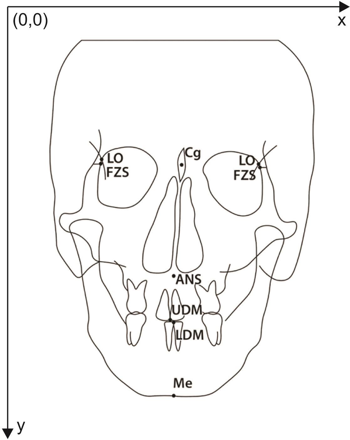
Figure 1 The posteroanterior cephalometric landmarks used in this study. LO, latero-orbitale; FZS, frontozygomatic suture point; Cg, crista galli; ANS, anterior nasal spine; UDM, upper dental midpoint; LDM, lower dental midpoint; Me, menton.
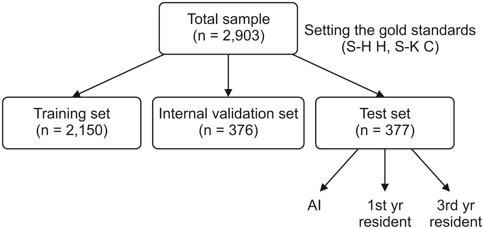
Figure 2 Flow chart showing sample allocation and study design. AI, artificial intelligence.

Figure 3 Cascaded convolutional neural network algorithm used in this study. Stage 1, the region of interest detection to propose the area of interest; stage 2, the landmark prediction to find the exact location of landmarks. W, width; H, height; K, number of object classes; A, number of anchors.
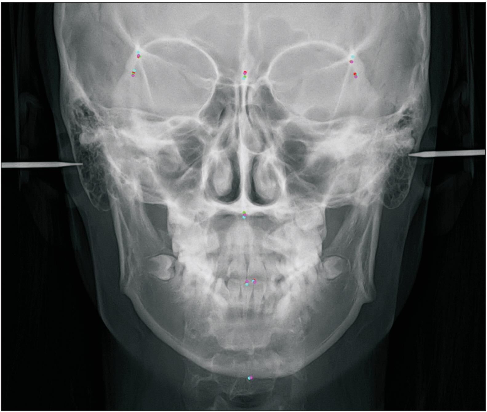
Figure 4 Examples of superimposition of the identified posteroanterior cephalometric landmarks. Red, gold standard; green, auto-identification by cascaded convolutional neural network algorithm; pink, Examiner-1; sky blue, Examiner-2.
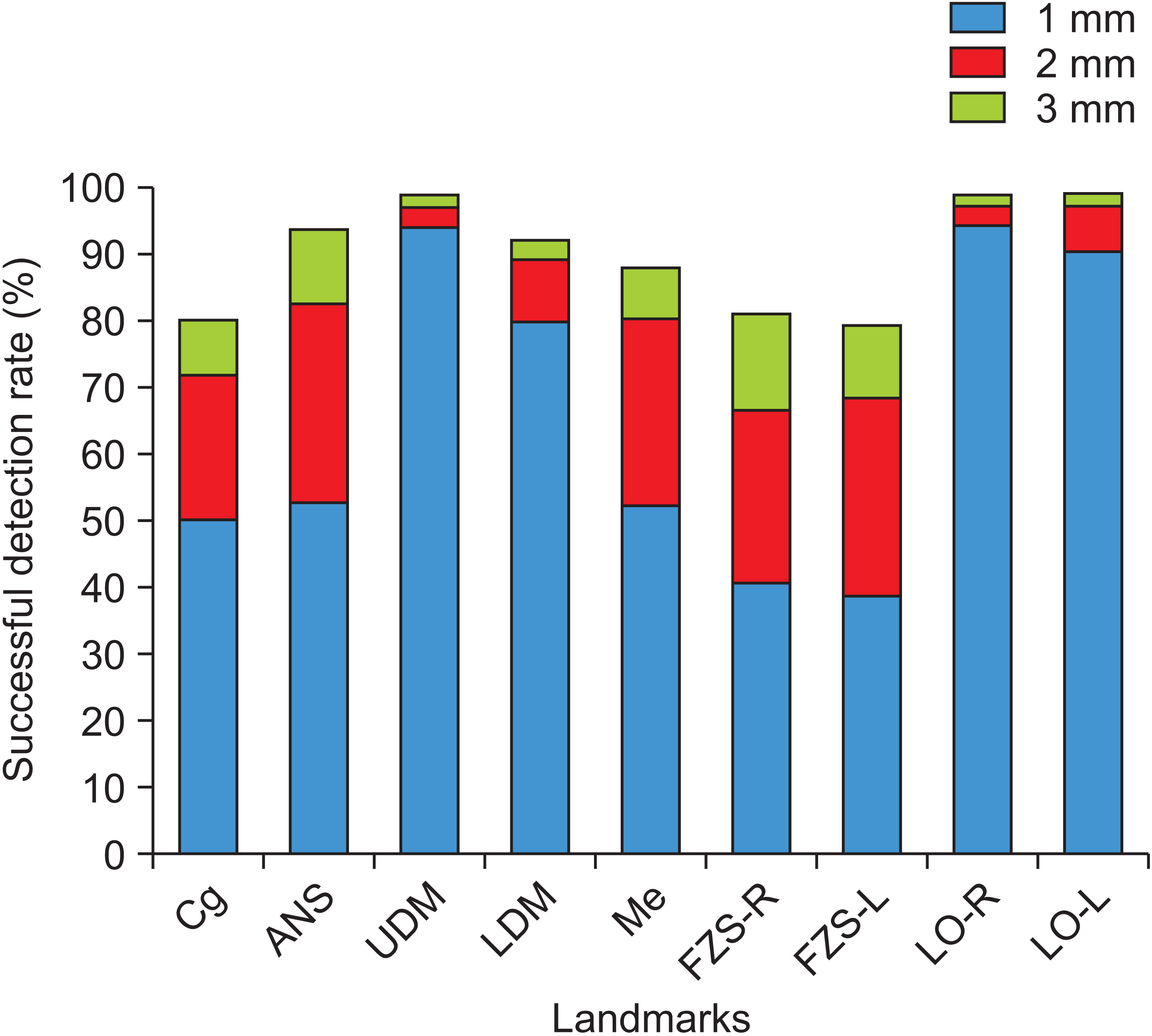
Figure 5 Comparison of the successful detection rate within the range of 1.0 mm, 2.0 mm, and 3.0 mm in each landmark. Cg, crista galli; ANS, anterior nasal spine; UDM, upper dental midpoint; LDM, lower dental midpoint; Me, menton; FZP-R, frontozygomatic suture point right; FZP-L, frontozygomatic suture point left; LO-R, latero-orbitale right; LO-L, latero-orbitale left.

Figure 6 Landmarks and the midsagittal reference line for measurements of the distance and direction of landmark deviation from the midsagittal line (ANS-mid, UDM-mid, LDM-mid, and Me-mid) on posteroanterior cephalogram images. LO, latero-orbitale; Cg, crista galli; ANS, anterior nasal spine; UDM, upper dental midpoint; LDM, lower dental midpoint; Me, menton; mid, midsagittal line.
Reference
-
1. McCarthy J. Thomason RH, editor. 1989. Artificial intelligence, logic and formalizing common sense. Philosophical logic and artificial intelligence. Springer;Dordrecht: p. 161–90. https://doi.org/10.1007/978-94-009-2448-2_6. DOI: 10.1007/978-94-009-2448-2_6.2. Hamet P, Tremblay J. 2017; Artificial intelligence in medicine. Metabolism. 69S:S36–40. https://doi.org/10.1016/j.metabol.2017.01.011. DOI: 10.1016/j.metabol.2017.01.011. PMID: 28126242.3. LeCun Y, Bengio Y, Hinton G. 2015; Deep learning. Nature. 521:436–44. https://doi.org/10.1038/nature14539. DOI: 10.1038/nature14539. PMID: 26017442.4. Yasaka K, Akai H, Kunimatsu A, Kiryu S, Abe O. 2018; Deep learning with convolutional neural network in radiology. Jpn J Radiol. 36:257–72. https://doi.org/10.1007/s11604-018-0726-3. DOI: 10.1007/s11604-018-0726-3. PMID: 29498017.5. Broadbent BH. 1931; A new x-ray technique and its application to orthodontia. Angle Orthod. 1:45–66. https://meridian.allenpress.com/angle-orthodontist/article/1/2/45/55162/A-NEW-X-RAY-TECHNIQUE-and-ITS-APPLICATION-TO.6. Kazandjian S, Kiliaridis S, Mavropoulos A. 2006; Validity and reliability of a new edge-based computerized method for identification of cephalometric landmarks. Angle Orthod. 76:619–24. https://pubmed.ncbi.nlm.nih.gov/16808568/.7. Yu HJ, Cho SR, Kim MJ, Kim WH, Kim JW, Choi J. 2020; Automated skeletal classification with lateral cephalometry based on artificial intelligence. J Dent Res. 99:249–56. https://doi.org/10.1177/0022034520901715. DOI: 10.1177/0022034520901715. PMID: 31977286.8. Kim IH, Kim YG, Kim S, Park JW, Kim N. 2021; Comparing intra-observer variation and external variations of a fully automated cephalometric analysis with a cascade convolutional neural net. Sci Rep. 11:7925. https://doi.org/10.1038/s41598-021-87261-4. DOI: 10.1038/s41598-021-87261-4. PMID: 33846506. PMCID: PMC8041841. PMID: da86601e38c44724a513d0a20be5513c.9. Wang CW, Huang CT, Hsieh MC, Li CH, Chang SW, Li WC, et al. 2015; Evaluation and comparison of anatomical landmark detection methods for cephalometric x-ray images: a grand challenge. IEEE Trans Med Imaging. 34:1890–900. https://doi.org/10.1109/TMI.2015.2412951. DOI: 10.1109/TMI.2015.2412951. PMID: 25794388.10. Park JH, Hwang HW, Moon JH, Yu Y, Kim H, Her SB, et al. 2019; Automated identification of cephalometric landmarks: Part 1-Comparisons between the latest deep-learning methods YOLOV3 and SSD. Angle Orthod. 89:903–9. https://doi.org/10.2319/022019-127.1. DOI: 10.2319/022019-127.1. PMID: 31282738. PMCID: PMC8109157.11. Hwang HW, Park JH, Moon JH, Yu Y, Kim H, Her SB, et al. 2020; Automated identification of cephalometric landmarks: Part 2-Might it be better than human? Angle Orthod. 90:69–76. https://doi.org/10.2319/022019-129.1. DOI: 10.2319/022019-129.1. PMID: 31335162. PMCID: PMC8087057.12. Song Y, Qiao X, Iwamoto Y, Chen YW. 2020; Automatic cephalometric landmark detection on x-ray images using a deep-learning method. Appl Sci. 10:2547. https://doi.org/10.3390/app10072547. DOI: 10.3390/app10072547. PMID: 4e8c7fbd4e9e4041896c8f71decd45f1.13. Kunz F, Stellzig-Eisenhauer A, Zeman F, Boldt J. 2020; Artificial intelligence in orthodontics: evaluation of a fully automated cephalometric analysis using a customized convolutional neural network. J Orofac Orthop. 81:52–68. https://doi.org/10.1007/s00056-019-00203-8. DOI: 10.1007/s00056-019-00203-8. PMID: 31853586.14. Hong M, Kim I, Cho JH, Kang KH, Kim M, Kim SJ, et al. 2022; Accuracy of artificial intelligence-assisted landmark identification in serial lateral cephalograms of Class III patients who underwent orthodontic treatment and two-jaw orthognathic surgery. Korean J Orthod. 52:287–97. https://doi.org/10.4041/kjod21.248. DOI: 10.4041/kjod21.248. PMID: 35719042. PMCID: PMC9314217.15. Muraev AA, Tsai P, Kibardin I, Oborotistov N, Shirayeva T, Ivanov S, et al. 2020; Frontal cephalometric landmarking: humans vs artificial neural networks. Int J Comput Dent. 23:139–48. https://pubmed.ncbi.nlm.nih.gov/32555767/.16. Gil SM, Kim I, Cho JH, Hong M, Kim M, Kim SJ, et al. 2022; Accuracy of auto-identification of the posteroanterior cephalometric landmarks using cascade convolution neural network algorithm and cephalometric images of different quality from nationwide multiple centers. Am J Orthod Dentofacial Orthop. 161:e361–71. https://doi.org/10.1016/j.ajodo.2021.11.011. DOI: 10.1016/j.ajodo.2021.11.011. PMID: 35074216.17. Lin TY, Goyal P, Girshick R, He K, Dollar P. 2020; Focal loss for dense object detection. IEEE Trans Pattern Anal Mach Intell. 42:318–27. https://doi.org/10.1109/TPAMI.2018.2858826. DOI: 10.1109/TPAMI.2018.2858826. PMID: 30040631.18. Ronneberger O, Fischer P, Brox T. Navab N, Hornegger J, Wells W, Frangi A, editors. 2015. U-Net: convolutional networks for biomedical image segmentation. Medical image computing and computer-assisted intervention - MICCAI 2015. Springer;Cham: p. 234–41. https://doi.org/10.1007/978-3-319-24574-4_28. DOI: 10.1007/978-3-319-24574-4_28.19. He K, Zhang X, Ren S, Sun J. 2016. Deep residual learning for image recognition. Paper presented at: 2016 IEEE Conference on Computer Vision and Pattern Recognition (CVPR). 2016 Jun 27-30; Las Vegas, USA. Institute of Electrical and Electronics Engineers (IEEE);Piscataway: https://doi.org/10.1109/CVPR.2016.90. DOI: 10.1109/CVPR.2016.90. PMID: 26180094.20. Lee HJ, Lee S, Lee EJ, Song IJ, Kang BC, Lee JS, et al. 2016; A comparative study of the deviation of the menton on posteroanterior cephalograms and three-dimensional computed tomography. Imaging Sci Dent. 46:33–8. https://doi.org/10.5624/isd.2016.46.1.33. DOI: 10.5624/isd.2016.46.1.33. PMID: 27051637. PMCID: PMC4816769.21. Major PW, Johnson DE, Hesse KL, Glover KE. 1994; Landmark identification error in posterior anterior cephalometrics. Angle Orthod. 64:447–54. https://pubmed.ncbi.nlm.nih.gov/7864466/.22. Na ER, Aljawad H, Lee KM, Hwang HS. 2019; A comparative study of the reproducibility of landmark identification on posteroanterior and anteroposterior cephalograms generated from cone-beam computed tomography scans. Korean J Orthod. 49:41–8. https://doi.org/10.4041/kjod.2019.49.1.41. DOI: 10.4041/kjod.2019.49.1.41. PMID: 30603624. PMCID: PMC6306316.
- Full Text Links
-
- Actions
-
Cited
- CITED
-
- Close
- Share
-
- Similar articles
-
- Evaluation of a multi-stage convolutional neural network-based fully automated landmark identification system using cone-beam computed tomographysynthesized posteroanterior cephalometric images
- Benefits of lateral cephalogram during landmark identification on posteroanterior cephalograms
- Comparative Analysis of Accuracy between Computerized Tomography and Cephalogram for 3-Dimensional Measurement of Maxillofacial Structure
- The use of posteroanterior cephalogram in orthodontics
- Comparison of midsagittal reference plane in PA cephalogram and 3D CT
