Clin Endosc.
2024 Jan;57(1):1-10. 10.5946/ce.2023.031.
Advanced endoscopic imaging for detection of Barrett’s esophagus
- Affiliations
-
- 1Division of Digestive Diseases and Nutrition, Department of Internal Medicine, Rush University Medical Center, Chicago, IL, USA
- KMID: 2551184
- DOI: http://doi.org/10.5946/ce.2023.031
Abstract
- Barrett’s esophagus (BE) is the precursor to esophageal adenocarcinoma (EAC), and is caused by chronic gastroesophageal reflux. BE can progress over time from metaplasia to dysplasia, and eventually to EAC. EAC is associated with a poor prognosis, often due to advanced disease at the time of diagnosis. However, if BE is diagnosed early, pharmacologic and endoscopic treatments can prevent progression to EAC. The current standard of care for BE surveillance utilizes the Seattle protocol. Unfortunately, a sizable proportion of early EAC and BE-related high-grade dysplasia (HGD) are missed due to poor adherence to the Seattle protocol and sampling errors. New modalities using artificial intelligence (AI) have been proposed to improve the detection of early EAC and BE-related HGD. This review will focus on AI technology and its application to various endoscopic modalities such as high-definition white light endoscopy, narrow-band imaging, and volumetric laser endomicroscopy.
Keyword
Figure
-
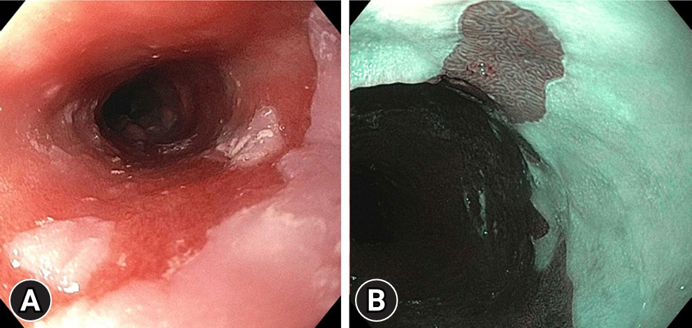
Fig. 1. High-quality endoscopic Barrett’s esophagus examination using high-definition white light endoscopy (A) and narrow-band imaging (B) to identify dysplastic lesions.
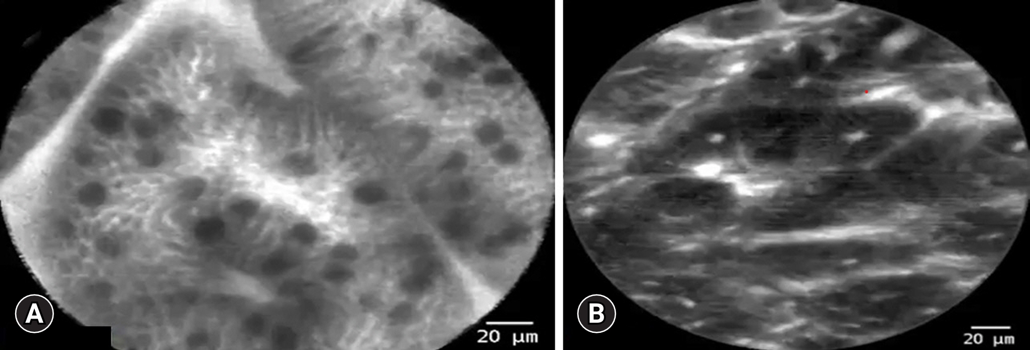
Fig. 2. Images of confocal laser endomicroscopy using a through-the-scope probe showing. (A) Non-dysplastic Barrett’s esophagus. (B) Dysplastic Barrett’s esophagus.

Fig. 3. Volumetric laser endomicroscopy (VLE) images showing a classic VLE image with Barrett’s esophagus dysplasia (A) and magnified image of A showing the dysplastic area (B).
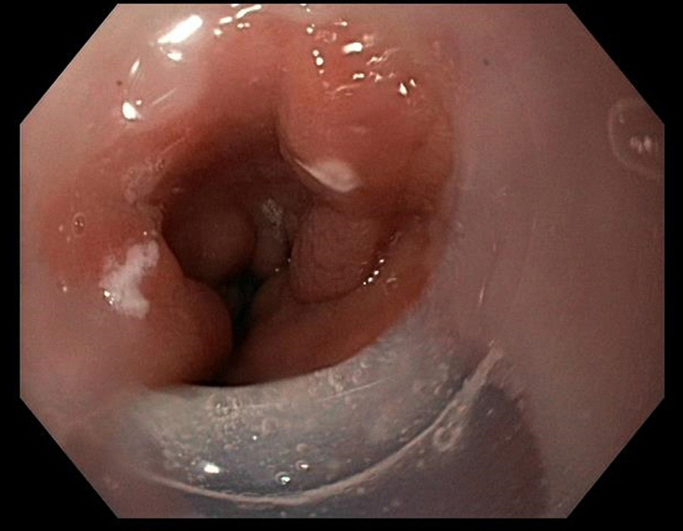
Fig. 4. Laser markings of esophageal mucosa during volumetric laser endomicroscopy examination.
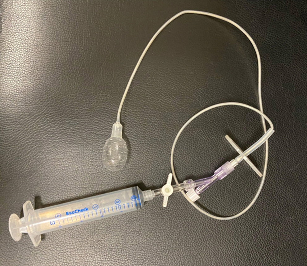
Fig. 5. EsoCheck device (Lucid Dx Labs) with an inflatable silicone balloon attached to a syringe.
Reference
-
1. Edgren G, Adami HO, Weiderpass E, et al. A global assessment of the oesophageal adenocarcinoma epidemic. Gut. 2013; 62:1406–1414.2. Bray F, Ferlay J, Soerjomataram I, et al. Global cancer statistics 2018: GLOBOCAN estimates of incidence and mortality worldwide for 36 cancers in 185 countries. CA Cancer J Clin. 2018; 68:394–424.3. Coleman HG, Xie SH, Lagergren J. The epidemiology of esophageal adenocarcinoma. Gastroenterology. 2018; 154:390–405.4. Konda VJ, Ellison A, Codipilly DC, et al. Quality in Barrett’s esophagus: diagnosis and management. Tech Innov Gastrointest Endosc. 2022; 24:364–380.5. Thein HH, Jembere N, Thavorn K, et al. Estimates and predictors of health care costs of esophageal adenocarcinoma: a population-based cohort study. BMC Cancer. 2018; 18:694.6. Then EO, Lopez M, Saleem S, et al. Esophageal cancer: an updated surveillance epidemiology and end results database analysis. World J Oncol. 2020; 11:55–64.7. Muthusamy VR, Wani S, Gyawali CP, et al. AGA clinical practice update on new technology and innovation for surveillance and screening in Barrett’s esophagus: expert review. Clin Gastroenterol Hepatol. 2022; 20:2696–2706.8. Wani S, Williams JL, Komanduri S, et al. Endoscopists systematically undersample patients with long-segment Barrett’s esophagus: an analysis of biopsy sampling practices from a quality improvement registry. Gastrointest Endosc. 2019; 90:732–741.9. Peters FP, Curvers WL, Rosmolen WD, et al. Surveillance history of endoscopically treated patients with early Barrett’s neoplasia: nonadherence to the Seattle biopsy protocol leads to sampling error. Dis Esophagus. 2008; 21:475–479.10. ASGE Technology Committee, Thosani N, Abu Dayyeh BK, et al. ASGE Technology Committee systematic review and meta-analysis assessing the ASGE Preservation and Incorporation of Valuable Endoscopic Innovations thresholds for adopting real-time imaging-assisted endoscopic targeted biopsy during endoscopic surveillance of Barrett's esophagus. Gastrointest Endosc. 2016; 83:684–698.11. Struyvenberg MR, de Groof AJ, van der Putten J, et al. A computer-assisted algorithm for narrow-band imaging-based tissue characterization in Barrett's esophagus. Gastrointest Endosc. 2021; 93:89–98.12. Boeriu A, Boeriu C, Drasovean S, et al. Narrow-band imaging with magnifying endoscopy for the evaluation of gastrointestinal lesions. World J Gastrointest Endosc. 2015; 7:110–120.13. ASGE Standards of Practice Committee, Qumseya B, Sultan S, et al. ASGE guideline on screening and surveillance of Barrett’s esophagus. Gastrointest Endosc. 2019; 90:335–359.14. Canto MI. Acetic-acid chromoendoscopy for Barrett’s esophagus: the “pros”. Gastrointest Endosc. 2006; 64:13–16.15. Amamra N, Touzet S, Colin C, et al. Current practice compared with the international guidelines: endoscopic surveillance of Barrett’s esophagus. J Eval Clin Pract. 2007; 13:789–794.16. Abrams JA, Kapel RC, Lindberg GM, et al. Adherence to biopsy guidelines for Barrett’s esophagus surveillance in the community setting in the United States. Clin Gastroenterol Hepatol. 2009; 7:736–742.17. Odze RD, Goldblum J, Kaul V. Role of wide-area transepithelial sampling with 3D computer-assisted analysis in the diagnosis and management of Barrett’s esophagus. Clin Transl Gastroenterol. 2021; 12:e00422.18. Suresh Kumar VC, Harne P, Patthipati VS, et al. Wide-area transepithelial sampling in adjunct to forceps biopsy increases the absolute detection rates of Barrett’s oesophagus and oesophageal dysplasia: a meta-analysis and systematic review. BMJ Open Gastroenterol. 2020; 7:e000494.19. Shaheen NJ, Falk GW, Iyer PG, et al. Diagnosis and management of Barrett’s esophagus: an updated ACG guideline. Am J Gastroenterol. 2022; 117:559–587.20. Kataria R, Smith M. Wide area transepithelial sampling (WATS-3D) improves detection of Barrett’s esophagus following endoscopic ablation. Am J Gastroenterol. 2012; 107:S22.21. de Groof AJ, Struyvenberg MR, van der Putten J, et al. Deep-learning system detects neoplasia in patients with Barrett’s esophagus with higher accuracy than endoscopists in a multistep training and validation study with benchmarking. Gastroenterology. 2020; 158:915–929.22. Swager AF, van der Sommen F, Klomp SR, et al. Computer-aided detection of early Barrett’s neoplasia using volumetric laser endomicroscopy. Gastrointest Endosc. 2017; 86:839–846.23. Struyvenberg MR, van der Sommen F, Swager AF, et al. Improved Barrett’s neoplasia detection using computer-assisted multiframe analysis of volumetric laser endomicroscopy. Dis Esophagus. 2020; 33:doz065.24. Struyvenberg MR, de Groof AJ, Fonollà R, et al. Prospective development and validation of a volumetric laser endomicroscopy computer algorithm for detection of Barrett’s neoplasia. Gastrointest Endosc. 2021; 93:871–879.25. Kahn A, McKinley MJ, Stewart M, et al. Artificial intelligence-enhanced volumetric laser endomicroscopy improves dysplasia detection in Barrett’s esophagus in a randomized cross-over study. Sci Rep. 2022; 12:16314.26. Wang KK, Carr-Locke DL, Singh SK, et al. Use of probe-based confocal laser endomicroscopy (pCLE) in gastrointestinal applications: a consensus report based on clinical evidence. United European Gastroenterol J. 2015; 3:230–254.27. ASGE Technology Committee. Confocal laser endomicroscopy. Gastrointest Endosc. 2014; 80:928–938.28. Pohl H, Rösch T, Vieth M, et al. Miniprobe confocal laser microscopy for the detection of invisible neoplasia in patients with Barrett’s oesophagus. Gut. 2008; 57:1648–1653.29. Sharma P, Meining AR, Coron E, et al. Real-time increased detection of neoplastic tissue in Barrett’s esophagus with probe-based confocal laser endomicroscopy: final results of an international multicenter, prospective, randomized, controlled trial. Gastrointest Endosc. 2011; 74:465–472.30. Song J, Zhang J, Wang J, et al. Meta-analysis of the effects of endoscopy with narrow band imaging in detecting dysplasia in Barrett’s esophagus. Dis Esophagus. 2015; 28:560–566.31. Wu J, Pan YM, Wang TT, et al. Confocal laser endomicroscopy for detection of neoplasia in Barrett’s esophagus: a meta-analysis. Dis Esophagus. 2014; 27:248–254.32. Xiong YQ, Ma SJ, Zhou JH, et al. A meta-analysis of confocal laser endomicroscopy for the detection of neoplasia in patients with Barrett's esophagus. J Gastroenterol Hepatol. 2016; 31:1102–1110.33. Becker V, von Delius S, Bajbouj M, et al. Intravenous application of fluorescein for confocal laser scanning microscopy: evaluation of contrast dynamics and image quality with increasing injection-to-imaging time. Gastrointest Endosc. 2008; 68:319–323.34. Kiesslich R, Anagnostopoulos GK, Axon A, et al. Interobserver variation and standardized training for confocal laser endomicroscopy image interpretation in the upper and lower GI tract. Gastrointest Endosc. 2007; 65:AB354.35. Elsbernd BL, Dunbar KB. Volumetric laser endomicroscopy in Barrett’s esophagus. Tech Innov Gastrointest Endosc. 2021; 23:69–76.36. Wallace MB. Detecting dysplasia with optical coherence tomography. Clin Gastroenterol Hepatol. 2006; 4:36–37.37. Evans JA, Poneros JM, Bouma BE, et al. Optical coherence tomography to identify intramucosal carcinoma and high-grade dysplasia in Barrett's esophagus. Clin Gastroenterol Hepatol. 2006; 4:38–43.38. Leggett CL, Gorospe EC, Chan DK, et al. Comparative diagnostic performance of volumetric laser endomicroscopy and confocal laser endomicroscopy in the detection of dysplasia associated with Barrett's esophagus. Gastrointest Endosc. 2016; 83:880–888.39. Swager AF, Tearney GJ, Leggett CL, et al. Identification of volumetric laser endomicroscopy features predictive for early neoplasia in Barrett’s esophagus using high-quality histological correlation. Gastrointest Endosc. 2017; 85:918–926.40. Alshelleh M, Inamdar S, McKinley M, et al. Incremental yield of dysplasia detection in Barrett’s esophagus using volumetric laser endomicroscopy with and without laser marking compared with a standardized random biopsy protocol. Gastrointest Endosc. 2018; 88:35–42.41. Trindade AJ, McKinley MJ, Fan C, et al. Endoscopic surveillance of Barrett’s esophagus using volumetric laser endomicroscopy with artificial intelligence image enhancement. Gastroenterology. 2019; 157:303–305.42. Smith MS, Cash B, Konda V, et al. Volumetric laser endomicroscopy and its application to Barrett’s esophagus: results from a 1,000 patient registry. Dis Esophagus. 2019; 32:doz029.43. Trindade AJ, Inamdar S, Smith MS, et al. Learning curve and competence for volumetric laser endomicroscopy in Barrett’s esophagus using cumulative sum analysis. Endoscopy. 2018; 50:471–478.44. Gora MJ, Quénéhervé L, Carruth RW, et al. Tethered capsule endomicroscopy for microscopic imaging of the esophagus, stomach, and duodenum without sedation in humans (with video). Gastrointest Endosc. 2018; 88:830–840.45. Dong J, Grant C, Vuong B, et al. Feasibility and safety of tethered capsule endomicroscopy in patients with Barrett’s esophagus in a multi-center study. Clin Gastroenterol Hepatol. 2022; 20:756–765.46. Fitzgerald RC, di Pietro M, O'Donovan M, et al. Cytosponge-trefoil factor 3 versus usual care to identify Barrett’s oesophagus in a primary care setting: a multicentre, pragmatic, randomised controlled trial. Lancet. 2020; 396:333–344.
- Full Text Links
-
- Actions
-
Cited
- CITED
-
- Close
- Share
-
- Similar articles
-
- Diagnosis of Barrett's Esophagus
- Confocal Laser Endomicroscopy and Molecular Imaging in Barrett Esophagus and Stomach
- Barrett's Esophagus-With Emphasis on Endoscopic Disgnosis
- Laser Imaging Facilitates Early Detection of Synchronous Adenocarcinomas in Patients with Barrett's Esophagus
- Endoscopic Imaging in Barrett's Oesophagus: Applications in Routine Clinical Practice and Future Outlook
