Push-out bond strength and intratubular biomineralization of a hydraulic root-end filling material premixed with dimethyl sulfoxide as a vehicle
- Affiliations
-
- 1Department of Conservative Dentistry, School of Dentistry, Jeonbuk National University, Jeonju, Korea
- 2Department of Dentistry, College of Medicine, Kosin University, Busan, Korea
- 3Research Institute of Clinical Medicine of Jeonbuk National University, Jeonju, Korea
- 4Biomedical Research Institute of Jeonbuk National University Hospital, Jeonju, Korea
- KMID: 2548176
- DOI: http://doi.org/10.5395/rde.2023.48.e8
Abstract
Objectives
This study was designed to evaluate the parameters of bonding performance to root dentin, including push-out bond strength and dentinal tubular biomineralization, of a hydraulic bioceramic root-end filling material premixed with dimethyl sulfoxide (Endocem MTA Premixed) in comparison to a conventional powder-liquid–type cement (ProRoot MTA).
Materials and Methods
The root canal of a single-rooted premolar was filled with either ProRoot MTA or Endocem MTA Premixed (n = 15). A slice of dentin was obtained from each root. Using the sliced specimen, the push-out bond strength was measured, and the failure pattern was observed under a stereomicroscope. The apical segment was divided into halves; the split surface was observed under a scanning electron microscope, and intratubular biomineralization was examined by observing the precipitates formed in the dentinal tubule. Then, the chemical characteristics of the precipitates were evaluated with energy-dispersive X-ray spectroscopic (EDS) analysis. The data were analyzed using the Student’s t-test followed by the Mann-Whitney U test (p < 0.05).
Results
No significant difference was found between the 2 tested groups in push-out bond strength, and cohesive failure was the predominant failure type. In both groups, flake-shaped precipitates were observed along dentinal tubules. The EDS analysis indicated that the mass percentage of calcium and phosphorus in the precipitate was similar to that found in hydroxyapatite.
Conclusions
Regarding bonding to root dentin, Endocem MTA Premixed may have potential for use as an acceptable root-end filling material.
Keyword
Figure
-
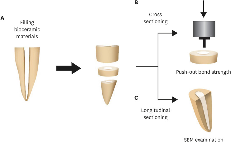
Figure 1 Illustration of the experimental procedure. (A) The tooth filled with the tested materials was sectioned horizontally to obtain a sliced specimen and an apical segment. (B) Push-out bond strength was measured with the sliced specimen using a universal testing machine. (C) The apical segment was sectioned longitudinally, and the intratubular biomineralization was observed under scanning electron microscopy.SEM, scanning electron microscope.

Figure 2 Push-out bond strength and the failure patterns of the tested materials. (A) Bar chart showing the mean bond strength of the 2 tested material groups. (B) Failure mode distribution according to filling material.PR, ProRoot MTA; EP, Endocem MTA Premixed.
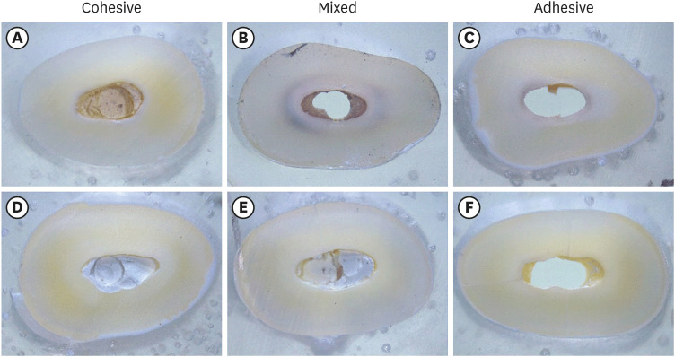
Figure 3 Failure mode analysis using a stereomicroscope at ×30 magnification. (A-C) Representative images of the ProRoot MTA group. (D-F) Representative images of the Endocem MTA Premixed group.
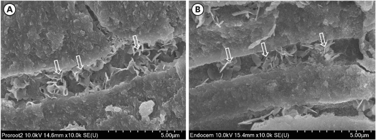
Figure 4 Representative scanning electron microscopy images of intratubular biomineralization. (A) ProRoot MTA-filled root canal. (B) Endocem MTA Premixed-filled root canal. Arrows indicate the flake-shaped intratubular precipitates.
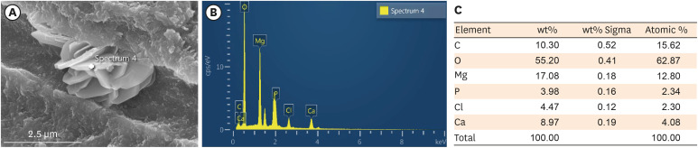
Figure 5 EDS analysis of the chemical characteristics of intratubular precipitate. (A) A scanning electron microscope image showing the precipitate (white cross). (B) Representative graph of EDS analysis of the precipitate. (C) Semiquantitative chemical composition showing the calcium/phosphorus ratio of the crystalline area denoted with a white cross.EDS, energy-dispersive X-ray spectroscopic.
Reference
-
1. Saxena P, Gupta SK, Newaskar V. Biocompatibility of root-end filling materials: recent update. Restor Dent Endod. 2013; 38:119–127. PMID: 24010077.
Article2. Bernabé PF, Gomes-Filho JE, Bernabé DG, Nery MJ, Otoboni-Filho JA, Dezan E Jr, Cintra LT. Sealing ability of MTA used as a root end filling material: effect of the sonic and ultrasonic condensation. Braz Dent J. 2013; 24:107–110. PMID: 23780366.
Article3. Torabinejad M, Hong CU, McDonald F, Pitt Ford TR. Physical and chemical properties of a new root-end filling material. J Endod. 1995; 21:349–353. PMID: 7499973.
Article4. Alsubait SA, Hashem Q, AlHargan N, AlMohimeed K, Alkahtani A. Comparative evaluation of push-out bond strength of ProRoot MTA, bioaggregate and Biodentine. J Contemp Dent Pract. 2014; 15:336–340. PMID: 25307817.
Article5. Nagas E, Cehreli ZC, Uyanik MO, Vallittu PK, Lassila LV. Effect of several intracanal medicaments on the push-out bond strength of ProRoot MTA and Biodentine. Int Endod J. 2016; 49:184–188. PMID: 25631153.
Article6. Buldur B, Oznurhan F, Kaptan A. The effect of different chelating agents on the push-out bond strength of ProRoot MTA and EndoSequence root repair material. Eur Oral Res. 2019; 53:88–93. PMID: 31309199.
Article7. Saghiri MA, Shokouhinejad N, Lotfi M, Aminsobhani M, Saghiri AM. Push-out bond strength of mineral trioxide aggregate in the presence of alkaline pH. J Endod. 2010; 36:1856–1859. PMID: 20951300.
Article8. Vivan RR, Guerreiro-Tanomaru JM, Bosso-Martelo R, Costa BC, Duarte MA, Tanomaru-Filho M. Push-out bond strength of root-end filling materials. Braz Dent J. 2016; 27:332–335. PMID: 27224569.
Article9. Shokouhinejad N, Razmi H, Fekrazad R, Asgary S, Neshati A, Assadian H, Kheirieh S. Push-out bond strength of two root-end filling materials in root-end cavities prepared by Er,Cr:YSGG laser or ultrasonic technique. Aust Endod J. 2012; 38:113–117. PMID: 23211070.
Article10. Ashofteh Yazdi K, Bolhari B, Sabetmoghaddam T, Meraji N, Kharazifard MJ. Effect of blood exposure on push-out bond strength of four calcium silicate based cements. Iran Endod J. 2017; 12:196–200. PMID: 28512485.11. Tuncel B, Nagas E, Cehreli Z, Uyanik O, Vallittu P, Lassila L. Effect of endodontic chelating solutions on the bond strength of endodontic sealers. Braz Oral Res. 2015; 29:S1806-83242015000100256.
Article12. Araújo CC, Brito-Júnior M, Faria-E-Silva AL, Pereira RD, Silva-Sousa YT, Cruz-Filho AM, Sousa-Neto MD. Root filling bond strength using reciprocating file-matched single-cones with different sealers. Braz Oral Res. 2016; 30:S1806-83242016000100251.
Article13. Prado MC, Martiniano K, Pereira AC, Cortellazzi KL, Marciano MA, Abuna G, de-Jesus-Soares A. Do intracanal medications used in regenerative endodontics affect the bond strength of powder-to-liquid and ready-to-use cervical sealing materials? J Conserv Dent. 2021; 24:464–469. PMID: 35399766.
Article14. Al-Hiyasat AS, Yousef WA. Push-out bond strength of calcium silicate-based cements in the presence or absence of a smear layer. Int J Dent. 2022; 2022:7724384. PMID: 35910089.
Article15. Salem Milani A, Froughreyhani M, Charchi Aghdam S, Pournaghiazar F, Asghari Jafarabadi M. Mixing with propylene glycol enhances the bond strength of mineral trioxide aggregate to dentin. J Endod. 2013; 39:1452–1455. PMID: 24139273.
Article16. Retana-Lobo C, Tanomaru-Filho M, Guerreiro-Tanomaru JM, Benavides-García M, Hernández-Meza E, Reyes-Carmona J. Push-out bond strength, characterization, and ion release of premixed and powder-liquid bioceramic sealers with or without gutta-percha. Scanning. 2021; 2021:6617930. PMID: 34040690.
Article17. Salim Al-Ani AA, Mutluay M, Stape THS, Tjäderhane L, Tezvergil-Mutluay A. Effect of various dimethyl sulfoxide concentrations on the durability of dentin bonding and hybrid layer quality. Dent Mater J. 2018; 37:501–505. PMID: 29593164.
Article18. Tjäderhane L, Mehtälä P, Scaffa P, Vidal C, Pääkkönen V, Breschi L, Hebling J, Tay FR, Nascimento FD, Pashley DH, Carrilho MR. The effect of dimethyl sulfoxide (DMSO) on dentin bonding and nanoleakage of etch-and-rinse adhesives. Dent Mater. 2013; 29:1055–1062. PMID: 23942144.
Article19. Lindblad RM, Lassila LVJ, Vallittu PK, Tjäderhane L. The effect of chlorhexidine and dimethyl sulfoxide on long-term sealing ability of two calcium silicate cements in root canal. Dent Mater. 2021; 37:328–335. PMID: 33341245.
Article20. Reyes-Carmona JF, Felippe MS, Felippe WT. Biomineralization ability and interaction of mineral trioxide aggregate and white Portland cement with dentin in a phosphate-containing fluid. J Endod. 2009; 35:731–736. PMID: 19410094.
Article21. Yoo YJ, Lee YS, Yoo JS, Perinpanayagam H, Yoo CS, Kang HS, Oh S, Chang SW, Kum KY. Intratubular biomineralization in a root canal filled with calcium-enriched material over 8 years. Materials (Basel). 2017; 10:1388. PMID: 29206138.
Article22. Reyes-Carmona JF, Felippe MS, Felippe WT. A phosphate-buffered saline intracanal dressing improves the biomineralization ability of mineral trioxide aggregate apical plugs. J Endod. 2010; 36:1648–1652. PMID: 20850670.
Article
- Full Text Links
-
- Actions
-
Cited
- CITED
-
- Close
- Share
-
- Similar articles
-
- Effect of dimethyl sulfoxide on bond durability of fiber posts cemented with etch-and-rinse adhesives
- Effect of ultrasonic cleaning on the bond strength of fiber posts in oval canals filled with a premixed bioceramic root canal sealer
- Push-out bond strength of a self-adhesive resin cement used as endodontic sealer
- Effect of Dimethyl Sulfoxide (DMSO) Monotherapy in Treatment of Interstitial Cystitis
- Effect of ultrasonic agitation on push-out bond strength and adaptation of root-end filling materials
