Combination of a new ultrasonic tip with rotary systems for the preparation of flattened root canals
- Affiliations
-
- 1Department of Restorative Dentistry, São Paulo State University (UNESP), School of Dentistry, Araraquara, SP, Brazil
- KMID: 2548100
- DOI: http://doi.org/10.5395/rde.2021.46.e56
Abstract
Objectives
This study evaluated 2 nickel-titanium rotary systems and a complementary protocol with an ultrasonic tip and a small-diameter instrument in flattened root canals.
Materials and Methods
Thirty-two human maxillary second premolars with flattened canals (buccolingual diameter ≥4 times larger than the mesiodistal diameter) at 9 mm from the radiographic apex were selected. The root canals were prepared by ProDesign Logic (PDL) 30/0.01 and 30/0.05 or Hyflex EDM (HEDM) 10/0.05 and 25/0.08 (n = 16), followed by application of the Flatsonic ultrasonic tip in the cervical and middle thirds and a PDL 25/0.03 file in the apical third (FPDL). The teeth were scanned using micro-computed tomography before and after the procedures. The percentage of volume increase, debris, and uninstrumented surface area were analyzed using the Kruskal-Wallis, Dunn, Wilcoxon, analysis of variance/Tukey, and paired and unpaired t-tests (α = 0.05).
Results
No significant difference was found in the volume increase and uninstrumented surface area between PDL and HEDM (p > 0.05). PDL had a higher percentage of debris than HEDM in the middle and apical thirds (p < 0.05). The FPDL protocol resulted in less debris and uninstrumented surface area for PDL and HEDM (p < 0.05). This protocol, with HEDM, reduced debris in the middle and apical thirds and uninstrumented surface area in the apical third (p < 0.05).
Conclusions
High percentages of debris and uninstrumented surface area were observed after preparation of flattened root canals. The HEDM, Flatsonic tip, and 25/0.03 instrument protocol enhanced cleaning in flattened root canals.
Figure
-
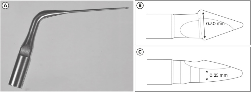
Figure 1 Representative image of the ultrasonic tip. (A) Flatsonic ultrasonic tip, (B) lateral view of the tip showing the inverted flattened arrow design, (C) front view of the tip showing the reduced diameter.
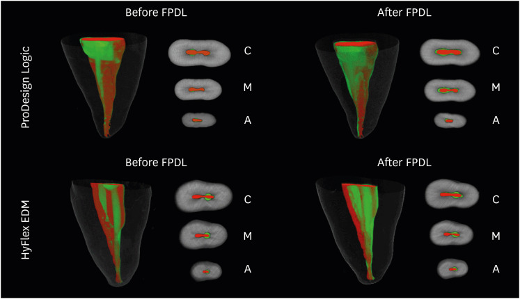
Figure 2 Three-dimensional reconstructions and cross-sectional views of the cervical (C), middle (M), and apical (A) thirds of representative images of flattened root canals of the maxillary second premolars prepared by ProDesign Logic or HyFlex EDM before and after complementary preparation with Flatsonic and PDL 25.03 (FPDL), showing uninstrumented surfaces (red) and instrumented surfaces (green).
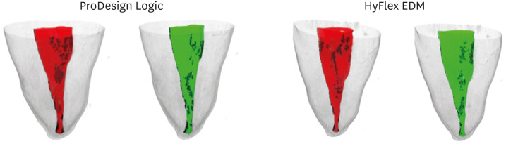
Figure 3 Three-dimensional reconstructions of representative images of flattened root canals of the maxillary second premolars prepared by ProDesign Logic or HyFlex EDM before (red) and after (green) complementary preparation with Flatsonic and PDL 25.03, showing accumulated debris (black).
Reference
-
1. Barbizam JV, Fariniuk LF, Marchesan MA, Pecora JD, Sousa-Neto MD. Effectiveness of manual and rotary instrumentation techniques for cleaning flattened root canals. J Endod. 2002; 28:365–366. PMID: 12026920.
Article2. Versiani MA, Pécora JD, de Sousa-Neto MD. Flat-oval root canal preparation with self-adjusting file instrument: a micro-computed tomography study. J Endod. 2011; 37:1002–1007. PMID: 21689560.
Article3. Vasconcelos LRSM, Midena RZ, Minotti PG, Pereira TC, Duarte MAH, Andrade FB. Effect of ultrasound streaming on the disinfection of flattened root canals prepared by rotary and reciprocating systems. J Appl Oral Sci. 2017; 25:477–482. PMID: 29069144.
Article4. Filpo-Perez C, Bramante CM, Villas-Boas MH, Húngaro Duarte MA, Versiani MA, Ordinola-Zapata R. Micro-computed tomographic analysis of the root canal morphology of the distal root of mandibular first molar. J Endod. 2015; 41:231–236. PMID: 25447505.
Article5. Lacerda MFLS, Marceliano-Alves MF, Pérez AR, Provenzano JC, Neves MAS, Pires FR, Gonçalves LS, Rôças IN, Siqueira JF Jr. Cleaning and shaping oval canals with 3 instrumentation systems: a correlative micro-computed tomographic and histologic study. J Endod. 2017; 43:1878–1884. PMID: 28951035.
Article6. Siqueira JF Jr, Pérez AR, Marceliano-Alves MF, Provenzano JC, Silva SG, Pires FR, Vieira GCS, Rôças IN, Alves FRF. What happens to unprepared root canal walls: a correlative analysis using micro-computed tomography and histology/scanning electron microscopy. Int Endod J. 2018; 51:501–508. PMID: 28196289.
Article7. Lopes RMV, Marins FC, Belladonna FG, Souza EM, De-Deus G, Lopes RT, Silva EJNL. Untouched canal areas and debris accumulation after root canal preparation with rotary and adaptive systems. Aust Endod J. 2018; 44:260–266. PMID: 28940891.
Article8. Moura-Netto C, Palo RM, Pinto LF, Mello-Moura AC, Daltoé G, Wilhelmsen NS. CT study of the performance of reciprocating and oscillatory motions in flattened root canal areas. Braz Oral Res. 2015; 29:1–6.
Article9. Arias A, Paqué F, Shyn S, Murphy S, Peters OA. Effect of canal preparation with TRUShape and Vortex rotary instruments on three-dimensional geometry of oval root canals. Aust Endod J. 2018; 44:32–39. PMID: 28418157.
Article10. Keskin C, Demiral M, Sarıyılmaz E. Comparison of the shaping ability of novel thermally treated reciprocating instruments. Restor Dent Endod. 2018; 43:e15. PMID: 29765896.
Article11. De-Deus G, Marins J, Silva EJ, Souza E, Belladonna FG, Reis C, Machado AS, Lopes RT, Versiani MA, Paciornik S, Neves AA. Accumulated hard tissue debris produced during reciprocating and rotary nickel-titanium canal preparation. J Endod. 2015; 41:676–681. PMID: 25670245.
Article12. De-Deus G, Belladonna FG, de Siqueira Zuolo A, Perez R, Carvalho MS, Souza EM, Lopes RT, Silva EJNL. Micro-CT comparison of XP-endo Finisher and passive ultrasonic irrigation as final irrigation protocols on the removal of accumulated hard-tissue debris from oval shaped-canals. Clin Oral Investig. 2019; 23:3087–3093.
Article13. Üreyen Kaya B, Erik CE, Sesli Çetin E, Köle M, Maden M. Mechanical reduction in intracanal Enterococcus faecalis when using three different single-file systems: an ex vivo comparative study. Int Endod J. 2019; 52:77–85. PMID: 29985531.
Article14. Bedier MM, Hashem AAR, Hassan YM. Improved dentin disinfection by combining different-geometry rotary nickel-titanium files in preparing root canals. Restor Dent Endod. 2018; 43:e46. PMID: 30483470.
Article15. Brasil SC, Marceliano-Alves MF, Marques ML, Grillo JP, Lacerda MFLS, Alves FRF, Siqueira JF Jr, Provenzano JC. Canal transportation, unprepared areas, and dentin removal after preparation with BT-RaCe and ProTaper next systems. J Endod. 2017; 43:1683–1687. PMID: 28712638.
Article16. Gagliardi J, Versiani MA, de Sousa-Neto MD, Plazas-Garzon A, Basrani B. Evaluation of the shaping characteristics of ProTaper Gold, ProTaper NEXT, and ProTaper universal in curved canals. J Endod. 2015; 41:1718–1724. PMID: 26321062.
Article17. Goo HJ, Kwak SW, Ha JH, Pedullà E, Kim HC. Mechanical properties of various heat-treated nickel-titanium rotary instruments. J Endod. 2017; 43:1872–1877. PMID: 28951028.
Article18. Stringheta CP, Bueno CES, Kato AS, Freire LG, Iglecias EF, Santos M, Pelegrine RA. Micro-computed tomographic evaluation of the shaping ability of four instrumentation systems in curved root canals. Int Endod J. 2019; 52:908–916. PMID: 30688377.
Article19. Venino PM, Citterio CL, Pellegatta A, Ciccarelli M, Maddalone M. A micro-computed tomography evaluation of the shaping ability of two nickel-titanium instruments, HyFlex EDM and ProTaper Next. J Endod. 2017; 43:628–632. PMID: 28215346.
Article20. Coelho BS, Amaral RO, Leonardi DP, Marques-da-Silva B, Silva-Sousa YTC, de Carvalho FMA, Baratto-Filho F. Performance of three single instrument systems in the preparation of long oval canals. Braz Dent J. 2016; 27:217–222. PMID: 27058387.
Article21. Tanomaru-Filho M, Galletti Espir C, Carolina Venção A, Macedo-Serrano N, Camilo-Pinto J, Guerreiro-Tanomaru J. Cyclic fatigue resistance of heat-treated nickel-titanium instruments. Iran Endod J. 2018; 13:312–317. PMID: 30083199.22. Gavini G, Santos MD, Caldeira CL, Machado MEL, Freire LG, Iglecias EF, Peters OA, Candeiro GTM. Nickel-titanium instruments in endodontics: a concise review of the state of the art. Braz Oral Res. 2018; 32:e67. PMID: 30365608.
Article23. Pirani C, Iacono F, Generali L, Sassatelli P, Nucci C, Lusvarghi L, Gandolfi MG, Prati C. HyFlex EDM: superficial features, metallurgical analysis and fatigue resistance of innovative electro discharge machined NiTi rotary instruments. Int Endod J. 2016; 49:483–493. PMID: 26011181.
Article24. Yılmaz K, Uslu G, Özyürek T. In vitro comparison of the cyclic fatigue resistance of HyFlex EDM, One G, and ProGlider nickel titanium glide path instruments in single and double curvature canals. Restor Dent Endod. 2017; 42:282–289. PMID: 29142876.
Article25. Versiani MA, Alves FR, Andrade-Junior CV, Marceliano-Alves MF, Provenzano JC, Rôças IN, Sousa-Neto MD, Siqueira JF Jr. Micro-CT evaluation of the efficacy of hard-tissue removal from the root canal and isthmus area by positive and negative pressure irrigation systems. Int Endod J. 2016; 49:1079–1087. PMID: 26459183.
Article26. Rivera-Peña ME, Duarte MAH, Alcalde MP, Furlan RD, Só MVR, Vivan RR. Ultrasonic tips as an auxiliary method for the instrumentation of oval-shaped root canals. Braz Oral Res. 2019; 33:e011. PMID: 30758408.
Article27. Rivera-Peña ME, Duarte MAH, Alcalde MP, DE Andrade FB, Vivan RR. A novel ultrasonic tip for removal of filling material in flattened/oval-shaped root canals: a microCT study. Braz Oral Res. 2018; 32:e88. PMID: 30110086.
Article28. Santos-Junior AO, Tanomaru-Filho M, Pinto JC, Tavares KI, Pivoto-João MM, Guerreiro-Tanomaru JM. New ultrasonic tip decreases uninstrumented surface and debris in flattened canals: a micro-computed tomographic study. J Endod. 2020; DOI: 10.1016/j.joen.2020.07.012. [Epub ahead of print].29. Espir CG, Nascimento-Mendes CA, Guerreiro-Tanomaru JM, Freire LG, Gavini G, Tanomaru-Filho M. Counterclockwise or clockwise reciprocating motion for oval root canal preparation: a micro-CT analysis. Int Endod J. 2018; 51:541–548. PMID: 28375575.
Article30. Espir CG, Nascimento-Mendes CA, Guerreiro-Tanomaru JM, Cavenago BC, Hungaro Duarte MA, Tanomaru-Filho M. Shaping ability of rotary or reciprocating systems for oval root canal preparation: a micro-computed tomography study. Clin Oral Investig. 2018; 22:3189–3194.
Article31. Jou YT, Karabucak B, Levin J, Liu D. Endodontic working width: current concepts and techniques. Dent Clin North Am. 2004; 48:323–335. PMID: 15066519.
Article32. Espir CG, Nascimento CA, Guerreiro-Tanomaru JM, Bonetti-Filho I, Tanomaru-Filho M. Radiographic and micro-computed tomography classification of root canal morphology and dentin thickness of mandibular incisors. J Conserv Dent. 2018; 21:57–62. PMID: 29628649.33. Wu MK, Dummer PM, Wesselink PR. Consequences of and strategies to deal with residual post-treatment root canal infection. Int Endod J. 2006; 39:343–356. PMID: 16640632.
Article34. De-Deus G, Simões-Carvalho M, Belladonna FG, Cavalcante DM, Portugal LS, Prado CG, Souza EM, Lopes RT, Silva EJNL. Arrowhead design ultrasonic tip as a supplementary tool for canal debridement. Int Endod J. 2020; 53:410–420. PMID: 31613994.
Article35. Siqueira Junior JF, Rôças IDN, Marceliano-Alves MF, Pérez AR, Ricucci D. Unprepared root canal surface areas: causes, clinical implications, and therapeutic strategies. Braz Oral Res. 2018; 32:e65. PMID: 30365606.
Article36. Paqué F, Balmer M, Attin T, Peters OA. Preparation of oval-shaped root canals in mandibular molars using nickel-titanium rotary instruments: a micro-computed tomography study. J Endod. 2010; 36:703–707. PMID: 20307747.
Article37. Pinheiro SR, Alcalde MP, Vivacqua-Gomes N, Bramante CM, Vivan RR, Duarte MAH, Vasconcelos BC. Evaluation of apical transportation and centring ability of five thermally treated NiTi rotary systems. Int Endod J. 2018; 51:705–713. PMID: 29178173.
Article38. Plotino G, Özyürek T, Grande NM, Gündoğar M. Influence of size and taper of basic root canal preparation on root canal cleanliness: a scanning electron microscopy study. Int Endod J. 2019; 52:343–351. PMID: 30129186.
Article39. Pinto JC, Pivoto-João MMB, Espir CG, Ramos MLG, Guerreiro-Tanomaru JM, Tanomaru-Filho M. Micro-CT evaluation of apical enlargement of molar root canals using rotary or reciprocating heat-treated NiTi instruments. J Appl Oral Sci. 2019; 27:e20180689. PMID: 31411264.
Article40. Pivoto-João MM, Tanomaru-Filho M, Pinto JC, Espir CG, Guerreiro-Tanomaru JM. Root canal preparation and enlargement using thermally-treated nickel-titanium rotary systems in curved canals. J Endod. 2020; DOI: 10.1016/j.joen.2020.08.007. [Epub ahead of print].
- Full Text Links
-
- Actions
-
Cited
- CITED
-
- Close
- Share
-
- Similar articles
-
- Root surface roughness following mechanical instrumentation in vivo and in vitro SEM study
- Relative efficacy of three Ni-Ti file systems used by undergraduates
- Comparison of screw-in effect for several nickel-titanium rotary instruments in simulated resin root canal
- Shaping ability of four rotary nickel-titanium instruments to prepare root canal at danger zone
- Evaluation of canal preparation with Ni-Ti rotary files by micro computed tomography
