Shaping ability of four rotary nickel-titanium instruments to prepare root canal at danger zone
- Affiliations
-
- 1Department of Conservative Dentistry, School of Dentistry, Kyungpook National University, Korea. skykim@knu.ac.kr
- 2Department of Dentistry, College of Medicine, Konkuk University, Korea.
- KMID: 1987124
- DOI: http://doi.org/10.5395/JKACD.2004.29.5.446
Abstract
- The aim of this study was to evaluate the shaping abilities of four different rotary nickel-titanium instruments with anticurvature motion to prepare root canal at danger zone by measuring the change of dentin thickness in order to have techniques of safe preparation of canals with nickel-titanium files. Mesiobuccal and mesiolingual canals of forty mesial roots of extracted human lower molars were instrumented using the crown-down technique with ProFile, GT(TM) Rotary file, Quantec file and ProTaper(TM). In each root, one canal was prepared with a straight up-and-down motion and the other canal was with an anticurvature motion. Canals were instrumented until apical foramens were up to size of 30 by one operator. The muffle system was used to evaluate the root canal preparation. After superimposing the pre- and post-instrumentation canal, change in root dentin thickness was measured at the inner and outer sides of the canal at 1, 3, and 5 mm levels from the furcation. Data were analyzed using two-way ANOVA. Root dentin thickness at danger zone was significantly thinner than that at safe zone at all levels (p < 0.05). There was no significant difference in the change of root dentin thickness between the straight up-and-down and the anticurvature motions at both danger and safe zones in all groups (p > 0.05). ProTaper removed significantly more dentin than other files especially at furcal 3 mm level of danger and safe zones (p < 0.05) Therefore, it was concluded that anticurvature motion with nickel-titanium rotary instruments does not seem to be effective in danger zone of lower molars.
Keyword
Figure
-
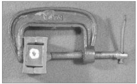
Figure 1 Both halves of the muffle with C clamp to maintain muffle-to-muffle and muffle-to-resin relationships. A tooth is embedded in acrylic resin in the muffle. C clamp also serves as a convenient handle during experimental procedure.

Figure 2 Levels of cross-section. Roots were sectioned horizontally 1, 3, and 5 mm levels from the furcation.
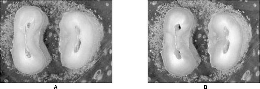
Figure 3 Computer captured images. A, before root canal instrumentation; B, after canal instrumentation.
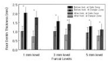
Figure 4 Thickness of root dentin (mm) at danger and safe zones before and after root canal instrumentation (mean ± S.D., n = 80 each). *Root dentin thickness at danger zone was significantly thinner than that at safe zone (p < 0.05).
Cited by 2 articles
-
Effect of anticurvature filing method on preparation of the curved root canal using ProFile
Hyun-Ji Song, Juhea Chang, Kyung-Mo Cho, Jin-Woo Kim
J Korean Acad Conserv Dent. 2005;30(4):327-334. doi: 10.5395/JKACD.2005.30.4.327.Change of working length in curved canals by various instrumentation techniques
Jeong-Im Jo, Myoung-Uk Jin, Young Kyung Kim, Sung Kyo Kim
J Korean Acad Conserv Dent. 2006;31(1):30-35. doi: 10.5395/JKACD.2006.31.1.030.
Reference
-
1. Schilder H. Cleaning and shaping the root canal. Dent Clin North Am. 1974. 18:269–296.2. Buchanan LS. Paradigm shifts in cleaning and shaping. J Calif Dent Assoc. 1991. 19:23–26. 28–33.3. Kessler JR, Peters DD, Lorton L. Comparison of the relative risk of molar root perforations using various endodontic instrumentation techniques. J Endod. 1983. 9:439–447.
Article4. Goerig AC, Michelich RJ, Schultz H. Instrumentation of root canals in molar using step-down technique. J Endod. 1982. 8:550–554.5. Bramante CM, Berber A, Borges RP. A methodology for evaluation of root canal instrumentation. J Endod. 1987. 13:243–245.
Article6. McCann JT, Keller DL, LaBounty GL. A modification of the muffle model system to study root canal morphology. J Endod. 1990. 16:114–115.
Article7. Ruddle C. Cohen S, Burns R, editors. Cleaning and shaping the root canal system. Pathways of the pulp. 2002. 8th ed. St Louis: Mosby;231–291.8. Bower RC. Furcation morphology relative to periodontal treatment. J Periodontol. 1979. 50:23–27.9. Abou-Rass M, Frank AL, Glick DH. The anticurvature filing method to prepare the curved canal. J Am Dent Assoc. 1980. 101:792–794.10. Walia HM, Brantley WA, Gerstein H. An initial investigation of the bending and torsional properties of Nitinol root canal files. J Endod. 1988. 14:346–351.
Article11. Kavanagh D, Lumley PJ. An in vitro evaluation of canal preparation using ProFile .04 and .06 taper instruments. Endod Dent Traumatol. 1998. 14:16–20.
Article12. Bryant ST, Dummer PM, Pitoni C, Bourba M, Moghal S. Shaping ability of .04 and .06 taper ProFile rotary nickel-titanium instruments in simulated root canals. Int Endod J. 1999. 32:155–164.
Article13. Park HS, Lee MK, Kim JJ, Lee JY. A study on the shape of a canal prepared with ProFiles in curved canal. J Korean Acad Conserv Dent. 1999. 24:633–638.14. Lee JH, Cho YB. The effect of some canal preparation techniques on the shape of root canals. J Korean Acad Conserv Dent. 1999. 24:337–345.15. Kum KY, Spangberg L, Cha BY, Jung IY, Lee SJ, Lee CY. Shaping ability of three ProFile rotary instrumentation techniques in simulated resin root canals. J Endod. 2000. 26:719–723.
Article16. Park H. A comparison of Greater Taper files, ProFiles, and stainless steel files to shape curved root canals. Oral Surg Oral Med Oral Pathol Oral Radiol Endod. 2001. 91:715–718.
Article17. Short JA, Morgan LA, Baumgartner JC. A comparison of canal centering ability of four instrumentation techniques. J Endod. 1997. 23:503–507.
Article18. Isom TL, Marshall JG, Baumgartner JC. Evaluation of root thickness in curved canals after flaring. J Endod. 1995. 21:368–371.
Article19. Glossen CR, Haller RH, Dove SB, Del Rio CE. A comparison of root canal preparation using NiTi hand, NiTi engine-driven, and K-Flex endodontic instruments. J Endod. 1995. 21:146–151.20. Yun HH, Kim SK. A comparison of the shaping abilities of four nickel-titanium rotary instruments in simulated root canals. Oral Surg Oral Med Oral Pathol Oral Radiol Endod. 2003. 95:228–233.
Article21. Portenier I, Barbakow F. Preparation of the apical part of the root canal by the Lightspeed and step-back techniques. Int Endod J. 1998. 31:103–111.
Article
- Full Text Links
-
- Actions
-
Cited
- CITED
-
- Close
- Share
-
- Similar articles
-
- Shaping ability of nickel-titaniumrotary files
- Influence of root canal curvature on the screw-in effect of nickel-titanium rotary files in simulated resin root canal
- Influence of taper on the screw-in effect of nickel-titanium rotary files in simulated resin root canal
- A comparison of the shaping ability of four rotary nickel-titanium files in simulated root canals
- Comparison of screw-in effect for several nickel-titanium rotary instruments in simulated resin root canal
