J Korean Acad Conserv Dent.
2010 Sep;35(5):374-379. 10.5395/JKACD.2010.35.5.374.
Influence of root canal curvature on the screw-in effect of nickel-titanium rotary files in simulated resin root canal
- Affiliations
-
- 1Department of Conservative Dentistry, Kyungpook National University School of Dentistry, Daegu, Korea. wisekim@knu.ac.kr
- KMID: 2176436
- DOI: http://doi.org/10.5395/JKACD.2010.35.5.374
Abstract
OBJECTIVES
Nickel-titanium (Ni-Ti) rotary instruments have some unexpected disadvantages including the tendency to screw-in to the canal. The purpose of this study was to evaluate the influence of root canal curvatures on the screw-in effect of Ni-Ti rotary files.
MATERIALS AND METHODS
A total of 80 simulated root canals in clear resin blocks were used in the study. Canals with curvature of 0, 10, 20 and 30 degrees were instrumented with ProTaper instruments SX, S1, S2 and a ProFile of #25/0.06 to 1.0-2.0 mm beyond the initial point of root curvature. The screw-in force was measured with a specially designed device while canal was instrumented with a ProFile of #30/0.06 at a constant speed of 300 rpm. The data were subjected to one-way ANOVA and Scheffe multiple range test for post-hoc test.
RESULTS
Larger degree of canal curvature generated significantly lesser screw-in forces in all groups (p < 0.001).
CONCLUSIONS
More attention needs to be paid when using rotary instruments in canals with less curvature than canals with more curvatures to prevent or reduce any accidental overinstrumentation.
Keyword
Figure
-

Figure 1 The custom-made device for the measurement of the screw-in effect. The device is composed of a part generating a single pecking movement of the handpiece (a) with a constant speed of 300 rpm, compression / tension sensor (b) and computer (c) for data storage, analysis.
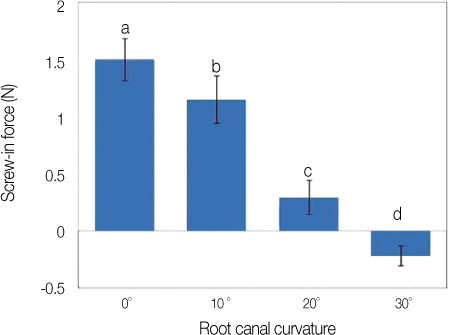
Figure 2 Screw-in force according to the root canal curvature of each resin block (mean ± S.D.). a, b, c, d: Letters indicate significant differences between materials by Scheffe multiple range test; p < 0.001.
Cited by 1 articles
-
Mechanical and geometric features of endodontic instruments and its clinical effect
Hyeon-Cheol Kim
J Korean Acad Conserv Dent. 2011;36(1):1-11. doi: 10.5395/JKACD.2011.36.1.1.
Reference
-
1. Pettiette MT, Olutayo Delano E, Trope M. Evaluation of success rate of endodontic treatment performed by students with stainless-steel K-files and Nickel-Titanium hand files. J Endod. 2001. 27:124–127.
Article2. Al-Omari MA, Dummer PM, Newcombe RG, Doller R. Comparison of six files to prepare simulated root canals. Part 2. Int Endod J. 1992. 25:67–81.
Article3. Nagy CD, Bartha K, Bernath M, Verdes E, Szabo J. The effect of root canal morphology on canal shape following instrumentation using different techniques. Int Endod J. 1997. 30:133–140.
Article4. Peters OA, Peters CI, Schönenberger K, Barbakow F. ProTaper rotary root canal preparation: assessment of torque and force in relation to canal anatomy. Int Endod J. 2003. 36:93–99.
Article5. Esposito PT, Cunningham CJ. A comparison of canal preparation with nickel-titanium and stainless steel instruments. J Endod. 1995. 21:173–176.
Article6. Glossen CR, Hallor RH, Dove SB, del Rio CE. A comparison of root canal preparations using nickel-titanium hand, nickel-titanium engine-driven, and K-flex endodontic instruments. J Endod. 1995. 21:146–151.7. Short JA, Morgan LA, Baumgartner JC. A comparison of canal centering ability of four instrumentation techniques. J Endod. 1997. 23:503–507.
Article8. Huh YJ, Kim SK. Change in root canal configuration using different file types and techniques. J Korean Acad Conserv Dent. 1997. 22:291–304.9. Sjogren U, Hägglund B, Sundqvist G, Wing K. Factors Affecting the Long-term Result of Endodontic Treatment. J Endod. 1990. 16:498–504.10. Park WK, Lee HJ, Hur B. Shaping ability of nickel-titanium rotary files. J Korean Acad Conserv Dent. 2004. 29:44–50.11. Walia H, Brantley WA, Gerstein H. An initial investigation of the bending and torsional properties of Nitinol root canal files. J Endod. 1988. 14:346–351.
Article12. Yun HH, Kim SK. A comparison of the shaping abilities of 4 nickel-titanium rotary instruments in simulated root canals. Oral Surg Oral Med Oral Pathol Oral Radiol Endod. 2003. 95:228–233.
Article13. Ha JH, Jin MU, Kim YK, Kim SK. Comparison of screw-in effect for several nickel-titanium rotary instruments in simulated root canal. J Korean Acad Conserv Dent. 2010. 35:267–272.
Article14. Oh SH, Park JK, Hur B, Kim HC. Comparison of screw-in effect of three NiTi file systems used by undergraduates. J Korean Acad Conserv Dent. 2006. 31:477–484.
Article15. Gutierrez JH, Brizuela C, Villota E. Human teeth with periapical pathosis after overinstrumentation and overfilling of the root canals: a scanning electron microscopic study. Int Endod J. 1999. 32:40–48.
Article16. Malueg LA, Wilcox LR, Johnson W. Examination of external apical root resorption with scanning electron microscopy. Oral Surg Oral Med Oral Pathol Oral Radiol Endod. 1996. 82:89–93.
Article17. Seltzer S, Soltanoff W, Sinai I, Goldenberg A, Bender IB. Biologic aspects of endodontics. Part III. Periapical tissue reaction to root canal instrumentation. Oral Surg Oral Med Oral Pathol. 1968. 26:534–546.18. Friedman S, Claus L, Malaekeh Z, Trope M. Evaluation of success and failure after endodontic therapy using a glass ionomer cement sealer. J Endod. 1995. 21:384–390.
Article19. Smith CS, Setchell DJ, Harty FJ. Factors influencing the success of conventional root canal therapy-a five-year retrospective study. Int Endod J. 1993. 26:321–333.
Article20. Diemer F, Calas P. Effect of Pitch Length on the Behavior of Rotary Triple Helix Root Canal Instruments. J Endod. 2004. 30:716–718.
Article21. Schrader C, Peter OA. Analysis of torque and force with differently tapered rotary endodontic instruments in vitro. J Endod. 2005. 31:120–123.
Article22. Sung HJ. Influence of taper on the screw-in effect of nickel-titanium rotary files. 2006. Daegu, Korea: Kyungpook National University;MSc thesis.23. Schilder H. Cleaning and shaping the root canal. Dent Clin North Am. 1974. 18:269–296.24. Weine FS, Kelly RF, Lio PJ. The effect of preparation procedures on original canal shape and on apical foramen shape. J Endod. 1975. 1:255–262.
Article25. Sabala CL, Roane JB, Southard LZ. Instrumentation of curved canals using a modofied tipped instrument: a comparison study. J Endod. 1988. 14:59–64.
Article26. Schneider SW. A comparison of canal preparations in straight and curved root canals. Oral Surg Oral Med Oral Pathol. 1971. 32:271–275.
Article27. Leeb J. Canal orifice enlargement as related to biomechanical preparation. J Endod. 1983. 9:463–470.
Article28. Contreras MAL, Zinman EH, Kaplan SK. Comparison of the first file that fits at the apex, before and after early flaring. J Endod. 2001. 27:113–116.
Article29. Tan BT, Messer H. The effect of instrument type and preflaring on apical file size determination. Int Endod J. 2002. 35:752–758.
Article30. Thompson SA, Dummer PM. Shaping ability of Hero 642 rotary nickel-titanium instruments in simulated root canals, part 1. Int Endod J. 2000. 33:248–254.
Article31. Johnson BW. Wei S, editor. Endodontics: what, when, and why. Contemporary endodontics Dentsply Asia. 2002. 1–6.32. Haïkel Y, Serfaty R, Bateman G, Senger B, Allemann C. Dynamic and cyclic fatigue of engine-driven rotary nickel-titanium endodontic instrument. J Endod. 1999. 25:434–440.
Article33. Roane JB, Sabala CL, Duncanson MG Jr. The "balanced force" concept for instrumentation of curved canals. J Endod. 1985. 11:203–211.
Article34. Kyomen SM, Caputo AA, White SN. Critical analysis of the balanced force technique in endodontics. J Endod. 1994. 20:332–337.
Article35. Wildey WL, Senia ES, Montgomery S. Another look at root canal instrumentation. Oral Surg Oral Med Oral Pathol. 1992. 74:499–507.
Article
- Full Text Links
-
- Actions
-
Cited
- CITED
-
- Close
- Share
-
- Similar articles
-
- Influence of taper on the screw-in effect of nickel-titanium rotary files in simulated resin root canal
- Comparison of screw-in effect for several nickel-titanium rotary instruments in simulated resin root canal
- Shaping ability of nickel-titaniumrotary files
- Effect of various canal preparation techniques using rotary nickel-titanium files on the maintenance of canal curvature
- The change of canal configuration after instrumentation by several nickel-titanium files in the simulated canal with abrupt curvature
