Is conservative treatment (enucleation using modified Carnoy’s solution) of odontogenic keratocyst in the maxilla good prognosis?
- Affiliations
-
- 1Department of Oral and Maxillofacial Surgery, School of Dentistry, Jeonbuk National University, Jeonju, Korea
- 2Research Institute of Clinical Medicine of Jeonbuk National University-Biomedical Research Institute of Jeonbuk National University Hospital, Jeonju, Korea
- KMID: 2547573
- DOI: http://doi.org/10.5125/jkaoms.2023.49.5.287
Abstract
- Odontogenic keratocysts (OKCs) located in the maxillae have rarely been reported in the literature. Standard treatment modalities for OKC range from marsupialization to marginal resection. However, most of the studies on OKC treatment have been related to mandibular OKCs. The anatomical structure and loose bone density of the maxillae and the empty space of the maxillary sinus could allow rapid growth of a lesion and the ability to tolerate tumor occupancy in the entire maxilla within a short period of time. Therefore, OKCs of the maxillae require more aggressive surgery, such as resection. As an alternative, this report introduces a modified Carnoy’s solution, a strong acid, as an adjuvant chemotherapy after cyst enucleation. This report describes the clinical outcomes of enucleation using a modified Carnoy’s solution in patients with large OKCs on the posterior maxillae. In three cases, application of a modified Carnoy’s solution had few side effects or morbidity. Each patient was followed for four to six years, and none showed any signs of recurrence. In conclusion, adjuvant treatment with a modified Carnoy’s solution can be considered a treatment option capable of reducing the recurrence rate of OKC in the maxillae.
Keyword
Figure
-
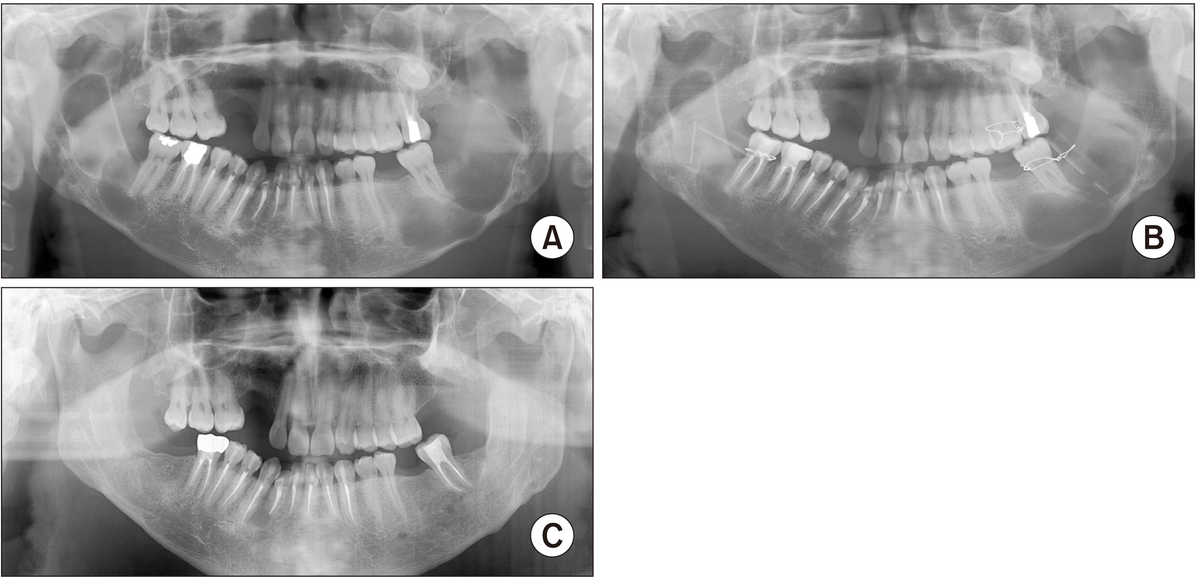
Fig. 1 Radiographs of Patient No. 1 (37-year-old male). A. A radiograph showing large radiolucent lesions on the left posterior maxilla and both posterior mandibles. B. A radiograph showing marsupialization to reduce size. C. A radiograph taken four years after surgery and showing no recurrence.
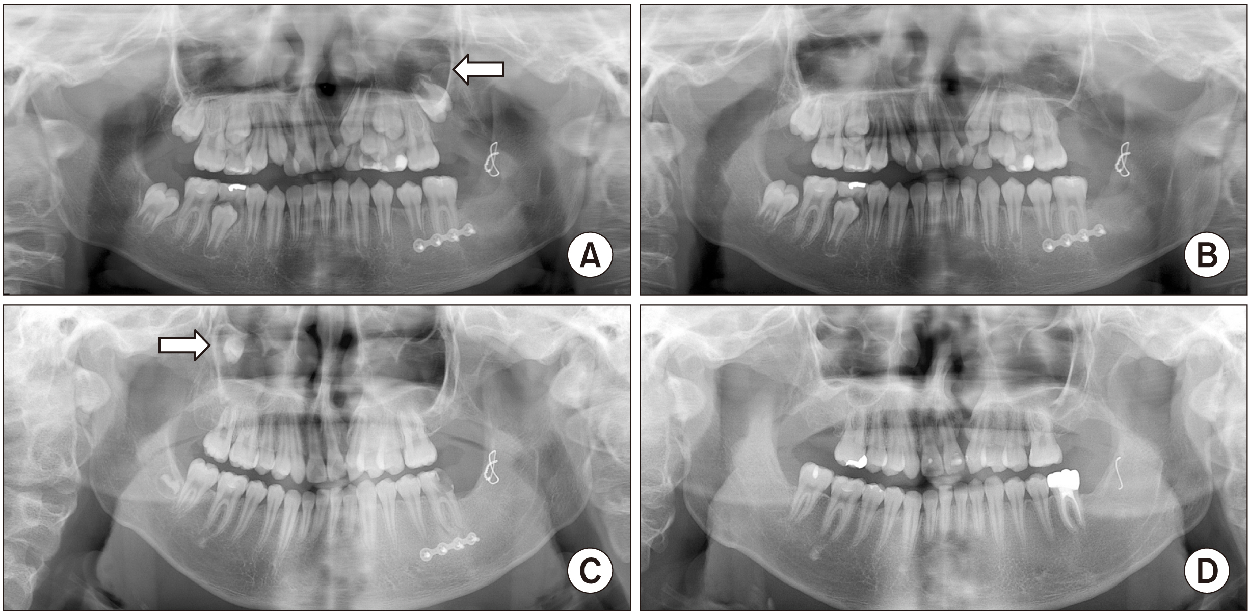
Fig. 2 Radiographs of Patient No. 3 (10-year-old female). A. A radiograph showing radiolucent lesions on both posterior maxillae (arrows). B. A radiograph taken six years after surgery, showing no recurrence.

Fig. 3 Radiographs of Patient No. 2 (10-year-old male). A. A radiograph showing a patient with odontogenic keratocyst (OKC) on the left posterior maxilla (arrow). B. A panoramic view after cyst enucleation and extraction of #27. C. A radiograph taken three years after surgery, showing an additional OKC on the right posterior maxilla (arrow). D. A radiograph showing no recurrence throughout the follow-up period of three years (mandible) and five years (maxilla).
Reference
-
References
1. Maurette PE, Jorge J, de Moraes M. 2006; Conservative treatment protocol of odontogenic keratocyst: a preliminary study. J Oral Maxillofac Surg. 64:379–83. https://doi.org/10.1016/j.joms.2005.11.007. DOI: 10.1016/j.joms.2005.11.007. PMID: 16487797.
Article2. Myoung H, Hong SP, Hong SD, Lee JI, Lim CY, Choung PH, et al. 2001; Odontogenic keratocyst: review of 256 cases for recurrence and clinicopathologic parameters. Oral Surg Oral Med Oral Pathol Oral Radiol Endod. 91:328–33. https://doi.org/10.1067/moe.2001.113109. DOI: 10.1067/moe.2001.113109. PMID: 11250631.
Article3. Chirapathomsakul D, Sastravaha P, Jansisyanont P. 2006; A review of odontogenic keratocysts and the behavior of recurrences. Oral Surg Oral Med Oral Pathol Oral Radiol Endod. 101:5–9. discussion 10. https://doi.org/10.1016/j.tripleo.2005.03.023. DOI: 10.1016/j.tripleo.2005.03.023. PMID: 16360602.
Article4. Khan AA, Qahtani SA, Dawasaz AA, Saquib SA, Asif SM, Ishfaq M, et al. 2019; Management of an extensive odontogenic keratocyst: a rare case report with 10-year follow-up. Medicine (Baltimore). 98:e17987. https://doi.org/10.1097/md.0000000000017987. DOI: 10.1097/MD.0000000000017987. PMID: 31860950. PMCID: PMC6940056.
Article5. Kaczmarzyk T, Mojsa I, Stypulkowska J. 2012; A systematic review of the recurrence rate for keratocystic odontogenic tumour in relation to treatment modalities. Int J Oral Maxillofac Surg. 41:756–67. https://doi.org/10.1016/j.ijom.2012.02.008. DOI: 10.1016/j.ijom.2012.02.008. PMID: 22445416.
Article6. Blanas N, Freund B, Schwartz M, Furst IM. 2000; Systematic review of the treatment and prognosis of the odontogenic keratocyst. Oral Surg Oral Med Oral Pathol Oral Radiol Endod. 90:553–8. https://doi.org/10.1067/moe.2000.110814. DOI: 10.1067/moe.2000.110814. PMID: 11077375.
Article7. Jo HJ, Kim HY, Kang DC, Leem DH, Baek JA, Ko SO. 2020; A clinical study of inferior alveolar nerve damage caused by Carnoy's solution used as a complementary therapeutic agent in a cystic lesion. Maxillofac Plast Reconstr Surg. 42:16. https://doi.org/10.1186/s40902-020-00257-4. DOI: 10.1186/s40902-020-00257-4. PMID: 32509707. PMCID: PMC7248162.
Article8. Donnelly LA, Simmons TH, Blitstein BJ, Pham MH, Saha PT, Phillips C, et al. 2021; Modified Carnoy's compared to Carnoy's solution is equally effective in preventing recurrence of odontogenic keratocysts. J Oral Maxillofac Surg. 79:1874–81. https://doi.org/10.1016/j.joms.2021.03.010. DOI: 10.1016/j.joms.2021.03.010. PMID: 33901451.
Article9. Alchalabi NJ, Merza AM, Issa SA. 2017; Using Carnoy's solution in treatment of keratocystic odontogenic tumor. Ann Maxillofac Surg. 7:51–6. https://doi.org/10.4103/ams.ams_127_16. DOI: 10.4103/ams.ams_127_16. PMID: 28713736. PMCID: PMC5502516.
Article10. Lal B, Kumar RD, Alagarsamy R, Shanmuga Sundaram D, Bhutia O, Roychoudhury A. 2021; Role of Carnoy's solution as treatment adjunct in jaw lesions other than odontogenic keratocyst: a systematic review. Br J Oral Maxillofac Surg. 59:729–41. https://doi.org/10.1016/j.bjoms.2020.12.019. DOI: 10.1016/j.bjoms.2020.12.019. PMID: 34272109.
Article11. Altas E, Karasen RM, Yilmaz AB, Aktan B, Kocer I, Erman Z. 1997; A case of a large dentigerous cyst containing a canine tooth in the maxillary antrum leading to epiphora. J Laryngol Otol. 111:641–3. https://doi.org/10.1017/s0022215100138198. DOI: 10.1017/S0022215100138198. PMID: 9282204.
Article12. Albright CR, Hennig GH. 1971; Large dentigerous cyst of the maxilla near the maxillary sinus: report of case. J Am Dent Assoc. 83:1112–5. https://doi.org/10.14219/jada.archive.1971.0414. DOI: 10.14219/jada.archive.1971.0414. PMID: 5286148.
Article13. Cho JY, Nam KY. 2012; Expansile dentigerous cyst invading the entire maxillary sinus: a case report. J Korean Assoc Oral Maxillofac Surg. 38:245–8. https://doi.org/10.5125/jkaoms.2012.38.4.245. DOI: 10.5125/jkaoms.2012.38.4.245.
Article
- Full Text Links
-
- Actions
-
Cited
- CITED
-
- Close
- Share
-
- Similar articles
-
- Carnoy's Solution Application for Patient Preliminarily Diagnosed with Keratocystic Odotogenic Tumor: Case Report
- Keratoameloblastoma of the maxilla: a case report
- Conservative management with Carnoy’s solution in ameloblastoma involving two unerupted teeth: a report of two cases
- Comparison of clinico-histopathologic findings before and after decompression of odontogenic cyst in the jaw
- Treatment of OKC on Ramus of Mandible by Sagittal Splitting Technique
