Middle superior and anterior superior alveolar nerve injury following trauma to the maxillary sinus: a prospective clinico-radiographic evaluation
- Affiliations
-
- 1Department of Oral and Maxillofacial Surgery, JSS Dental College and Hospital, JSS Academy of Higher Education and Research, Mysuru, India
- KMID: 2547570
- DOI: http://doi.org/10.5125/jkaoms.2023.49.5.262
Abstract
Objectives
Anterior maxillary sinus wall fractures are common in all types of maxillofacial trauma. They can result in various complications, including injury to the surrounding nerves. Owing to its anatomy, trauma to the maxillary antrum can result in injury to the middle superior alveolar nerve (MSAN) and the anterior superior alveolar nerve (ASAN). The purpose of this study is to evaluate neurosensory deficits (NSD) present in maxillary gingiva, incisors, and premolars after injury to the anterior wall of the maxillary antrum.
Materials and Methods
This prospective study was conducted among 39 patients sustaining unilateral fractures of the anterior maxillary sinus wall. Clinical neurosensory tests including two-point discrimination and fine touch discrimination were performed to classify the extent of nerve injuries as mild, moderate, severe, or anesthetic. Additional temperature discrimination and pulpal sensibility tests (electric pulp testing and cold testing) were carried out. A comparison of radiographic fracture patterns and severity of nerve injury was done. Testing was carried out immediately after trauma and at 2-month follow-up.
Results
More than half of the patients assessed in the study group presented with NSD of the teeth and gingiva after trauma. The incidence of deficits varied with the type of test used to measure them. Most frequently, patients presented with both loss of two point as well as fine touch discrimination thresholds. Severe nerve injuries were associated with loss of temperature discrimination clinically and displaced fractures radiographically. There was no significant relationship between the recovery of pulpal and gingival sensation. The patterns of injury and recovery in ASAN and MSAN were similar.
Conclusion
NSD after trauma to the maxillary antrum is relatively common. Clinical loss of temperature discrimination and radiographic signs of fracture lines passing through the canalis sinuosus are predictors of persistent and severe oral NSD.
Figure
-
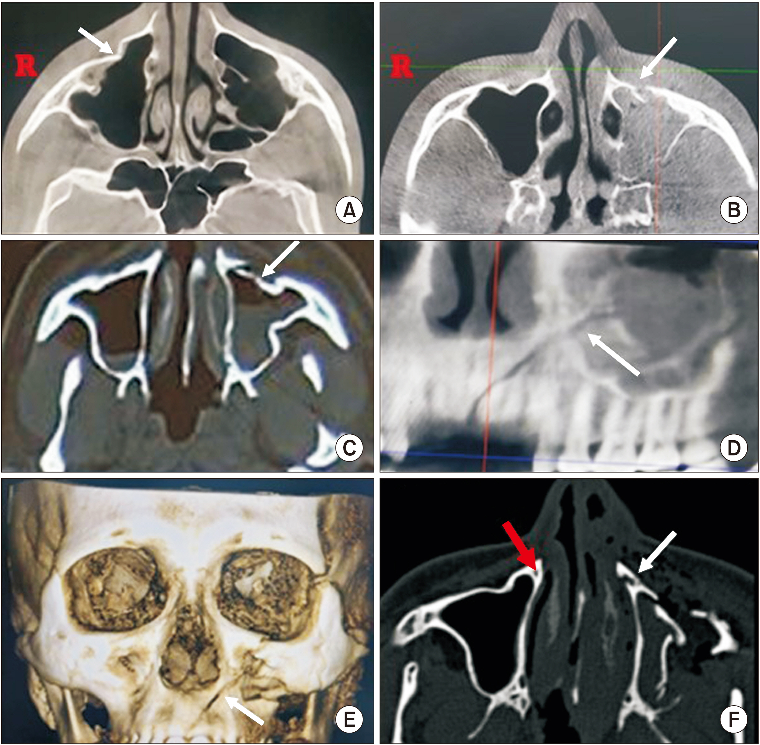
Fig. 1 Computed tomography (CT) and cone-beam CT (CBCT) images of patients with neurosensory deficits. A. Depressed fracture of sinus (arrow). B. Discontinuous fracture (arrow). C. Displaced fracture (arrow). D. CBCT formatted image of the patient with severe neurosensory deficit with fracture line passing through the canine fossa (arrow). E. Three-dimensional reformatted image of the same patient with fracture line (arrow). F. The increase of diameter of the canalis sinuosus (CS) (depicted by white arrow) as compared to the intact CS (depicted by red arrow) in a patient with anaesthesia.
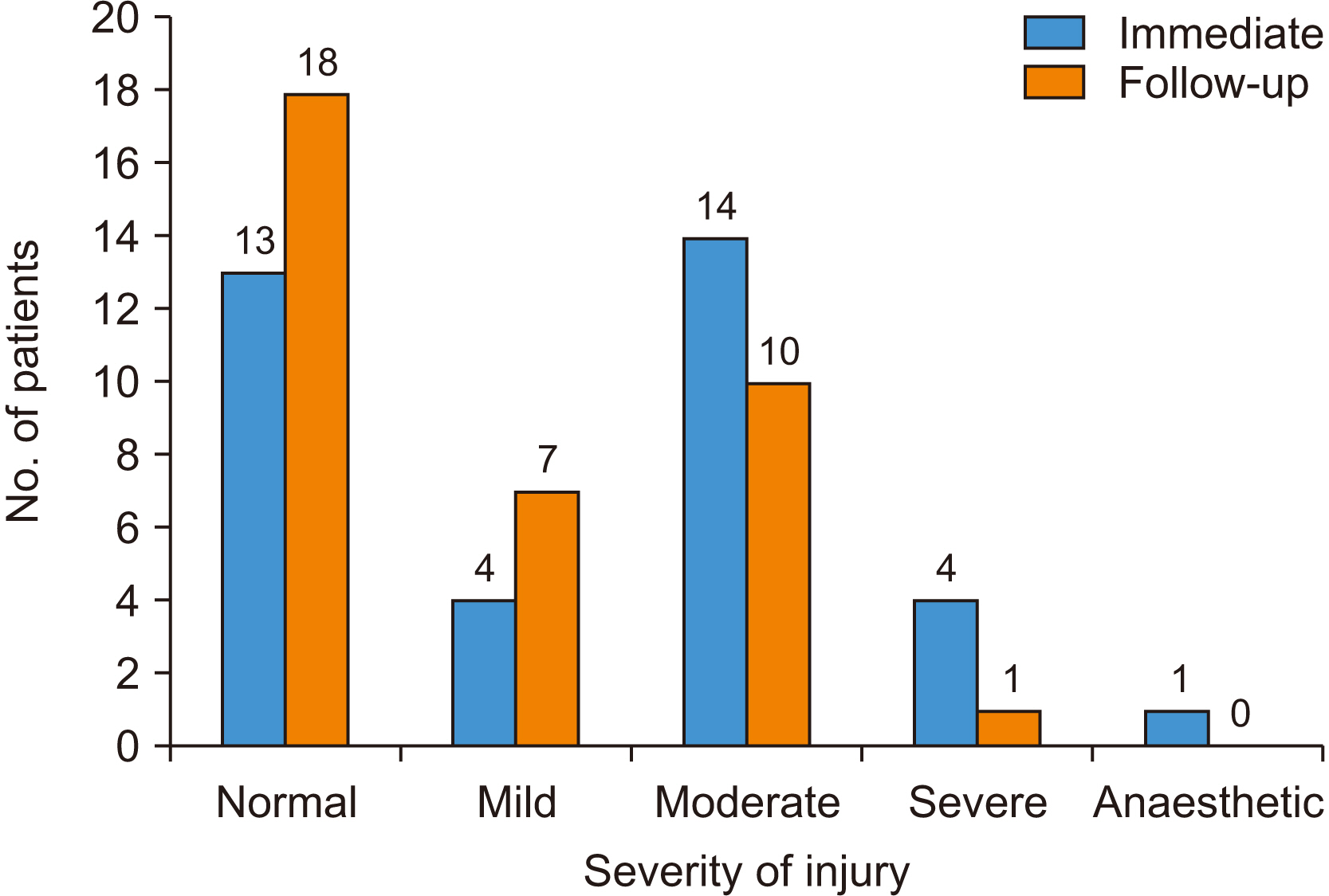
Fig. 2 Change in severity of nerve injury to the ASAN (anterior superior alveolar nerve) over two months.
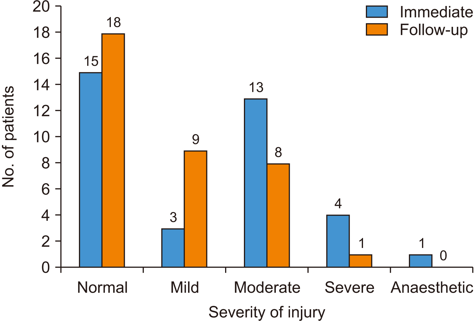
Fig. 3 Change in severity of nerve injury for MSAN (middle superior alveolar nerve) over two months.
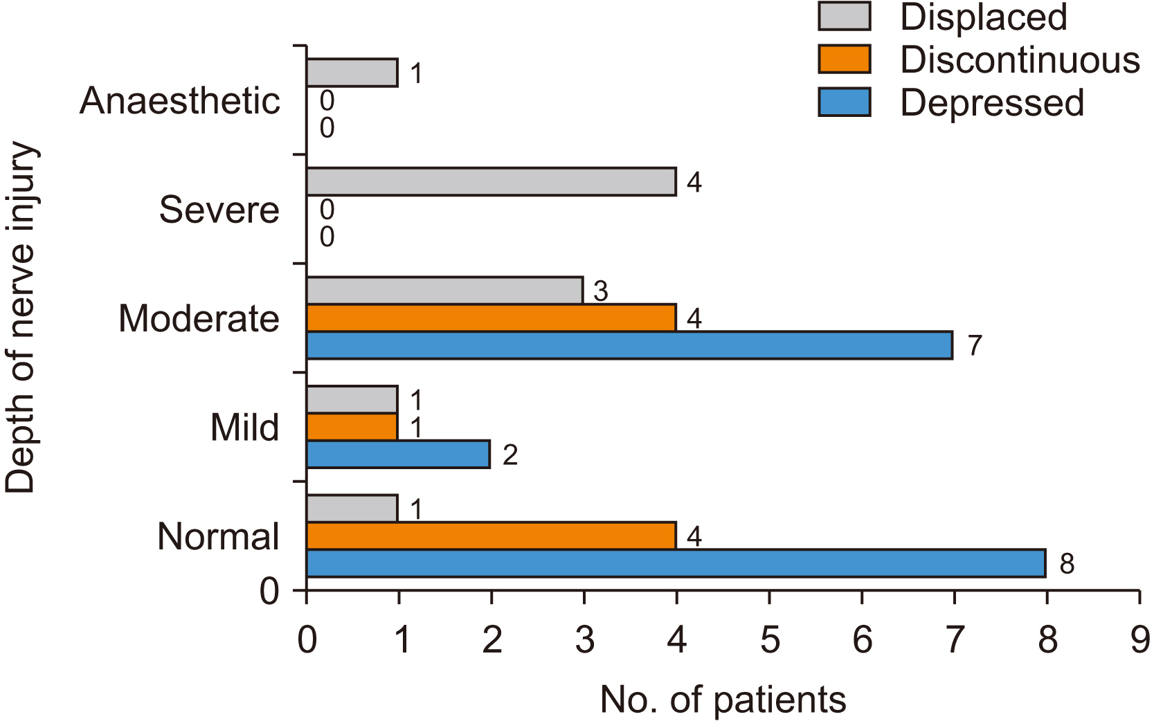
Fig. 4 Fracture patterns in patients with depth of nerve injury.
Reference
-
References
1. Joseph B, Vyloppilli S, Sayd S, Ummer N. 2015; Sinonasal sarcoidosis of the maxillary sinus and infraorbital nerve: a case report. J Korean Assoc Oral Maxillofac Surg. 41:217–21. https://doi.org/10.5125/jkaoms.2015.41.4.217. DOI: 10.5125/jkaoms.2015.41.4.217. PMID: 26339583. PMCID: PMC4558193.
Article2. Olenczak JB, Hui-Chou HG, Aguila DJ 3rd, Shaeffer CA, Dellon AL, Manson PN. 2015; Posttraumatic midface pain: clinical significance of the anterior superior alveolar nerve and Canalis sinuosus. Ann Plast Surg. 75:543–7. https://doi.org/10.1097/sap.0000000000000335. DOI: 10.1097/SAP.0000000000000335. PMID: 25710550.
Article3. Dorafshar AH, Dellon AL, Wan EL, Reddy S, Wong VW. 2017; Injured anterior superior alveolar nerve endoscopically resected within maxillary sinus. Craniomaxillofac Trauma Reconstr. 10:208–11. https://doi.org/10.1055/s-0036-1592088. DOI: 10.1055/s-0036-1592088. PMID: 28751945. PMCID: PMC5526682.
Article4. Musavi L, Macmillan A, Pedriera R, Lopez J, Dorafshar AH, Dellon AL. 2018; Resection of the posterior, middle, and anterior superior alveolar nerves and infraorbital nerve neurolysis for refractory maxillary pain. J Oral Maxillofac Surg. 76:1175–80. https://doi.org/10.1016/j.joms.2017.12.024. DOI: 10.1016/j.joms.2017.12.024. PMID: 29391162.
Article5. Zuniga JR, Essick GK. 1992; A contemporary approach to the clinical evaluation of trigeminal nerve injuries. Oral Maxillofac Surg Clin N Am. 4:353–67. https://doi.org/10.1016/S1042-3699(20)30593-8. DOI: 10.1016/S1042-3699(20)30593-8.
Article6. Jacobs R, Wu CH, Goossens K, Van Loven K, Van Hees J, Van Steenberghe D. 2002; Oral mucosal versus cutaneous sensory testing: a review of the literature. J Oral Rehabil. 29:923–50. https://doi.org/10.1046/j.1365-2842.2002.00960.x. DOI: 10.1046/j.1365-2842.2002.00960.x. PMID: 12421324.
Article7. Komiyama O, De Laat A. 2005; Tactile and pain thresholds in the intra- and extra-oral regions of symptom-free subjects. Pain. 115:308–15. https://doi.org/10.1016/j.pain.2005.03.006. DOI: 10.1016/j.pain.2005.03.006. PMID: 15911157.
Article8. Vriens JP, Moos KF. 1995; Morbidity of the infraorbital nerve following orbitozygomatic complex fractures. J Craniomaxillofac Surg. 23:363–8. https://doi.org/10.1016/s1010-5182(05)80131-3. DOI: 10.1016/S1010-5182(05)80131-3. PMID: 8839330.
Article9. Won SY, Kim HK, Kim ME, Kim KS. 2017; Two-point discrimination values vary depending on test site, sex and test modality in the orofacial region: a preliminary study. J Appl Oral Sci. 25:427–35. https://doi.org/10.1590/1678-7757-2016-0462. DOI: 10.1590/1678-7757-2016-0462. PMID: 28877282. PMCID: PMC5595116.
Article10. Peltomaa J, Rihkanen H. 2000; Infraorbital nerve recovery after minimally dislocated facial fractures. Eur Arch Otorhinolaryngol. 257:449–52. https://doi.org/10.1007/s004050000264. DOI: 10.1007/s004050000264. PMID: 11073197.
Article11. Pedemonte C, Basili A. 2005; Predictive factors in infraorbital sensitivity disturbances following zygomaticomaxillary fractures. Int J Oral Maxillofac Surg. 34:503–6. https://doi.org/10.1016/j.ijom.2004.10.026. DOI: 10.1016/j.ijom.2004.10.026. PMID: 16053869.
Article12. Tajima S. 1977; Malar bone fractures: experimental fractures on the dried skull and clinical sensory disturbances. J Maxillofac Surg. 5:150–6. https://doi.org/10.1016/s0301-0503(77)80094-5. DOI: 10.1016/S0301-0503(77)80094-5. PMID: 267707.
Article13. Robertson LT, Levy JH, Petrisor D, Lilly DJ, Dong WK. 2003; Vibration perception thresholds of human maxillary and mandibular central incisors. Arch Oral Biol. 48:309–16. https://doi.org/10.1016/s0003-9969(03)00006-2. DOI: 10.1016/S0003-9969(03)00006-2. PMID: 12663076.
Article14. Lv D, Zhang J, Gu X, Shen H, Shao S, Hou W, et al. 2016; Transient pain following orthodontic fixed appliances induces sensitization of gingival and periodontal tissues. J Oral Facial Pain Headache. 30:228–33. https://doi.org/10.11607/ofph.1646. DOI: 10.11607/ofph.1646. PMID: 27472525.
Article15. Ivy RH. 1923; LXXIX. Radical operations on the maxillary sinus and damage to the teeth. Ann Otol Rhinol Laryngol. 32:1197–202. https://doi.org/10.1177/000348942303200410. DOI: 10.1177/000348942303200410.
Article16. Din AR, Buckley N, Ali N, Millwaters M, Sharma PK. 2021; A prospective cohort study evaluating subjective and objective neurosensory changes following LeFort I osteotomy. Am J Orthod Dentofacial Orthop. 160:410–22. https://doi.org/10.1016/j.ajodo.2020.11.038. DOI: 10.1016/j.ajodo.2020.11.038. PMID: 33975747.
Article17. Chen E, Abbott PV. 2009; Dental pulp testing: a review. Int J Dent. 2009:365785. https://doi.org/10.1155/2009/365785. DOI: 10.1155/2009/365785. PMID: 20339575. PMCID: PMC2837315.
Article18. Rowe AH, Pitt Ford TR. 1990; The assessment of pulpal vitality. Int Endod J. 23:77–83. https://doi.org/10.1111/j.1365-2591.1990.tb00843.x. DOI: 10.1111/j.1365-2591.1990.tb00843.x. PMID: 2202687.
Article19. Jespersen JJ, Hellstein J, Williamson A, Johnson WT, Qian F. 2014; Evaluation of dental pulp sensibility tests in a clinical setting. J Endod. 40:351–4. https://doi.org/10.1016/j.joen.2013.11.009. DOI: 10.1016/j.joen.2013.11.009. PMID: 24565651.
Article20. Brajdić D, Virag M, Uglešić V, Aljinović-Ratković N, Zajc I, Macan D. 2011; Evaluation of sensitivity of teeth after mandibular fractures. Int J Oral Maxillofac Surg. 40:266–70. https://doi.org/10.1016/j.ijom.2010.11.016. DOI: 10.1016/j.ijom.2010.11.016. PMID: 21177072.
Article21. Murakami G, Ohtsuka K, Sato I, Moriyama H, Shimada K, Tomita H. 1994; The superior alveolar nerves: their topographical relationship and distribution to the maxillary sinus in human adults. Okajimas Folia Anat Jpn. 70:319–28. https://doi.org/10.2535/ofaj1936.70.6_319. DOI: 10.2535/ofaj1936.70.6_319. PMID: 8041567.
Article
- Full Text Links
-
- Actions
-
Cited
- CITED
-
- Close
- Share
-
- Similar articles
-
- A Study on the Possibility of Neurological Morbidity of Prelacrimal Recess Approach Depending on Anatomical Differences
- Study on the position of the posterior superior alveolar artery in relation to the performance of the maxillary sinus bone graft procedure in a Korean population
- Transient Neurologic Deterioration after Total Removal of Parasagittal Meningioma Including Completely Occluding Superior Sagittal Sinus
- Evaluation of the posterior superior alveolar artery canal by cone-beam computed tomography in a sample of the Egyptian population
- Correlation of Middle Meatus, Ethmoid Sinus and Maxillary Sinus Microbiology in Patients with Chronic Sinusitis
