Brain Tumor Res Treat.
2023 Oct;11(4):271-273. 10.14791/btrt.2023.0027.
Massive Hyperostotic Meningioma En Plaque Mimicking Fibrous Dysplasia
- Affiliations
-
- 1Department of Neurosurgery, Kyung Hee University Hospital, Seoul, Korea
- 2Department of Neurosurgery, College of Medicine, Kyung Hee University, Seoul, Korea
- KMID: 2547416
- DOI: http://doi.org/10.14791/btrt.2023.0027
Abstract
- The authors report an extremely rare case of a massive hyperostotic meningioma en plaque, which had characteristics of unique bony growth. A 34-year-old man presented with a palpable solid mass in the left cranial region that had gradually grown in size with a broad base on the calvarium for 8 years. Radiologically, the area involved by the mass ranged from the sphenoid bone to the frontal, parietal, temporal, and occipital bones. Three-dimensional CT revealed multiple growing spiculate features on the inner and outer cranial surface. Even though the radiologic features resembled fibrous dysplasia, it was histologically found to be a type of meningioma.
Figure
-
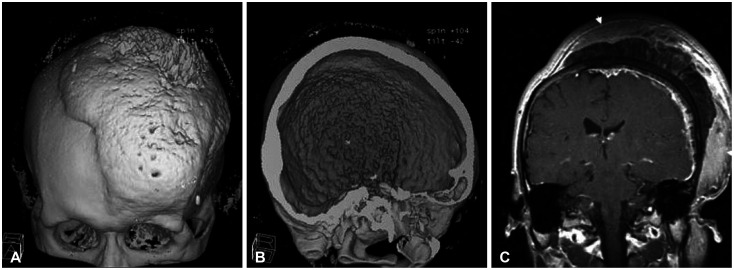
Fig. 1 Imaging findings of patient. A and B: Three-dimensional CT scan with bony reconstruction showing diffuse bony hyperostosis in the outer and inner surface of the cranial vault crossing the sagittal suture. C: Coronal image of a gadolinium-enhanced gradient-echo T1-weighted MRI showing mushroom-like growth combined with tumor under the dermis (arrow).
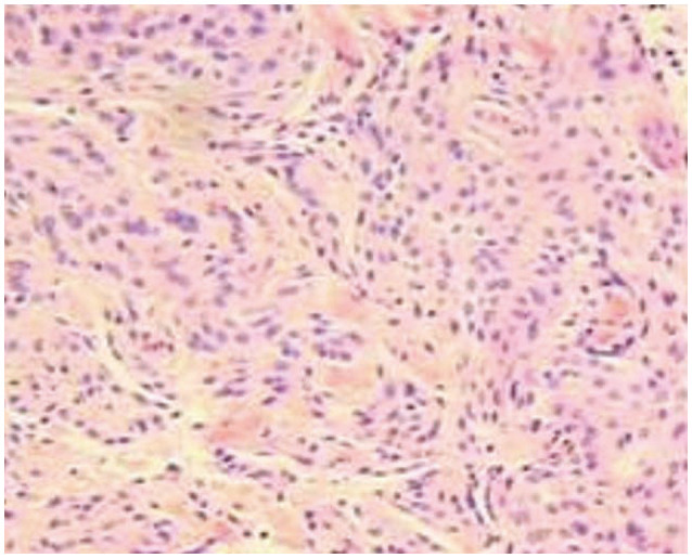
Fig. 2 Light microscopic findings demonstrate nodular tumor tissues, consisting of interlacing and whirling bundles of meningothelial cells without significant atypia (H&E, ×100). H&E, hematoxylin and eosin.
Reference
-
1. Bonnal J, Thibaut A, Brotchi J, Born J. Invading meningiomas of the sphenoid ridge. J Neurosurg. 1980; 53:587–599. PMID: 7431070.
Article2. Castellano F, Guidetti B, Olivecrona H. Pterional meningiomas en plaque. J Neurosurg. 1952; 9:188–196. PMID: 14908653.
Article3. Honeybul S, Neil-Dwyer G, Lang DA, Evans BT, Ellison DW. Sphenoid wing meningioma en plaque: a clinical review. Acta Neurochir (Wien). 2001; 143:749–757. discussion 758. PMID: 11678395.
Article4. Bikmaz K, Mrak R, Al-Mefty O. Management of bone-invasive, hyperostotic sphenoid wing meningiomas. J Neurosurg. 2007; 107:905–912. PMID: 17977259.
Article5. Elder TA, Yokoi H, Chugh AJ, Lagman C, Wu O, Wright CH, et al. En plaque meningiomas: a narrative review. J Neurol Surg B Skull Base. 2021; 82(Suppl 3):e33–e44. PMID: 34306915.
Article6. Muhsen BA, Aljariri AI, Hashem H, Alzoubi Q, Sarhan N, Al-Hussaini M, et al. En-plaque sphenoid wing grade II meningioma: case report and review of literature. Ann Med Surg (Lond). 2022; 74:103322. PMID: 35145681.
Article7. Fariselli L, Biroli A, Signorelli A, Broggi M, Marchetti M, Biroli F. The cavernous sinus meningiomas’ dilemma: surgery or stereotactic radiosurgery? Rep Pract Oncol Radiother. 2016; 21:379–385. PMID: 27330423.
Article8. Maroon JC, Kennerdell JS, Vidovich DV, Abla A, Sternau L. Recurrent spheno-orbital meningioma. J Neurosurg. 1994; 80:202–208. PMID: 8283257.
Article9. Li Y, Shi JT, An YZ, Zhang TM, Fu JD, Zhang JL, et al. Sphenoid wing meningioma en plaque: report of 37 cases. Chin Med J (Engl). 2009; 122:2423–2427. PMID: 20079153.10. Boari N, Gagliardi F, Spina A, Bailo M, Franzin A, Mortini P. Management of spheno-orbital en plaque meningiomas: clinical outcome in a consecutive series of 40 patients. Br J Neurosurg. 2013; 27:84–90. PMID: 22905887.
Article11. Kashimura H, Beppu T, Wada T, Yoshida Y, Suzuki M, Ogawa A. [A case of meningioma en plaque: review of 73 cases]. No Shinkei Geka. 1997; 25:1097–1100. Japanese. PMID: 9430144.12. Akutsu H, Sugita K, Sonobe M, Matsumura A. Parasagittal meningioma en plaque with extracranial extension presenting diffuse massive hyperostosis of the skull. Surg Neurol. 2004; 61:165–169. discussion 169. PMID: 14751632.
Article13. Kim SM, Jang KS, Kim MC, Joo WI. Convexity meningioma en plaque presenting with diffuse hyperostosis of the skull. J Korean Neurosurg Soc. 2006; 39:159–161.14. Leeds N, Seaman WB. Fibrous dysplasia of the skull and its differential diagnosis. A clinical and roentgenographic study of 46 cases. Radiology. 1962; 78:570–582. PMID: 14463598.
Article15. Pérez-Santonja JJ, Bueno JL, Serrano de la Iglesia JM, Zato MA, Queimadelos V, Ibarburen C. [Sphenoidal hyperostosis. Problems of differential diagnosis: report of a case]. Rev Clin Esp. 1992; 191:422–425. Spanish. PMID: 1336861.
- Full Text Links
-
- Actions
-
Cited
- CITED
-
- Close
- Share
-
- Similar articles
-
- Extracranial Extension of Intracranial Atypical Meningioma En Plaque with Osteoblastic Change of the Skull
- Convexity Meningioma En Plaque Presenting with Diffuse Hyperostosis of the Skull
- A Case Report of Meningioma "en plaque"
- Hyperostotic Esthesioneuroblastoma: Rare Variant and Fibrous Dysplasia Mimicker
- En Plaque Meningioma in Thoracic Spine: Case Report
