Extracranial Extension of Intracranial Atypical Meningioma En Plaque with Osteoblastic Change of the Skull
- Affiliations
-
- 1Department of Neurosurgery, Seoul Medical Center, Seoul, Korea.
- 2Department of Neurosurgery, Hanyang University Guri Hospital, Guri, Korea. kch5142@hanyang.ac.kr
- KMID: 2191093
- DOI: http://doi.org/10.3340/jkns.2014.55.4.205
Abstract
- Meningioma is a common primary tumor of central nervous system. However, extracranial extension of the intracranial meningioma is unusual, and mostly accompanied the osteolytic change of the skull. We herein describe an atypical meningioma having extracranial extension with hyperostotic change of the skull. The patient was a 72-year-old woman who presented a large mass in the right frontal scalp and left hemiparesis. Brain magnetic resonance imaging and computed tomography scans revealed an intracranial mass, diffuse meningeal thickening, hyperostotic change of the skull with focal extension into the right frontal scalp. She underwent total removal of extracranial tumor, bifrontal craniectomy, and partial removal of intracranial tumor followed by cranioplasty. Tumor pathology was confirmed as atypical meningioma, and she received adjuvant radiotherapy. In this report, we present and discuss a meningioma en plaque of atypical histopathology having an extracranial extension with diffuse intracranial growth and hyperostotic change of the skull.
MeSH Terms
Figure
-
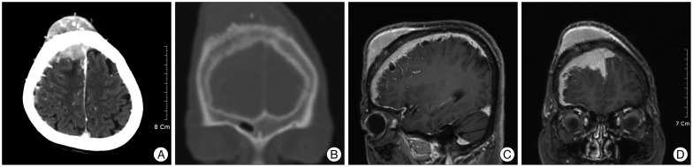
Fig. 1 Preoperative brain enhanced computed tomography (CT) scans and magnetic resonance (MR) image. A : Axial CT scan displaying the well-enhanced mass in the right frontal area extended through the skull diffusely. B : Coronal CT scan showing the frontal bulging with rough outer table by the hyperostotic change. C and D : Sagittal and coronal T2-weighted MR images revealing the ill-defined tumor compressing the right frontal lobe and the marginal multi-cystic mass in the right hemisphere.
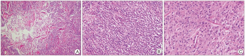
Fig. 2 Photomicrographs of the tumor specimens. H & E staining shows the findings of atypical meningioma characterized by geographic necrosis, sheet-like pattern (A, ×40), increased cellularity (B, ×100), mild to moderate pleomorphism and mitotic figures (C, ×400).
Reference
-
1. Basu K, Majumdar K, Chatterjee U, Ganguli M, Chatterjee S. En plaque meningioma with angioinvasion. Indian J Pathol Microbiol. 2010; 53:319–321. PMID: 20551544.
Article3. Daffner RH, Yakulis R, Maroon JC. Intraosseous meningioma. Skeletal Radiol. 1998; 27:108–111. PMID: 9526778.
Article4. De Jesús O, Toledo MM. Surgical management of meningioma en plaque of the sphenoid ridge. Surg Neurol. 2001; 55:265–269. PMID: 11516463.
Article5. Doyle WF, Rosegay H. Meningioma en plaque with hyperostosis : case report. Mil Med. 1972; 137:196–198. PMID: 4623534.6. Goyal LK, Suh JH, Mohan DS, Prayson RA, Lee J, Barnett GH. Local control and overall survival in atypical meningioma : a retrospective study. Int J Radiat Oncol Biol Phys. 2000; 46:57–61. PMID: 10656373.
Article7. Iglesias ME, Vázquez-Doval J, Idoate MA, Vanaclocha V, Idoate F, Quintanilla E. Intracranial osteolytic meningioma affecting the scalp. J Am Acad Dermatol. 1996; 35:641–642. PMID: 8859303.
Article8. Jo K, Park HJ, Nam DH, Lee JI, Kong DS, Park K, et al. Treatment of atypical meningioma. J Clin Neurosci. 2010; 17:1362–1366. PMID: 20800497.
Article9. Kashimura H, Beppu T, Wada T, Yoshida Y, Suzuki M, Ogawa A. [A case of meningioma en plaque : review of 73 cases]. No Shinkei Geka. 1997; 25:1097–1100. PMID: 9430144.10. Kim H, Jung TY, Kim IY, Lee JK. Two cases of primary osteolytic intraosseous meningioma of the skull metastasizing to whole skull and the spine. J Korean Neurosurg Soc. 2012; 51:151–154. PMID: 22639712.
Article11. Kim KS, Rogers LF, Goldblatt D. CT features of hyperostosing meningioma en plaque. AJR Am J Roentgenol. 1987; 149:1017–1023. PMID: 3118666.
Article12. Kumar S, Dhingra PL, Gondal R. Ectopic meningioma of the paranasal sinuses. Childs Nerv Syst. 1993; 9:483–484. PMID: 8124679.
Article13. Li Y, Shi JT, An YZ, Zhang TM, Fu JD, Zhang JL, et al. Sphenoid wing meningioma en plaque : report of 37 cases. Chin Med J (Engl). 2009; 122:2423–2427. PMID: 20079153.14. Maroon JC, Kennerdell JS, Vidovich DV, Abla A, Sternau L. Recurrent spheno-orbital meningioma. J Neurosurg. 1994; 80:202–208. PMID: 8283257.
Article15. Park HJ, Kang HC, Kim IH, Park SH, Kim DG, Park CK, et al. The role of adjuvant radiotherapy in atypical meningioma. J Neurooncol. 2013; 115:241–247. PMID: 23949108.
Article
