A single hepatic mass with two tales: hepatic tuberculosis and hepatocellular carcinoma
- Affiliations
-
- 1Lee Kong Chian School of Medicine, Nanyang Technological University, Singapore
- 2Department of Anatomical Pathology, Singapore General Hospital, Singapore
- 3Duke-NUS Medical School, Singapore
- 4School of Biological Sciences, Nanyang Technological University, Singapore
- KMID: 2546419
- DOI: http://doi.org/10.17998/jlc.2023.08.30
Abstract
- Hepatic tuberculosis (HTB) is an uncommon manifestation of tuberculous infections, and there has been no proven causal link between HTB and hepatocellular carcinoma (HCC). We herein present a rare case of a synchronous presentation of HTB and HCC within a single hepatic mass. A 57-year-old Chinese gentleman with recently diagnosed sigmoid adenocarcinoma was found to have a left lower lobe pulmonary nodule and solitary hepatic mass on staging computed tomography. Biopsies showed the hepatic mass to have both HTB and HCC components. This serves as a reminder that HTB is an important differential to consider for space-occupying lesions in the liver. Histological evaluation of suspected hepatic malignancies is recommended to exclude the presence of HTB in appropriate clinical settings.
Figure
-
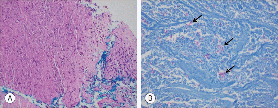
Figure 1. Histology of pulmonary nodule. (A) H&E stain shows caseating granuloma at 200× magnification. (B) Identification of acid-fast bacilli (arrows) on ZN staining at 600× magnification. H&E, hematoxylin and eosin; ZN, Ziehl-Neelsen.

Figure 2. Core biopsy of hepatic nodule. (A) Core showing caseating granuloma (arrows) and area of HCC on low-powered view (40× magnification). (B) Caseating granuloma with surrounding multinucleated giant cells at 100× magnification. (C) Area with HCC at 100× magnification. (D) High-powered view (200× magnification) of HCC displaying pseudo-glandular architecture. (E) Positive staining for CD34 marker, indicating capillarization of sinusoids. (F) Patchy staining for glypican-3. HCC, hepatocellular carcinoma.
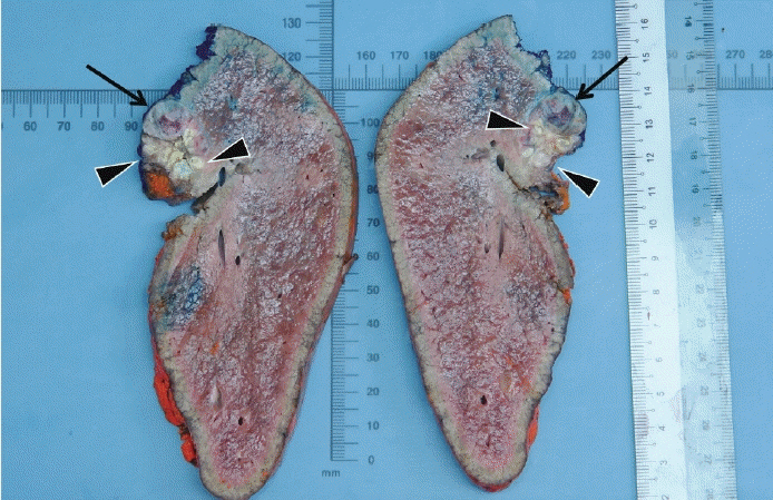
Figure 3. Sections of central hepatectomy specimen demonstrating tumor (arrows) with surrounding yellow necrotic nodules (arrowheads).
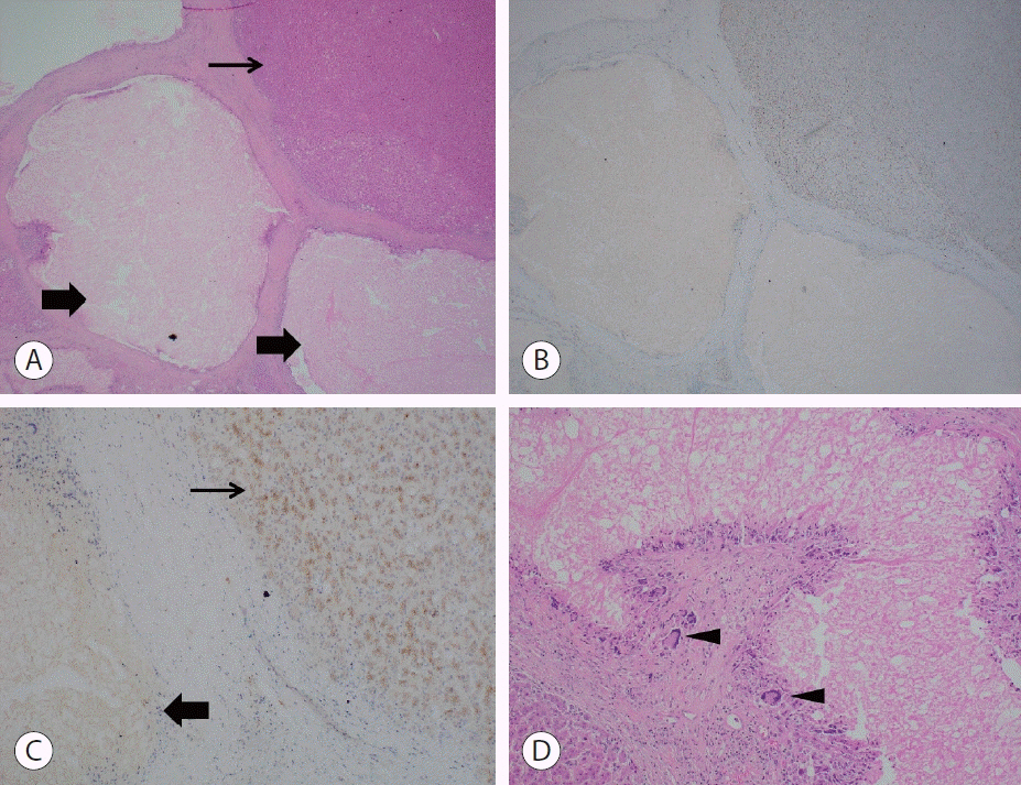
Figure 4. Histology of resected hepatic mass. (A) Caseating granulomas (bold arrows) located adjacent to area of HCC (thin arrow) at 20× magnification. (B) Area of HCC with positive CD34 staining (20× magnification). (C) Caseating granuloma (bold arrow) adjacent to area of HCC (thin arrow) staining positive for glypican-3 on high-powered view (100× magnification). (D) Caseating granuloma with surrounding multinucleated giant cells (arrowheads) and central area of necrosis at 100× magnification. HCC, hepatocellular carcinoma.
Reference
-
References
1. Akıcı M, Bozkurt E, Özdemir Ç, F Kaya. Primary hepatic tuberculosis mimicking malignancy. Int Surg J. 2019; 6:3078–3082.2. Falagas ME, Kouranos VD, Athanassa Z, Kopterides P. Tuberculosis and malignancy. QJM. 2010; 103:461–487.3. Tatco VR, Mejia-Santos MMA, Uy JAU. The many faces of hepatic tuberculosis. TB Corner. 2015; 1:1–6.4. Levine C. Primary macronodular hepatic tuberculosis: US and CT appearances. Gastrointest Radiol. 1990; 15:307–309.5. Limaiem F, Gargouri F, Bouraoui S, Lahmar A, Mzabi S. Co-existence of hepatocellular carcinoma and hepatic tuberculosis. Surg Infect (Larchmt). 2014; 15:437–440.6. Shah D, P G, S S, Koushik AK. Synchronous presentation of tuberculosis and hepatocellular carcinoma in a cirrhotic patient: a case report. Trop Doct. 2020; 50:71–74.7. Liao JR, Zhang D, Wu XL. Pulmonary tuberculosis combined with hepatic tuberculosis: a case report and literature review. Clin Respir J. 2015; 9:501–505.8. Hickey AJ, Gounder L, Moosa MY, Drain PK. A systematic review of hepatic tuberculosis with considerations in human immunodeficiency virus co-infection. BMC Infect Dis. 2015; 15:209.9. Zhang L, Yang NB, Ni SL, Zhang SN, Shen CB, Lu MQ. A case of multiple macronodular hepatic tuberculosis difficult to differentiate from hepatocellular carcinoma with intrahepatic metastasis: CTguided fine needle aspiration biopsy confirmed the diagnosis. Int J Clin Exp Pathol. 2014; 7:8240–8244.10. Yang C, Liu X, Ling W, Song B, Liu F. Primary isolated hepatic tuberculosis mimicking small hepatocellular carcinoma: a case report. Medicine (Baltimore). 2020; 99:e22580.
- Full Text Links
-
- Actions
-
Cited
- CITED
-
- Close
- Share
-
- Similar articles
-
- Primary Hepatic Tuberculosis Mimicking Hepatocelluar Carcinoma in Patient with Chronic Viral Hepatitis B and C
- A case report of hepatocellular carcinoma in common hepatic duct
- Efficacy of Hepatic Arterial Infusion Chemotherapy and Radiofrequency Ablation against Hepatocellular Carcinoma Refractory to Transarterial Chemoembolization and Vascular Variation: A Case Study
- Obstructive Jaundice Caused by the Fragment of Hepatocellular Carcinoma in the Common Hepatic Duct Confirmed by Peroral Choledochoscopy
- A case of spontaneous hepatic rupture in a patient with primary hepatocellular carcinoma during the puerperium
