Hepatic small vessel neoplasm: not totally benign, not yet malignant
- Affiliations
-
- 1Department of Pathology, Anatomy, and Laboratory Medicine, West Virginia University, Morgantown, WV, USA
- KMID: 2545970
- DOI: http://doi.org/10.4132/jptm.2023.06.19
Abstract
- Hepatic small vessel neoplasm (HSVN) is a rare vascular tumor with few reports in the literature. While imaging findings may show characteristic enhancement patterns, limited available literature may not reveal the full potential for image-based diagnosis. Histologically, HSVN mimics other entities, though certain morphologic and immunohistochemical findings provide clues for diagnosis. However, HSVN still provides diagnostic challenges, especially on core biopsies with limited material for morphologic and molecular evaluation. While current recommendations are surgical resection and close observation, the long-term course of the tumor is unknown. We report a case of HSVN in a liver with additional feature of organized lymphoid aggregates necessitating additional hematopathology consultation and workup to rule out concurrent entities.
Figure
-
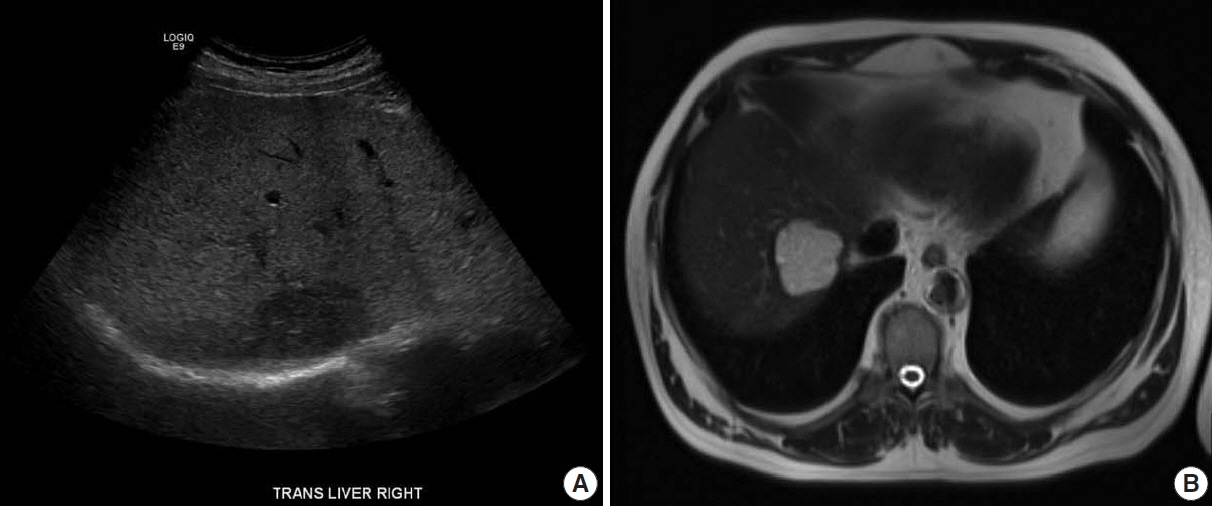
Fig. 1. (A) Liver ultrasound demonstrating a hypoechoic lesion in the right lobe of the liver. (B) Liver protocol abdominal magnetic resonance imaging showing a hyperintense lesion on T2 with heterogenous contrast enhancement.
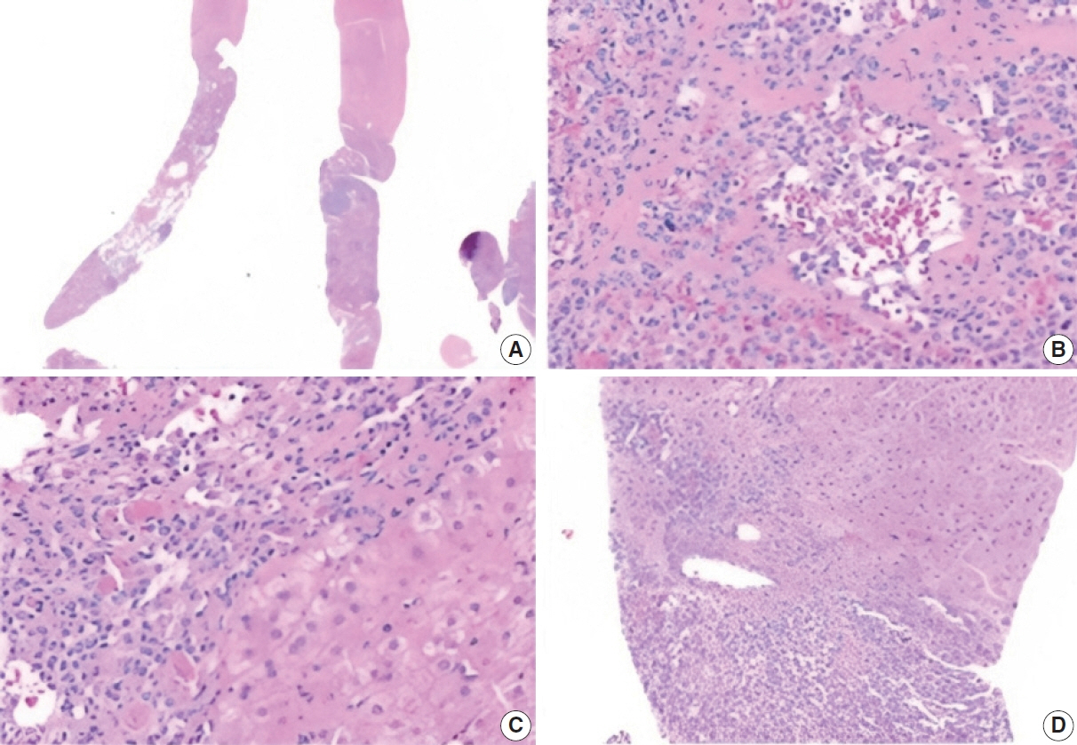
Fig. 2. Microscopic appearance of the biopsy specimen. (A) Multiple biopsy specimens with well-demarcated lesion. Multiple, various-sized lymphoid aggregates can be seen. (B) The lesion is composed of thin-walled small vascular channels lined by bland endothelial cells. (C) The lesion is well-demarcated from adjacent liver parenchyma in most areas. (D) Focal infiltration into portal tract.
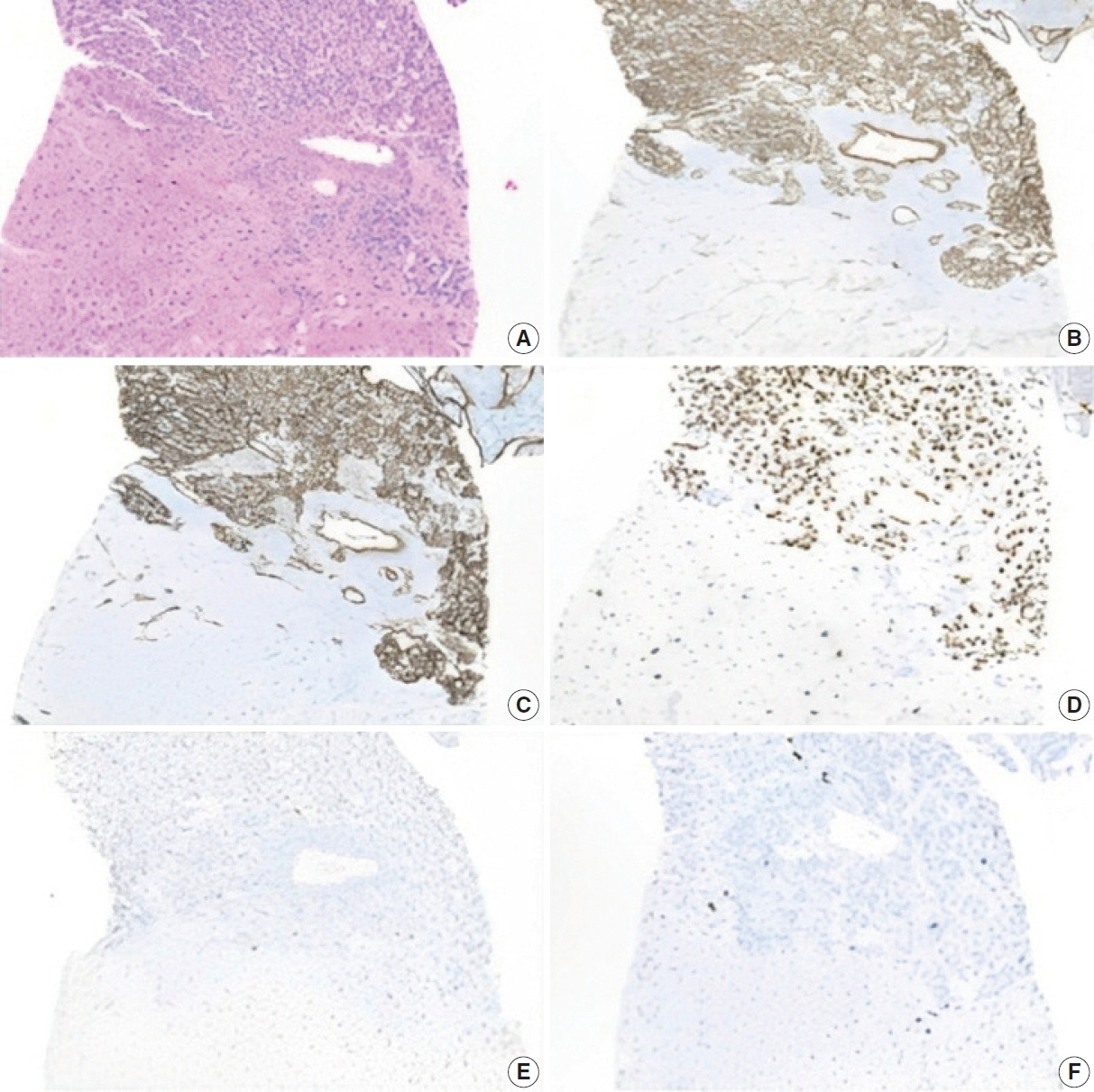
Fig. 3. Immunohistochemical staining. (A) Lesion next to normal hepatic parenchyma. (B) The lesion is positive for CD31. (C) Positive for CD34. (D) Positive for ERG. (E) p53 is patchy and weak. (F) Ki-67 proliferation index is 5%–10%.

Fig. 4. Immunohistochemical staining of lymphoid aggregate. (A) Varying sized aggregates of small lymphoid cells were present in the lesion. (B) CD3 stains a moderate number of small T cells. (C) CD20 stains a small number of small B cells. (D) CD8 did not demonstrate organized lymphoid tissue.
Reference
-
References
1. Gill RM, Buelow B, Mather C, et al. Hepatic small vessel neoplasm, a rare infiltrative vascular neoplasm of uncertain malignant potential. Hum Pathol. 2016; 54:143–51.2. Paisant A, Bellal S, Lebigot J, Canivet CM, Michalak S, Aube C. Imaging features of hepatic small vessel neoplasm: case series. Hepatology. 2021; 74:2894–6.3. Walcott-Sapp S, Tang E, Kakar S, Shen J, Hansen P. Resection of the largest reported hepatic small vessel neoplasm. Hum Pathol. 2018; 78:159–62.4. Cicala CM, Monaca F, Giustiniani MC, Di Salvatore M. Multifocal hepatic small vessel neoplasm with spleen dissemination. BMJ Case Rep. 2022; 15:e248785.5. Joseph NM, Brunt EM, Marginean C, et al. Frequent GNAQ and GNA14 mutations in hepatic small vessel neoplasm. Am J Surg Pathol. 2018; 42:1201–7.6. Akanuma N, Joseph NM, Stachler M, Behr S, Takahashi T, Gill RM. Malignant hepatic vascular neoplasm with novel RAF1 and GNA11 mutations: risk stratification considerations for hepatic small vessel neoplasm (HSVN). Hum Pathol Rep. 2022; 29:300671.7. Lappa E, Drakos E. Anastomosing hemangioma: short review of a benign mimicker of angiosarcoma. Arch Pathol Lab Med. 2020; 144:240–4.8. Lin J, Bigge J, Ulbright TM, Montgomery E. Anastomosing hemangioma of the liver and gastrointestinal tract: an unusual variant histologically mimicking angiosarcoma. Am J Surg Pathol. 2013; 37:1761–5.9. Bioulac-Sage P, Laumonier H, Laurent C, Blanc JF, Balabaud C. Benign and malignant vascular tumors of the liver in adults. Semin Liver Dis. 2008; 28:302–14.10. O’Neill AC, Craig JW, Silverman SG, Alencar RO. Anastomosing hemangiomas: locations of occurrence, imaging features, and diagnosis with percutaneous biopsy. Abdom Radiol (NY). 2016; 41:1325–32.11. Liu Z, Yi L, Chen J, et al. Comparison of the clinical and MRI features of patients with hepatic hemangioma, epithelioid hemangioendothelioma, or angiosarcoma. BMC Med Imaging. 2020; 20:71.12. Kampa KC, Guerra JA, Percicotte AP, Zaparolli M, Raeder Costa MA, Ramos EJ. Hepatic splenosis: a report of a very rare case. Gastroenterol Hepatol Open Access. 2019; 10:88–91.13. Shiozawa K, Watanabe M, Ikehara T, et al. A case of contiguous primary hepatic marginal zone B-cell lymphoma and hemangioma ultimately diagnosed using contrast-enhanced ultrasonography. Case Rep Oncol. 2015; 8:50–6.14. Goh IY, Mulholland P, Sokolova A, Liu C, Siriwardhane M. Hepatic small vessel neoplasm: a systematic review. Ann Med Surg (Lond). 2021; 72:103004.
- Full Text Links
-
- Actions
-
Cited
- CITED
-
- Close
- Share
-
- Similar articles
-
- Diagnosis of Focal Liver Masses on Ultrasonography
- The Imaging Findings of Small(< or =15mm) Portal Defects in the Liver on CT Arterial Portography: Evaluationwith CT Hepatic Arteriography and Lipiodol CT
- Vascular tumors of the liver: A brief review
- A case report of malignant paraganglioma with hepatic metastases
- Small Hepatic Cystic Lesions in Patients with Extrahepatic IVlalignancy: Incidence and Significance on CT
