Simultaneous Transcatheter Treatment for Combined Severe Degenerative Aortic Stenosis and Primary Mitral Regurgitation
- Affiliations
-
- 1Department of Cardiology, Asan Medical Center, University of Ulsan College of Medicine, Seoul, Korea
- KMID: 2545962
- DOI: http://doi.org/10.4070/kcj.2023.0088
Figure
-
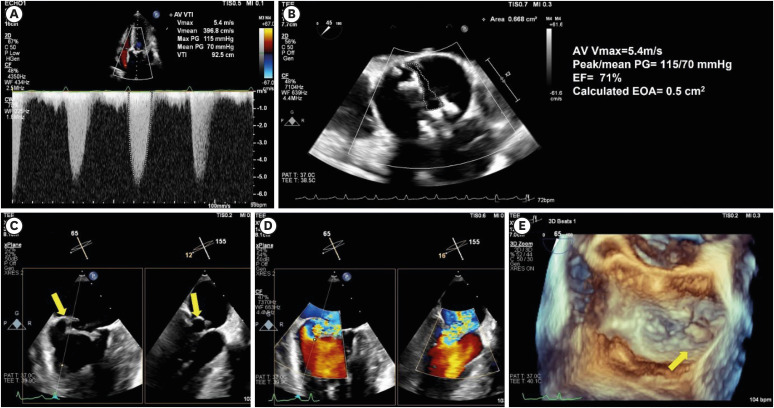
Figure 1 Echocardiographic images of the AVs and MVs. (A) Continuous wave doppler across the AV shows a mean gradient of 70 mmHg. (B) AV area by 2D planimetry is 0.5 cm2 in the mid-esophageal AV short-axis view of TEE. (C) The biplane images of TEE show a prolapse of the posterior medial scallop with coaptation gap. (D) Biplane images with color Doppler show a severe mitral regurgitation jet. (E) 3D en face view shows the reconstruction image of the MV in TEE. Yellow arrow: Prolapse of posterior medial scallop of the MV.AS = aortic stenosis; AV = aortic valve; MV = mitral valve; PG = pressure gradient; TEE = transesophageal echocardiography; VTI = velocity time integral.

Figure 2 Transfemoral transcatheter aortic valve replacement for severe bicuspid aortic stenosis. (A, B) Computed tomography shows severe calcification in the aortic valve. (C) After the first balloon valvuloplasty with an 18 mm balloon, dual left ventricle wiring is performed. (D) Additional predilation with a 20 mm balloon is done. (E) THV crosses the aortic valve after the second balloon valvuloplasty. (F) A dual wire is exchanged for a pig-tail catheter. (G) SAPIEN 3 23 mm valve is successfully implanted. (H) Post-dilation after THV was performed.THV = transcatheter heart valve.
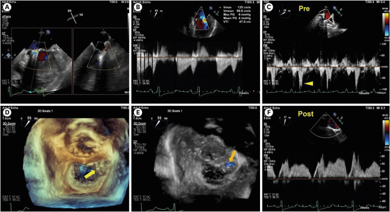
Figure 3 The images of intra-procedural transesophageal echocardiography. (A) Remnant mild MR after deploying the second clip. (B) Continuous wave Doppler across the mitral valve shows a mean gradient of 4mmHg after the TEER. (C) Pulmonary vein systolic reversal flow is shown before the procedure. (D) The 3D reconstruction image of mitral valve shows two deployed clips. (E) The 3D color Doppler image shows mild remnant MR. (F) Pulmonary vein systolic reversal flow disappeared after TEER. Yellow arrowhead: pulmonary systolic vein reversal before the procedure. Yellow arrow: deployed clip at medial leaflet of mitral valve. Orange arrow: remnant regurgitation flow after TEER.MR = mitral regurgitation; TEER = transcatheter edge-to-edge repair.

Figure 4 Final fluoroscopic image after performed concomitant transcatheter aortic valve replacement and transcatheter edge-to-edge repair.
- Full Text Links
-
- Actions
-
Cited
- CITED
-
- Close
- Share
-
- Similar articles
-
- Assessment of Mitral Stenosis by Doppler Echocardiography: Influence of Regurgitation on Doppler Pressure Half-Time
- Relationship between Degree of Aortic Regurgitation Graded by 2-D Color Doppler Echocardiography and Diastolic Fluttering of Anterior Mitral Leaflet
- A Case of Severe Aortic Stenosis Patient With High Operative Risk Treated by Transcatheter Aortic-Valve Implantation
- Recent Evidence and Initial Experiences of Transcatheter Edgeto-Edge Repair of the Mitral Valve in South Korea
- Silent Aortic Regurgitation
