Delayed post-ischemic leukoencephalopathy in emergent large-vessel occlusive stroke after mechanical thrombectomy: case reports
- Affiliations
-
- 1Department of Neurology, Asan Medical Center, University of Ulsan College of Medicine, Seoul, Korea
- 2Department of Neurology, Uijeongbu Eulji Medical Center, Eulji University School of Medicine, Uijeongbu, Korea
- 3Graduate School of Medicine, Kangwon National University, Chuncheon, Korea
- KMID: 2543389
- DOI: http://doi.org/10.18700/jnc.230011
Abstract
- Background
Delayed post-ischemic leukoencephalopathy (DPIL) is a rare complication after mechanical thrombectomy, with no well-established clinical characteristics and patho-mechanism. We explored the characteristics and possible mechanisms in three patients with DPIL.
Case Report
Based on the clinical manifestations and laboratory findings including magnetic-resonance imaging, magnetic resonance spectroscopy, electroencephalography, and lumbar puncture, DPIL was diagnosed in three patients. We administered antiplatelet agents and conservative treatment. Cardioembolism, successful recanalization using a balloon guiding catheter, and fluctuating or gradually worsening neurological symptoms in delayed phase between 13 and 70 days were common features of DPIL. Diffusion-weighted imaging and fluid-attenuated inversion recovery showed high-signal intensity in the affected subcortical white matter. Laboratory findings provided no evidence of an epileptic disorder, inflammatory demyelination, or tumorous conditions.
Conclusion
This report shows the characteristics and neuroradiologic images of DPIL. Among the various hypotheses, regional hypoxic-ischemic leukoencephalopathy and delayed reperfusion injury might be the patho-mechanism underlying DPIL.
Keyword
Figure
-
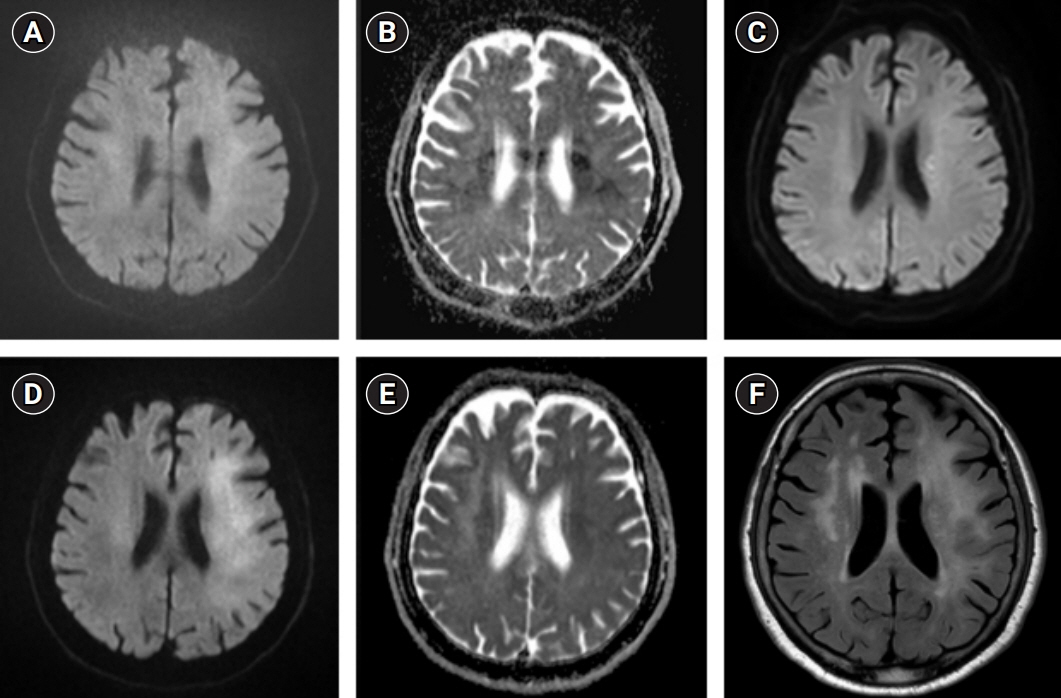
Fig. 1. Magnetic resonance imaging findings in case 1. Diffusion-weighted imaging (DWI) and apparent diffusion coefficient (A, B) on admission revealed no definite acute lesions. (C) DWI after mechanical thrombectomy showed some infarcts in the left corona radiata. On day 28, DWI (D) revealed high-signal intensities in the entire subcortex of left middle cerebral artery territory without signal changes on apparent diffusion coefficient (E). Fluid-attenuated inversion recovery (F) showed diffuse subcortical white matter lesions in the same territory.
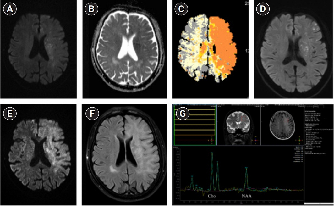
Fig. 2. Magnetic resonance imaging findings in case 2. Diffusion-weighted imaging (DWI) and apparent diffusion coefficient (A, B) on admission revealed new infarcts in the left corona radiata and subcortical area. (C) Magnetic resonance perfusion showed severe diffusion-perfusion mismatch. (D) DWI on day 6 showed new infarcts after mechanical thrombectomy. (E, F) On day 30, DWI and fluid-attenuated inversion recovery demonstrated the presence of diffuse subcortical white matter lesions. (G) Magnetic resonance spectroscopy showed a mildly increased choline/N-acetyl aspartate ratio.
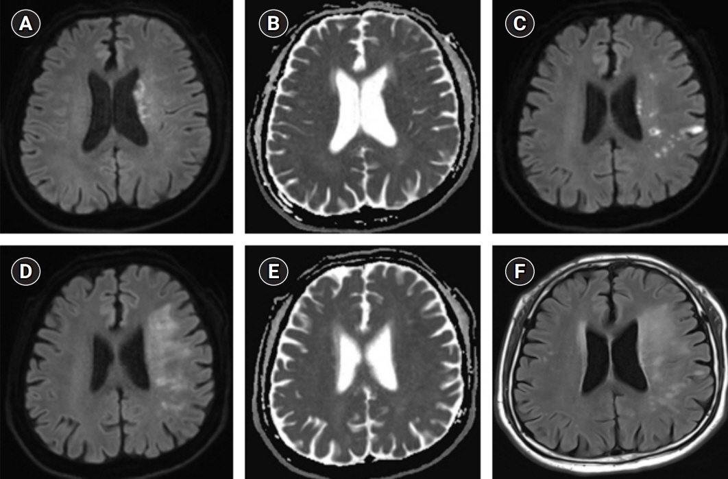
Fig. 3. Magnetic resonance imaging findings in case 3. (A, B) Diffusion-weighted imaging (DWI) and apparent diffusion coefficient on admission showed diffusion-restriction lesions in the left basal ganglia and corona radiata. (C) DWI after mechanical thrombectomy revealed new scattered infarcts in the left middle cerebral artery territory. On day 29, DWI and fluid-attenuated inversion recovery (D, F) demonstrated the presence of diffuse subcortical white matter lesions, without signal changes on apparent diffusion coefficient (E).
Reference
-
1. Sasaki T, Tomura N, Okada H, Tsuji E, Hayashi N, Kuwata T. A case of delayed white matter lesion after mechanical thrombectomy for middle cerebral artery occlusion. Jpn J Stroke. 2017; 40:270–4.2. Singu T, Inatomi Y, Yonehara T, Ando Y. Delayed leukoencephalopathy after recanalized cardioembolic stroke: two case reports. J Neurol Sci. 2017; 379:81–3.3. Nehme A, Pistono AA, Guilbert F, Stumpf É. Post-ischemic leukoencephalopathy after endovascular treatment for acute ischemic stroke. Can J Neurol Sci. 2019; 46:363–5.4. Iadecola C, Park L, Capone C. Threats to the mind: aging, amyloid, and hypertension. Stroke. 2009; 40(3 Suppl):S40–4.5. Zamora CA, Nauen D, Hynecek R, Ilica AT, Izbudak I, Sair HI, et al. Delayed posthypoxic leukoencephalopathy: a case series and review of the literature. Brain Behav. 2015; 5:e00364.6. Guglielmi V, LeCouffe NE, Zinkstok SM, Compagne KC, Eker R, Treurniet KM, et al. Collateral circulation and outcome in atherosclerotic versus cardioembolic cerebral large vessel occlusion. Stroke. 2019; 50:3360–8.7. Ferro JM. Cardioembolic stroke: an update. Lancet Neurol. 2003; 2:177–88.8. Lieb M, Shah U, Hines GL. Cerebral hyperperfusion syndrome after carotid intervention: a review. Cardiol Rev. 2012; 20:84–9.9. Lin YH, Liu HM. Update on cerebral hyperperfusion syndrome. J Neurointerv Surg. 2020; 12:788–93.10. Petersen NH, Silverman A, Strander SM, Kodali S, Wang A, Sansing LH, et al. Fixed compared with autoregulation-oriented blood pressure thresholds after mechanical thrombectomy for ischemic stroke. Stroke. 2020; 51:914–21.
- Full Text Links
-
- Actions
-
Cited
- CITED
-
- Close
- Share
-
- Similar articles
-
- Mechanical Thrombectomy in Strokes with Large-Vessel Occlusion Beyond 6 Hours: A Pooled Analysis of Randomized Trials
- Mechanical Thrombectomy for Large Vessel Occlusion via the Transbrachial Approach: Case Series
- Differences in mechanical thrombectomy for acute ischemic stroke on weekdays versus nights/ weekends in a Japanese primary stroke core center
- Delayed Development of Symptomatic Arterial Stenosis after a Mechanical Thrombectomy for an Acute Embolic Occlusion of the Middle Cerebral Artery
- Suction thrombectomy of distal medium vessel occlusion using microcatheter during mechanical thrombectomy for acute ischemic stroke: A case series
