Korean J Orthod.
2023 May;53(3):185-193. 10.4041/kjod22.208.
Covariance patterns between ramus morphology and the rest of the face: A geometric morphometric study
- Affiliations
-
- 1Clinic of Orthodontics and Pediatric Dentistry, Center of Dental Medicine, University of Zurich, Zurich, Switzerland
- 2Department of Orthodontics, School of Dentistry, National and Kapodistrian University of Athens, Athens, Greece
- KMID: 2542813
- DOI: http://doi.org/10.4041/kjod22.208
Abstract
Objective
The growth and development of the mandible strongly depend on modeling changes occurring at its ramus. Here, we investigated covariance patterns between the morphology of the ramus and the rest of the face.
Methods
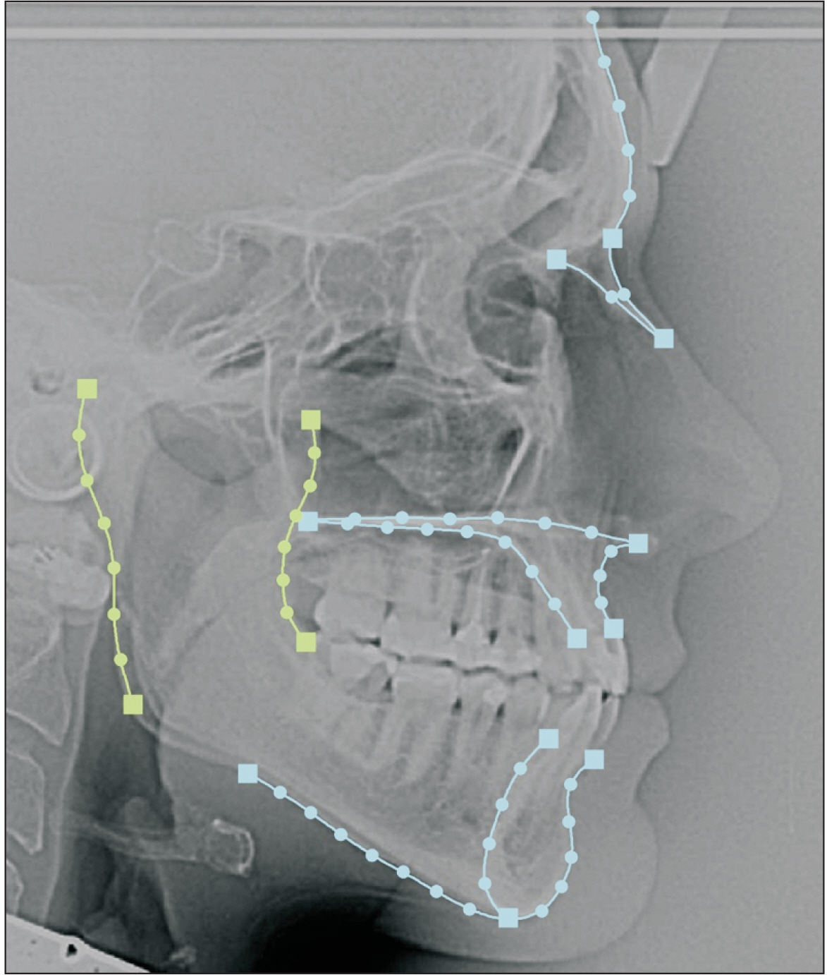
Lateral cephalograms of 159 adults (55 males and 104 females) with no history of orthodontic treatment were collected. Geometric morphometrics with sliding semi-landmarks was used. The covariance between the ramus and face was investigated using a two-block partial least squares analysis (PLS). Sexual dimorphism and allometry were also assessed.
Results
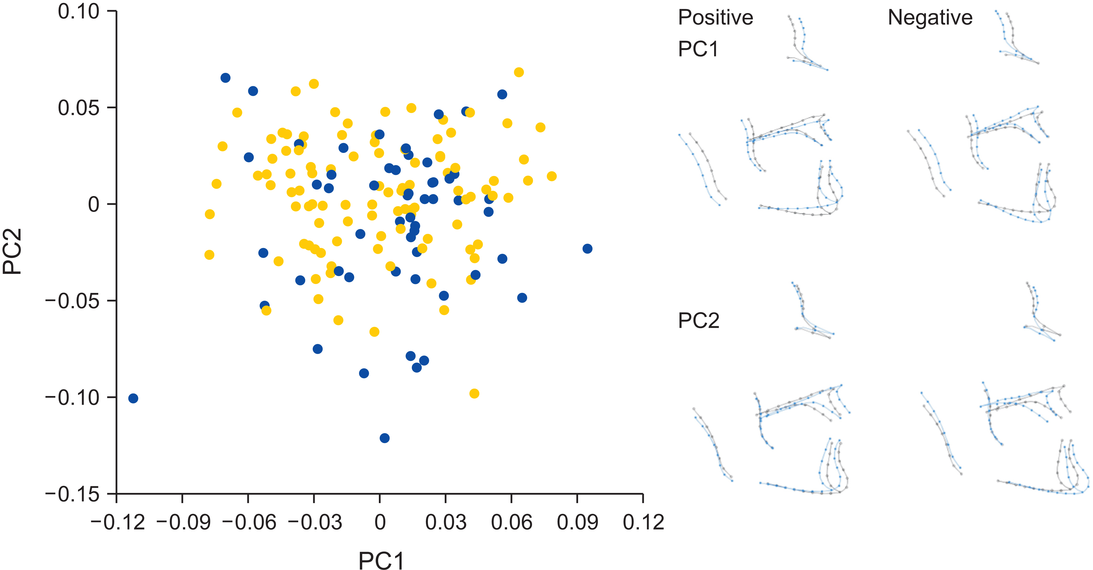
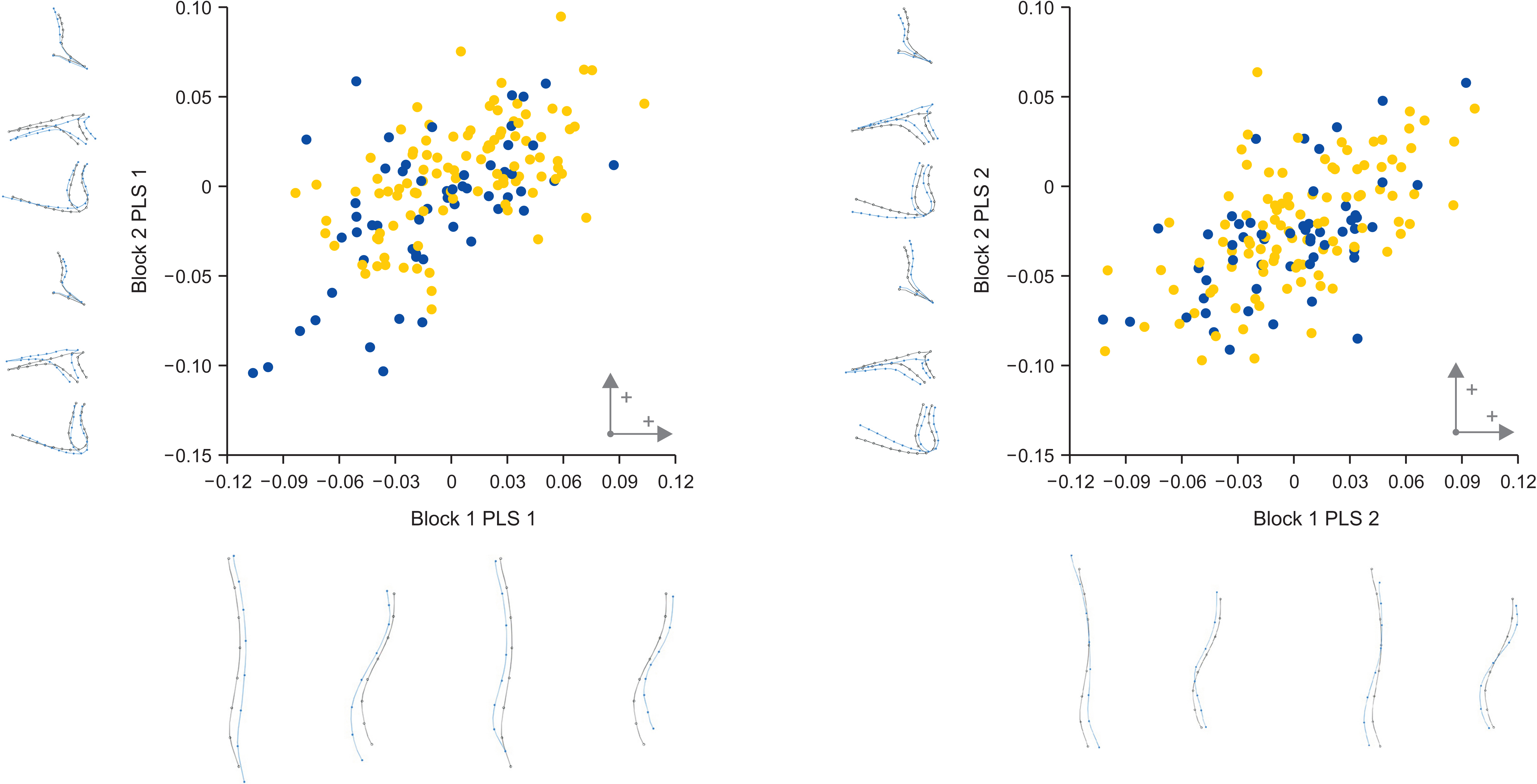
Differences in the divergence of the face and anteroposterior relationship of the jaws accounted for 24.1% and 21.6% of shape variation in the sample, respectively. Shape variation was greater in the sagittal plane for males than for females (30.7% vs. 17.4%), whereas variation in the vertical plane was similar for both sexes (23.7% for males and 25.4% for females). Size-related allometric differences between the sexes accounted for the shape variation to a maximum of 6% regarding the face. Regarding the covariation between the shapes of the ramus and the rest of the face, wider and shorter rami were associated with a decreased lower anterior facial height as well as a prognathic mandible and maxilla (PLS 1, 45.5% of the covariance). Additionally, a more posteriorly inclined ramus in the lower region was correlated with a Class II pattern and flat mandibular plane.
Conclusions
The width, height, and inclination of the ramus were correlated with facial shape changes in the vertical and sagittal planes.
Keyword
Figure
Cited by 1 articles
-
READER’S FORUM
G. Dave Singh
Korean J Orthod. 2023;53(5):287-288. doi: 10.4041/kjod23.121RF.
Reference
-
1. Charles SW. 1925; The temporomandibular joint and its influence on the growth of the mandible. Br Dent J. 46:845–55. https://cir.nii.ac.jp/crid/1573950400256289024.2. Brodie AG. 1941; On the growth pattern of the human head. From the third month to the eighth year of life. Am J Anat. 68:209–62. https://doi.org/10.1002/aja.1000680204. DOI: 10.1002/aja.1000680204.
Article3. Sicher H. 1947; The growth of the mandible. Am J Orthod. 33:30–5. https://doi.org/10.1016/0096-6347(47)90259-7. DOI: 10.1016/0096-6347(47)90259-7. PMID: 20278244.
Article4. Moss ML. 1960; Functional analysis of human mandibular growth. J Prosthet Dent. 10:1149–59. https://doi.org/10.1016/0022-3913(60)90228-6. DOI: 10.1016/0022-3913(60)90228-6.
Article5. Moss ML, Salentijn L. 1969; The primary role of functional matrices in facial growth. Am J Orthod. 55:566–77. https://doi.org/10.1016/0002-9416(69)90034-7. DOI: 10.1016/0002-9416(69)90034-7. PMID: 5253955.
Article6. Humphry GM. 1864. On the growth of the jaws. Cambridge University;Cambridge: https://books.google.co.kr/books/about/On_the_Growth_of_the_Jaws.html?id=iy0lzQEACAAJ&redir_esc=y. DOI: 10.1093/gmo/9781561592630.article.j601900.7. Brash JC. 1924. Growth of the Jaws and Palate. The growth of the jaws, normal and abnormal, in health and disease: five lectures. Dental Board of the United Kingdom;London: p. 23–66. https://books.google.co.kr/books/about/The_Growth_of_the_Jaws_Normal_and_Abnorm.html?id=Z5QPzQEACAAJ&redir_esc=y. DOI: 10.1093/gmo/9781561592630.article.j601900.8. Enlow DH, Harris DB. 1964; A study of the postnatal growth of the human mandible. Am J Orthod. 50:25–50. https://doi.org/10.1016/S0002-9416(64)80016-6. DOI: 10.1016/S0002-9416(64)80016-6.
Article9. Bang S, Enlow DH. 1967; Postnatal growth of the rabbit mandible. Arch Oral Biol. 12:993–8. https://doi.org/10.1016/0003-9969(67)90094-5. DOI: 10.1016/0003-9969(67)90094-5. PMID: 5231276.
Article10. Proffit WR, Fields HW, Sarver DM. 2007. Contemporary orthodontics. Mosby Elsevier;St. Louis: https://www.worldcat.org/ko/title/contemporary-orthodontics/oclc/769189433. DOI: 10.1002/j.0022-0337.2007.71.12.tb04437.x.11. Enlow DH, Hans MG. 1996. Essentials of facial growth. Saunders;Philadelphia: https://www.amazon.com/Essentials-Facial-Growth-Donald-Enlow/dp/0721661068. DOI: 10.1097/00008505-199600540-00041.12. Moyers RE, Bookstein FL. 1979; The inappropriateness of conventional cephalometrics. Am J Orthod. 75:599–617. https://doi.org/10.1016/0002-9416(79)90093-9. DOI: 10.1016/0002-9416(79)90093-9. PMID: 287374.
Article13. McIntyre GT, Mossey PA. 2003; Size and shape measurement in contemporary cephalometrics. Eur J Orthod. 25:231–42. https://doi.org/10.1093/ejo/25.3.231. DOI: 10.1093/ejo/25.3.231. PMID: 12831212.
Article14. Gunz P, Mitteroecker P. 2013; Semilandmarks: a method for quantifying curves and surfaces. Hystrix It J Mamm. 24:103–9. https://doi.org/10.4404/hystrix-24.1-6292.
Article15. Halazonetis DJ. 2004; Morphometrics for cephalometric diagnosis. Am J Orthod Dentofacial Orthop. 125:571–81. https://doi.org/10.1016/j.ajodo.2003.05.013. DOI: 10.1016/j.ajodo.2003.05.013. PMID: 15127026.
Article16. Enlow DH. 1983; JCO/interviews Dr. Donald H. Enlow on craniofacial growth. J Clin Orthod. 17:669–79. https://pubmed.ncbi.nlm.nih.gov/6586731/.17. Klingenberg CP. 2009; Morphometric integration and modularity in configurations of landmarks: tools for evaluating a priori hypotheses. Evol Dev. 11:405–21. https://doi.org/10.1111/j.1525-142X.2009.00347.x. DOI: 10.1111/j.1525-142X.2009.00347.x. PMID: 19601974. PMCID: PMC2776930.
Article18. Fruciano C, Franchini P, Meyer A. 2013; Resampling-based approaches to study variation in morphological modularity. PLoS One. 8:e69376. https://doi.org/10.1371/journal.pone.0069376. DOI: 10.1371/journal.pone.0069376. PMID: 23874956. PMCID: PMC3712944. PMID: 3636493e64c9478da1df331b4ab3be8b.
Article19. Halazonetis DJ. 2004; At what resolution should I scan cephalometric radiographs? Am J Orthod Dentofacial Orthop. 125:118–9. https://doi.org/10.1016/j.ajodo.2003.11.004. DOI: 10.1016/j.ajodo.2003.11.004. PMID: 14718889.
Article20. Bookstein FL. 1991. Morphometric tools for landmark data: geometry and biology. Cambridge University Press;Cambridge: https://doi.org/10.1017/CBO9780511573064. DOI: 10.1017/CBO9780511573064.
Article21. Klingenberg CP, Monteiro LR. 2005; Distances and directions in multidimensional shape spaces: implications for morphometric applications. Syst Biol. 54:678–88. https://doi.org/10.1080/10635150590947258. DOI: 10.1080/10635150590947258. PMID: 16126663.
Article22. Klingenberg CP. 2011; MorphoJ: an integrated software package for geometric morphometrics. Mol Ecol Resour. 11:353–7. https://doi.org/10.1111/j.1755-0998.2010.02924.x. DOI: 10.1111/j.1755-0998.2010.02924.x. PMID: 21429143.
Article23. Velemínská J, Bigoni L, Krajíček V, Borský J, Šmahelová D, Cagáňová V, et al. 2012; Surface facial modelling and allometry in relation to sexual dimorphism. Homo. 63:81–93. https://doi.org/10.1016/j.jchb.2012.02.002. DOI: 10.1016/j.jchb.2012.02.002. PMID: 22425585.
Article24. Klingenberg CP. 2016; Size, shape, and form: concepts of allometry in geometric morphometrics. Dev Genes Evol. 226:113–37. https://doi.org/10.1007/s00427-016-0539-2. DOI: 10.1007/s00427-016-0539-2. PMID: 27038023. PMCID: PMC4896994.
Article25. Rohlf FJ, Corti M. 2000; Use of two-block partial least-squares to study covariation in shape. Syst Biol. 49:740–53. https://doi.org/10.1080/106351500750049806. DOI: 10.1080/106351500750049806. PMID: 12116437.
Article26. Gkantidis N, Halazonetis DJ. 2011; Morphological integration between the cranial base and the face in children and adults. J Anat. 218:426–38. https://doi.org/10.1111/j.1469-7580.2011.01346.x. DOI: 10.1111/j.1469-7580.2011.01346.x. PMID: 21323666. PMCID: PMC3077525.
Article27. Wellens HL, Kuijpers-Jagtman AM, Halazonetis DJ. 2013; Geometric morphometric analysis of craniofacial variation, ontogeny and modularity in a cross-sectional sample of modern humans. J Anat. 222:397–409. https://doi.org/10.1111/joa.12027. DOI: 10.1111/joa.12027. PMID: 23425043. PMCID: PMC3610033.
Article28. Katsadouris A, Halazonetis DJ. 2017; Geometric morphometric analysis of craniofacial growth between the ages of 12 and 14 in normal humans. Eur J Orthod. 39:386–94. https://doi.org/10.1093/ejo/cjw070. DOI: 10.1093/ejo/cjw070. PMID: 27940444.
Article29. Zollikofer CP, Ponce De León MS. 2002; Visualizing patterns of craniofacial shape variation in Homo sapiens. Proc Biol Sci. 269:801–7. https://doi.org/10.1098/rspb.2002.1960. DOI: 10.1098/rspb.2002.1960. PMID: 11958711. PMCID: PMC1690963.
Article30. Bastir M, Rosas A. 2004; Facial heights: evolutionary relevance of postnatal ontogeny for facial orientation and skull morphology in humans and chimpanzees. J Hum Evol. 47:359–81. https://doi.org/10.1016/j.jhevol.2004.08.009. DOI: 10.1016/j.jhevol.2004.08.009. PMID: 15530353.
Article31. Nielsen IL. 1991; Vertical malocclusions: etiology, development, diagnosis and some aspects of treatment. Angle Orthod. 61:247–60. https://pubmed.ncbi.nlm.nih.gov/1763835/. Erratum in: Angle Orthod 1992;62:87. DOI: 10.1043/0003-3219(1991)061<0247:VMEDDA>2.0.CO;2. PMID: 1763835.32. Manlove AE, Romeo G, Venugopalan SR. 2020; Craniofacial growth: current theories and influence on management. Oral Maxillofac Surg Clin North Am. 32:167–75. https://doi.org/10.1016/j.coms.2020.01.007. DOI: 10.1016/j.coms.2020.01.007. PMID: 32151371.
Article33. Enlow DH, Pfister C, Richardson E, Kuroda T. 1982; An analysis of Black and Caucasian craniofacial patterns. Angle Orthod. 52:279–87. https://pubmed.ncbi.nlm.nih.gov/6961829/. DOI: 10.1043/0003-3219(1982)052<0279:AAOBAC>2.0.CO;2. PMID: 6961829.34. Bastir M, Rosas A, Kuroe K. 2004; Petrosal orientation and mandibular ramus breadth: evidence for an integrated petroso-mandibular developmental unit. Am J Phys Anthropol. 123:340–50. https://doi.org/10.1002/ajpa.10313. DOI: 10.1002/ajpa.10313. PMID: 15022362.
Article35. Smith RJ, Josell SD. 1984; The plan of the human face: a test of three general concepts. Am J Orthod. 85:103–8. https://doi.org/10.1016/0002-9416(84)90001-0. DOI: 10.1016/0002-9416(84)90001-0. PMID: 6594050.
Article36. Bastir M, Rosas A. 2005; Hierarchical nature of morphological integration and modularity in the human posterior face. Am J Phys Anthropol. 128:26–34. https://doi.org/10.1002/ajpa.20191. DOI: 10.1002/ajpa.20191. PMID: 15778978.
Article37. Mehta S, Arqub SA, Sharma R, Patel N, Tadinada A, Upadhyay M, et al. 2022; Variability associated with mandibular ramus area thickness and depth in subjects with different growth patterns, gender, and growth status. Am J Orthod Dentofacial Orthop. 161:e223–34. https://doi.org/10.1016/j.ajodo.2021.10.006. DOI: 10.1016/j.ajodo.2021.10.006. PMID: 34802867.
Article38. Knigge RP, McNulty KP, Oh H, Hardin AM, Leary EV, Duren DL, et al. 2021; Geometric morphometric analysis of growth patterns among facial types. Am J Orthod Dentofacial Orthop. 160:430–41. https://doi.org/10.1016/j.ajodo.2020.04.038. DOI: 10.1016/j.ajodo.2020.04.038. PMID: 34175161. PMCID: PMC8405563.
Article39. Aki T, Nanda RS, Currier GF, Nanda SK. 1994; Assessment of symphysis morphology as a predictor of the direction of mandibular growth. Am J Orthod Dentofacial Orthop. 106:60–9. https://doi.org/10.1016/S0889-5406(94)70022-2. DOI: 10.1016/S0889-5406(94)70022-2. PMID: 8017351.
Article40. Alarcón JA, Bastir M, García-Espona I, Menéndez-Núñez M, Rosas A. 2014; Morphological integration of mandible and cranium: orthodontic implications. Arch Oral Biol. 59:22–9. https://doi.org/10.1016/j.archoralbio.2013.10.005. DOI: 10.1016/j.archoralbio.2013.10.005. PMID: 24192112.
Article41. Enlow DH, DiGangi D, McNamara JA Jr, Mina M. 1988; An evaluation of the morphogenic and anatomic effects of the functional regulator utilizing the counterpart analysis. Eur J Orthod. 10:192–202. https://doi.org/10.1093/ejo/10.3.192. DOI: 10.1093/ejo/10.3.192. PMID: 3181298.
Article42. Fan Y, Penington A, Kilpatrick N, Hardiman R, Schneider P, Clement J, et al. 2019; Quantification of mandibular sexual dimorphism during adolescence. J Anat. 234:709–17. https://doi.org/10.1111/joa.12949. DOI: 10.1111/joa.12949. PMID: 30834524. PMCID: PMC6481415.
Article43. Sharma M, Gorea RK, Gorea A, Abuderman A. 2016; A morphometric study of the human mandible in the Indian population for sex determination. Egypt J Forensic Sci. 6:165–9. https://doi.org/10.1016/j.ejfs.2015.01.002. DOI: 10.1016/j.ejfs.2015.01.002. PMID: df5aacfc90f04f129c073c794b316c23.
Article
- Full Text Links
- Actions
-
Cited
- CITED
-
- Close
- Share
- Similar articles
-
- Academic Effectiveness of Non-face-to-face Classes in Deciduous Tooth Morphology Practice during COVID-19 Pandemic
- Prenatal Development of Sebaceous Gland: Morphologic and Morphometric Observation
- A Study of Bilateral Asymmetry Pattern in the Eye Orbit and Nose of the Joseon Dynasty Skeletal Group Using Geometric Morphometrics
- A study on the morphologic differences between long-face adults and normal-face adults on the lateral and P-A cephalograms
- The Analysis of Covariance of Do(province) Population Variability


