Healthc Inform Res.
2023 Apr;29(2):145-151. 10.4258/hir.2023.29.2.145.
Automatic Method for Optic Disc Segmentation Using Deep Learning on Retinal Fundus Images
- Affiliations
-
- 1Department of Informatics, Faculty of Engineering, Mulawarman University, Samarinda, Indonesia
- 2Department of Computer, System Engineering, Institut Sains & Teknologi AKPRIND, Yogyakarta, Indonesia
- 3Departmen of Information Technology, Samarinda Polytechnic of Agriculture, Samarinda, Indonesia
- 4Computer Vision Research Group, Faculty of Computer Science, Brawijaya University, Malang, Indonesia
- KMID: 2542140
- DOI: http://doi.org/10.4258/hir.2023.29.2.145
Abstract
Objectives
The optic disc is part of the retinal fundus image structure, which influences the extraction of glaucoma features. This study proposes a method that automatically segments the optic disc area in retinal fundus images using deep learning based on a convolutional neural network (CNN).
Methods
This study used private and public datasets containing retinal fundus images. The private dataset consisted of 350 images, while the public dataset was the Retinal Fundus Glaucoma Challenge (REFUGE). The proposed method was based on a CNN with a single-shot multibox detector (MobileNetV2) to form images of the region-of-interest (ROI) using the original image resized into 640 × 640 input data. A pre-processing sequence was then implemented, including augmentation, resizing, and normalization. Furthermore, a U-Net model was applied for optic disc segmentation with 128 × 128 input data.
Results
The proposed method was appropriately applied to the datasets used, as shown by the values of the F1-score, dice score, and intersection over union of 0.9880, 0.9852, and 0.9763 for the private dataset, respectively, and 0.9854, 0.9838 and 0.9712 for the REFUGE dataset.
Conclusions
The optic disc area produced by the proposed method was similar to that identified by an ophthalmologist. Therefore, this method can be considered for implementing automatic segmentation of the optic disc area.
Figure
-
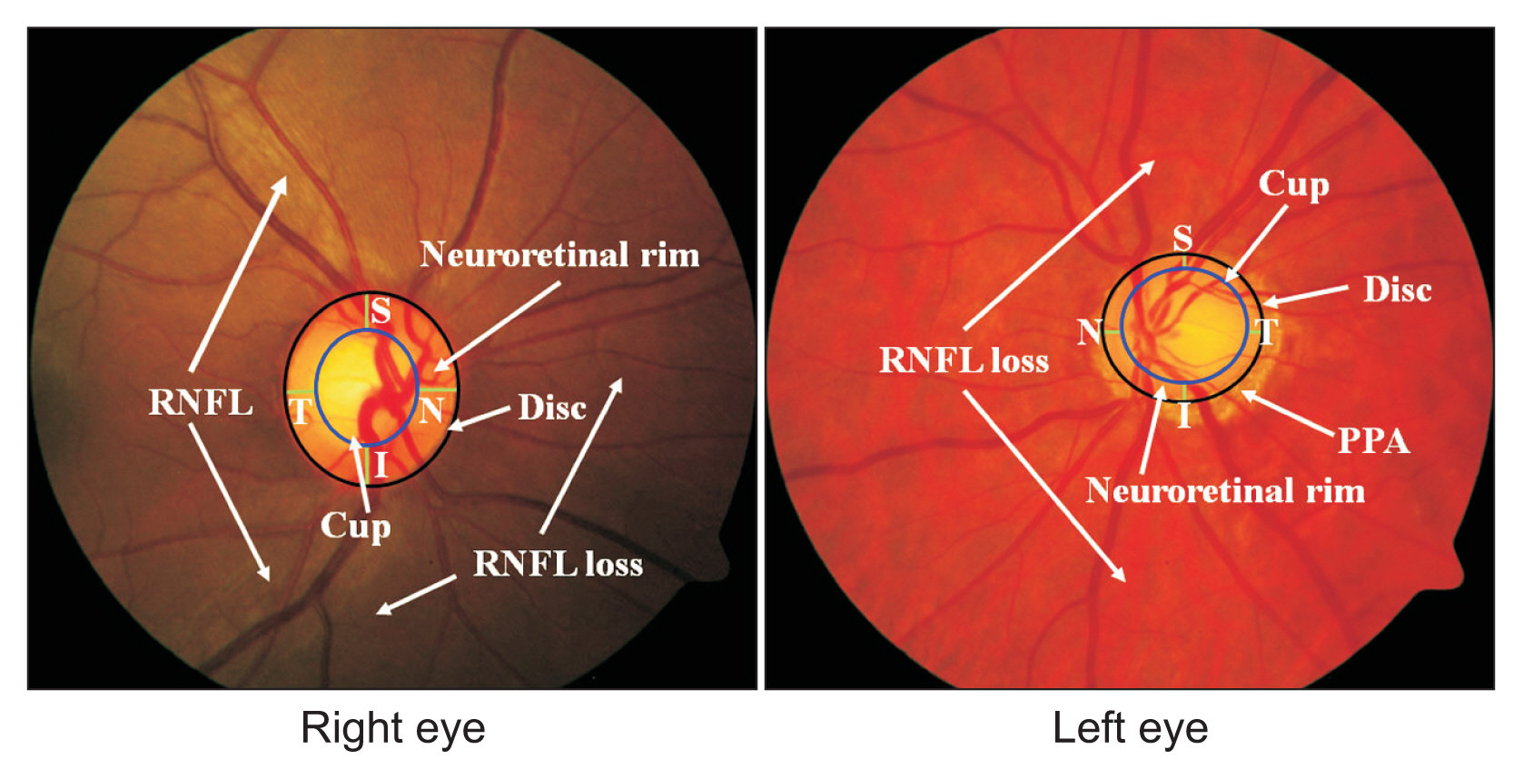
Figure 1 Structure of retinal fundus images from the right and left eyes. RNFL: retinal nerve fiber layer, PPA: peripapillary atrophy.

Figure 2 Main processes of the proposed method.
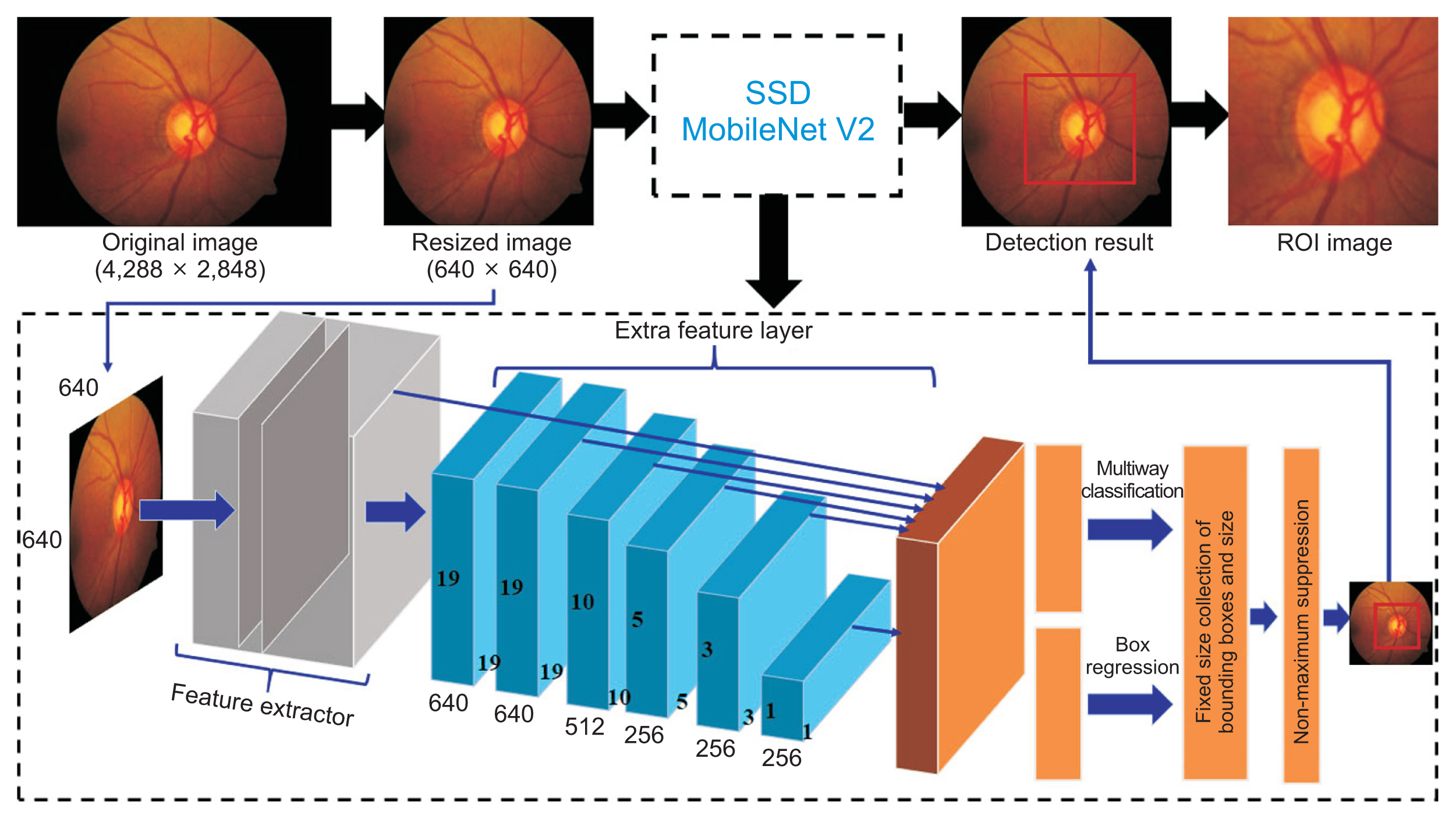
Figure 3 Overview of the process of forming a region-of-interest (ROI) image and the resulting image in each step. SSD: single-shot detector.
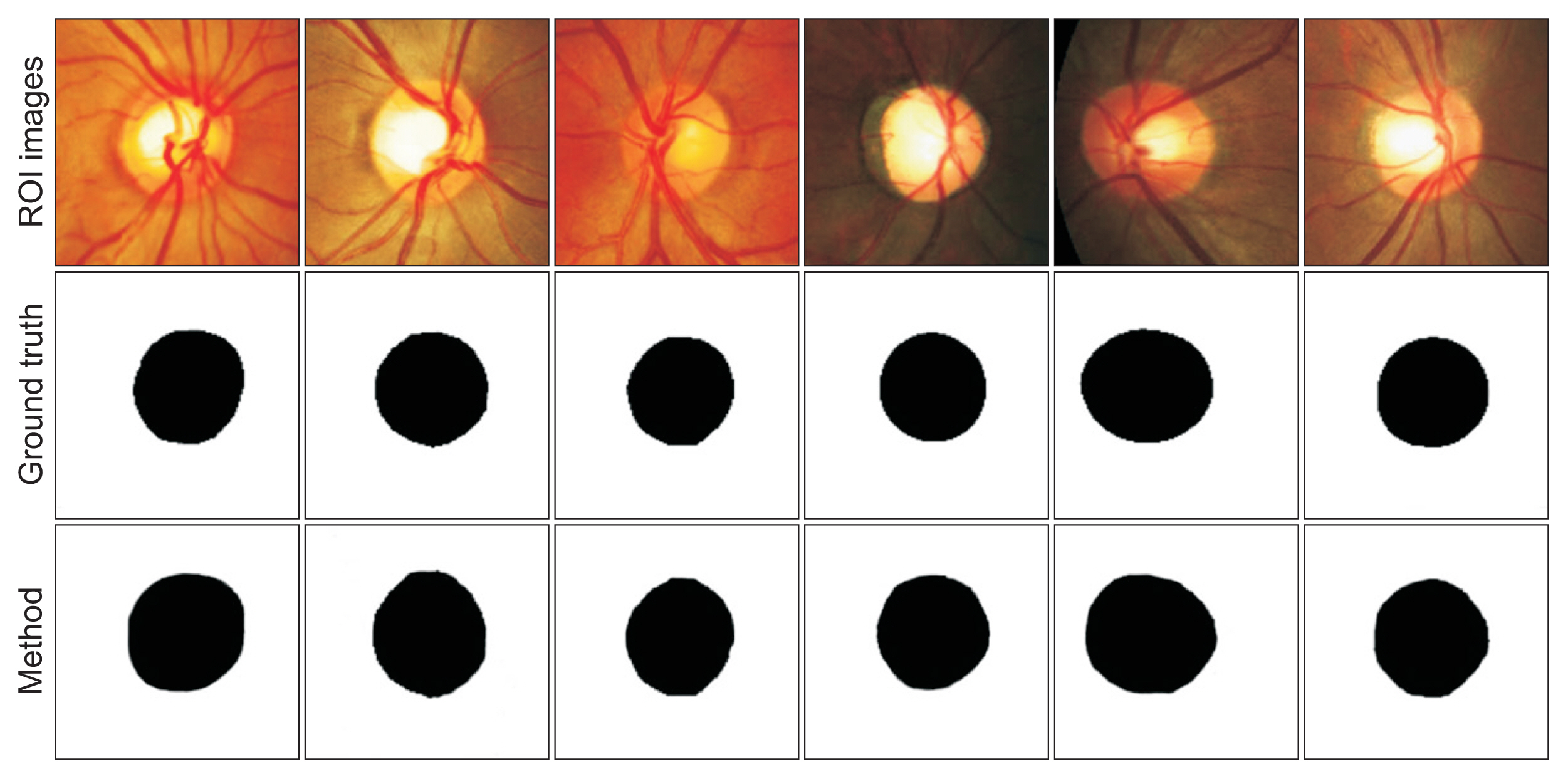
Figure 4 Examples of the optic disc segmentation results obtained by the ophthalmologist (ground truth) and the proposed method. ROI: region-of-interest.
Reference
-
References
1. Nugroho HA, Frannita EL, Ardiyanto I, Choridah L. Computer aided diagnosis for thyroid cancer system based on internal and external characteristics. J King Saud Univ-Comput Inf Sci. 2021; 33(3):329–39. https://doi.org/10.1016/j.jksuci.2019.01.007.
Article2. Kim YJ, Ganbold B, Kim KG. Web-based spine segmentation using deep learning in computed tomography images. Healthc Inform Res. 2020; 26(1):61–7. https://doi.org/10.4258/hir.2020.26.1.61.
Article3. Soulami KB, Kaabouch N, Saidi MN, Tamtaoui A. Breast cancer: one-stage automated detection, segmentation, and classification of digital mammograms using UNet model based-semantic segmentation. Biomed Signal Process Control. 2022; 66:102481. https://doi.org/10.1016/j.bspc.2021.102481.
Article4. Sela EI, Pulungan R, Widyaningrum R, Shantiningsih RR. Method for automated selection of the trabecular area in digital periapical radiographic images using morphological operations. Healthc Inform Res. 2019; 25(3):193–200. https://doi.org/10.4258/hir.2019.25.3.193.
Article5. Das P, Pal C, Acharyya A, Chakrabarti A, Basu S. Deep neural network for automated simultaneous intervertebral disc (IVDs) identification and segmentation of multi-modal MR images. Comput Methods Programs Biomed. 2021; 205:106074. https://doi.org/10.1016/j.cmpb.2021.106074.
Article6. Li X, Dou Q, Chen H, Fu CW, Qi X, Belavy DL, et al. 3D multi-scale FCN with random modality voxel dropout learning for intervertebral disc localization and segmentation from multi-modality MR images. Med Image Anal. 2018; 45:41–54. https://doi.org/10.1016/j.media.2018.01.004.
Article7. Makroum MA, Adda M, Bouzouane A, Ibrahim H. Machine learning and smart devices for diabetes management: systematic review. Sensors (Basel). 2022; 22(5):1843. https://doi.org/10.3390/s22051843.
Article8. Kumar S, Adarsh A, Kumar B, Singh AK. An automated early diabetic retinopathy detection through improved blood vessel and optic disc segmentation. Opt Laser Technol. 2020; 121:105815. https://doi.org/10.1016/j.optlastec.2019.105815.
Article9. Septiarini A, Hamdani , Khairina DM. The contour extraction of cup in fundus images for glaucoma detection. Int J Electr Comput Eng. 2016; 6(6):2797–804. http://doi.org/10.11591/ijece.v6i6.pp2797-2804.
Article10. Abdullah F, Imtiaz R, Madni HA, Khan HA, Khan TM, Khan MA, et al. A review on glaucoma disease detection using computerized techniques. IEEE Access. 2021; 9:37311–33. https://doi.org/10.1109/ACCESS.2021.3061451.
Article11. Thakur N, Juneja M. Optic disc and optic cup segmentation from retinal images using hybrid approach. Expert Syst Appl. 2019; 127:308–22. https://doi.org/10.1016/j.eswa.2019.03.009.
Article12. Veena HN, Muruganandham A, Kumaran TS. A novel optic disc and optic cup segmentation technique to diagnose glaucoma using deep learning convolutional neural network over retinal fundus images. J King Saud Univ-Comput Inf Sci. 2022; 34(8):6187–98. https://doi.org/10.1016/j.jksuci.2021.02.003.
Article13. Septiarini A, Pulungan R, Harjoko A, Ekantini R. Peripapillary atrophy detection in fundus images based on sectors with scan lines approach. In : Proceedings of 2018 3rd International Conference on Informatics and Computing (ICIC); 2018 Oct 17–18; Palembang, Indonesia. p. 1–6. https://doi.org/10.1109/IAC.2018.8780490.
Article14. Sharma A, Agrawal M, Roy SD, Gupta V, Vashisht P, Sidhu T. Deep learning to diagnose Peripapillary Atrophy in retinal images along with statistical features. Biomedical Signal Processing and Control. 2021; 64:102254. https://doi.org/10.1016/j.bspc.2020.102254.
Article15. Odstrcilik J, Kolar R, Tornow RP, Jan J, Budai A, Mayer M, et al. Thickness related textural properties of retinal nerve fiber layer in color fundus images. Comput Med Imaging Graph. 2014; 38(6):508–16. https://doi.org/10.1016/j.compmedimag.2014.05.005.
Article16. Septiarini A, Harjoko A, Pulungan R, Ekantini R. Automated detection of retinal nerve fiber layer by texture-based analysis for glaucoma evaluation. Healthc Inform Res. 2018; 24(4):335–45. https://doi.org/10.4258/hir.2018.24.4.335.
Article17. Rehman ZU, Naqvi SS, Khan TM, Arsalan M, Khan MA, Khalil MA. Multi-parametric optic disc segmentation using superpixel based feature classification. Expert Syst Appl. 2019; 120:461–73. https://doi.org/10.1016/j.eswa.2018.12.008.
Article18. Zulfira FZ, Suyanto S, Septiarini A. Segmentation technique and dynamic ensemble selection to enhance glaucoma severity detection. Comput Biol Med. 2021; 139:104951. https://doi.org/10.1016/j.compbiomed.2021.104951.
Article19. Septiarini A, Harjoko A, Pulungan R, Ekantini R. Optic disc and cup segmentation by automatic thresholding with morphological operation for glaucoma evaluation. Signal Image Video Process. 2017; 11:945–52. https://doi.org/10.1007/s11760-016-1043-x.
Article20. Fu Y, Chen J, Li J, Pan D, Yue X, Zhu Y. Optic disc segmentation by U-Net and probability bubble in abnormal fundus images. Pattern Recognit. 2021; 117:107971. https://doi.org/10.1016/j.patcog.2021.107971.
Article21. Hasan MK, Alam MA, Elahi MT, Roy S, Marti R. DRNet: segmentation and localization of optic disc and fovea from diabetic retinopathy image. Artif Intell Med. 2021; 111:102001. https://doi.org/10.1016/j.artmed.2020.102001.
Article22. Yu S, Xiao D, Frost S, Kanagasingam Y. Robust optic disc and cup segmentation with deep learning for glaucoma detection. Comput Med Imaging Graph. 2019; 74:61–71. https://doi.org/10.1016/j.compmedimag.2019.02.005.
Article23. Bian X, Luo X, Wang C, Liu W, Lin X. Optic disc and optic cup segmentation based on anatomy guided cascade network. Comput Methods Programs Biomed. 2020; 197:105717. https://doi.org/10.1016/j.cmpb.2020.105717.
Article24. Zilly J, Buhmann JM, Mahapatra D. Glaucoma detection using entropy sampling and ensemble learning for automatic optic cup and disc segmentation. Comput Med Imaging Graph. 2017; 55:28–41. https://doi.org/10.1016/j.compmedimag.2016.07.012.
Article25. Yuan X, Zhou L, Yu S, Li M, Wang X, Zheng X. A multi-scale convolutional neural network with context for joint segmentation of optic disc and cup. Artif Intell Med. 2021; 113:102035. https://doi.org/10.1016/j.artmed.2021.102035.
Article26. Orlando JI, Fu H, Barbosa Breda J, van Keer K, Bathula DR, Diaz-Pinto A, et al. REFUGE challenge: a unified framework for evaluating automated methods for glaucoma assessment from fundus photographs. Med Image Anal. 2020; 59:101570. https://doi.org/10.1016/j.media.2019.101570.
Article27. Zhang Y, Cai X, Zhang Y, Kang H, Ji X, Yuan X. TAU: transferable attention U-Net for optic disc and cup segmentation. Knowl Based Syst. 2021; 213:106668. https://doi.org/10.1016/j.knosys.2020.106668.
Article28. Zhang L, Lim CP. Intelligent optic disc segmentation using improved particle swarm optimization and evolving ensemble models. Appl Soft Comput. 2020; 92:106328. https://doi.org/10.1016/j.asoc.2020.106328.
Article29. Wang L, Gu J, Chen Y, Liang Y, Zhang W, Pu J, et al. Automated segmentation of the optic disc from fundus images using an asymmetric deep learning network. Pattern Recognit. 2021; 112:107810. https://doi.org/10.1016/j.patcog.2020.107810.
Article
- Full Text Links
-
- Actions
-
Cited
- CITED
-
- Close
- Share
-
- Similar articles
-
- Measurements of Diameter and Area of Optic Disc with the Scale of the Volk Lens
- Automatic Glaucoma Detection Method Applying a Statistical Approach to Fundus Images
- A Three-Dimensional Deep Convolutional Neural Network for Automatic Segmentation and Diameter Measurement of Type B Aortic Dissection
- Convolutional Neural Network-Based Automatic Segmentation of Substantia Nigra on Nigrosome and Neuromelanin Sensitive MR Images
- Web-Based Spine Segmentation Using Deep Learning in Computed Tomography Images
