Web-Based Spine Segmentation Using Deep Learning in Computed Tomography Images
- Affiliations
-
- 1Department of Biomedical Engineering, College of Health Science, Gachon University, Incheon, Korea. kimkg@gachon.ac.kr
- 2Department of Health Sciences and Technology, Gachon Advanced Institute for Health Sciences and Technology (GAIHST), Gachon University, Incheon, Korea.
- 3Department of Biomedical Engineering, College of Medicine, Gachon Uinversity, Incheon, Korea.
- 4Medical Device R&D Center, Biomedical & Convergence Institute, Gachon University Gil Hospital, Incheon, Korea.
- KMID: 2469401
- DOI: http://doi.org/10.4258/hir.2020.26.1.61
Abstract
OBJECTIVES
Back pain, especially lower back pain, is experienced in 60% to 80% of adults at some points during their lives. Various studies have found that lower back pain is a very common problem among adolescents, and the highest incidence rates are for adults in their 30s. There has been a remarkable increase in using computer-aided diagnosis to assist doctors in the interpretation of medical images. Spine segmentation in computed tomography (CT) scans using algorithmic methods allows improved diagnosis of back pain.
METHODS
In this study, we developed a web-based automatic spine segmentation method using deep learning and obtained the dice coefficient by comparison with the predicted image. Our method is based on convolutional neural networks for segmentation. More specifically, we train a hierarchical data format file using U-Net architecture and then insert the test data label to perform segmentation. Thus, we obtained more specific and detailed results. A total of 344 CT images were used in the experiment. Of these, 330 were used for learning, and the remaining 14 for testing.
RESULTS
Our method achieved an average dice coefficient of 90.4%, a precision of 96.81%, and an F1-score of 91.64%.
CONCLUSIONS
The proposed web-based deep learning approach can be very practical and accurate for spine segmentation as a diagnostic method.
MeSH Terms
Figure
-

Figure 1 Process of spine segmentation using the U-Net architecture. CT: computed tomography.
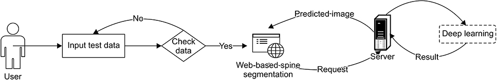
Figure 2 Web-based spine segmentation process.
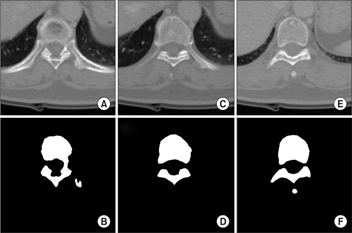
Figure 3 Examples of spinal area segmentation results based on deep learning: (A, C, E) computed tomography images and (B, D, F) deep learning-based spinal segmentation results.
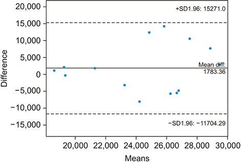
Figure 4 Bland-Altman plot between deep learning-based segmentation and manual segmentation results.
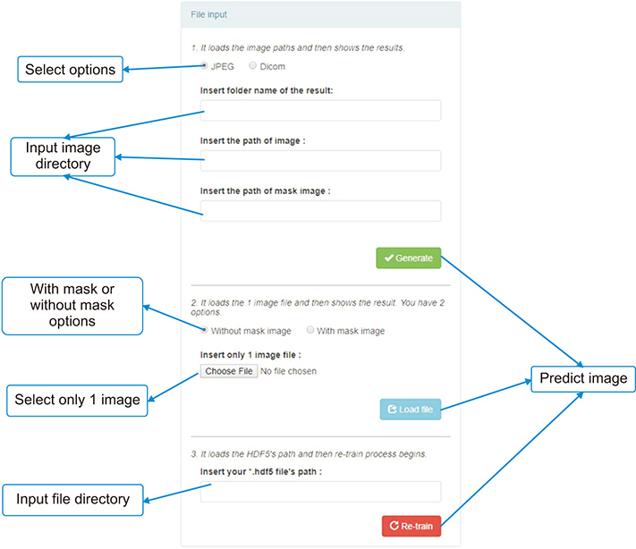
Figure 5 User interface of webpage for uploading files.
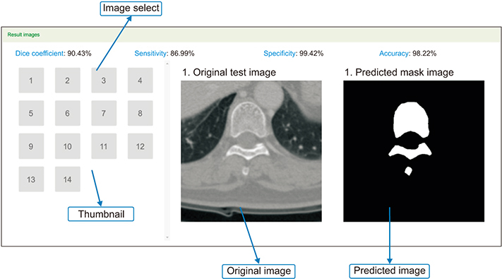
Figure 6 User interface of webpage to provide spinal segmentation results using deep learning.
Cited by 1 articles
-
Automatic Method for Optic Disc Segmentation Using Deep Learning on Retinal Fundus Images
Anindita Septiarini, Hamdani Hamdani, Emy Setyaningsih, Eko Junirianto, Fitri Utaminingrum
Healthc Inform Res. 2023;29(2):145-151. doi: 10.4258/hir.2023.29.2.145.
Reference
-
1. Mayo Clinic. Back pain [Internet]. Scottsdale (AZ): Mayo Clinic;cited at 2020 Jan 31. Available from: https://www.mayoclinic.org/diseases-conditions/backpain/symptoms-causes/syc-20369906.2. Abu-Naser SS, Aldahdooh R. Lower back pain expert system diagnosis and treatment. J Multidiscip Eng Sci Stud. 2016; 2(4):441–446.3. National Academies of Sciences, Engineering, and Medicine. Improving diagnosis in health care. Washington (DC): National Academies Press;2015.4. Shiraishi J, Li Q, Appelbaum D, Doi K. Computer-aided diagnosis and artificial intelligence in clinical imaging. Semin Nucl Med. 2011; 41(6):449–462.
Article5. Vania M, Mureja D, Lee D. Automatic spine segmentation using convolutional neural network via redundant generation of class labels for 3D spine modeling [Internet]. Ithaca (NY): arXiv.org;2017. cited at 2020 Jan 31. Available from: https://arxiv.org/abs/1712.01640.6. Kamboj A, Gupta A. Simulink model based image segmentation. Int J Adv Res Comput Sci Softw Eng. 2012; 2(6):146–149.7. Lathen G. Segmentation methods for medical image analysis: blood vessels, multi-scale filtering and level set methods [master's thesis]. Linkoping, Sweden: Linkoping University;2010.8. Nyul LG, Kanyo J, Mate E, Makay G, Balogh E, Fidrich M, et al. Method for automatically segmenting the spinal cord and canal from 3D CT images. In : Gagalowicz A, Philips W, editors. Computer analysis of images and patterns. Heidelberg, Germany: Springer;2005. p. 456–463.9. Folk M, Heber G, Koziol Q, Pourmal E, Robinson D. An overview of the HDF5 technology suite and its applications. In : Proceedings of the EDBT/ICDT 2011 Workshop on Array Databases; 2011 Mar 25; Uppsala, Sweden. p. 36–47.10. Collette A. Python and HDF5: unlocking scientific data. Sebastopol (CA): O'Reilly Media Inc.;2013.11. Ronneberger O, Fischer P, Brox T. U-net: convolutional networks for biomedical image segmentation. In : Navab N, Hornegger J, Wells W, Frangi A, editors. Medical image computing and computer-assisted intervention – MICCAI 2015. Cham: Springer;2015. p. 234–241.12. Dong H, Yang G, Liu F, Mo Y, Guo Y. Automatic brain tumor detection and segmentation using U-net based fully convolutional networks. In : Valdes Hernandez M, Gonzalez-Castro V, editors. Medical image understanding and analysis. Cham: Springer;2017. p. 506–517.13. Grinberg M. Flask web development: developing web applications with python. Sebastopol (CA): O'Reilly Media Inc.;2018.14. Mainland G, Morrisett G, Welsh M, Newton R. Sensor network programming with Flask. In : Proceedings of the 5th International Conference on Embedded Networked Sensor Systems; 2017 Nov 6-9; Sydney, Australia. p. 385–386.15. Van der Walt S, Colbert SC, Varoquaux G. The NumPy array: a structure for efficient numerical computation. Comput Sci Eng. 2011; 13(2):22–30.
Article16. Zou KH, Warfield SK, Bharatha A, Tempany CM, Kaus MR, Haker SJ, et al. Statistical validation of image segmentation quality based on a spatial overlap index1: scientific reports. Acad Radiol. 2004; 11(2):178–189.
Article17. Davis J, Goadrich M. The relationship between precision-recall and ROC curves. In : Proceedings of the 23rd International Conference on Machine Learning; 2006 Sep 10-13; Sofia, Bulgaria. p. 233–240.
- Full Text Links
-
- Actions
-
Cited
- CITED
-
- Close
- Share
-
- Similar articles
-
- Synthetic Computed Tomography Generation while Preserving Metallic Markers for Three-Dimensional Intracavitary Radiotherapy: Preliminary Study
- Temporomandibular Joint Segmentation Using Deep Learning for Automated Three-Dimensional Reconstruction
- Deep Learning-Based Computed Tomography Image Standardization to Improve Generalizability of Deep Learning-Based Hepatic Segmentation
- Deep Learning in Dental Radiographic Imaging
- Detection and Weak Segmentation of Masses in Gray-Scale Breast Mammogram Images Using Deep Learning
