Cerebral fat embolism syndrome: diagnostic challenges and catastrophic outcomes: a case series
- Affiliations
-
- 1Neurology Section, Department of Medicine, King Abdulaziz Medical City, Jeddah, Saudi Arabia
- 2Research Office King Abdullah International Medical Research Center, Jeddah, Saudi Arabia
- 3College of Medicine, King Saud bin Abdulaziz University for Health Sciences, Jeddah, Saudi Arabia
- 4Department of Neuroscience, King Faisal Specialist Hospital & Research Centre, Jeddah, Saudi Arabia
- 5Department of Medicine, Aseer Central Hospital, Abha, Saudi Arabia
- 6Neurology Department, Kasr Al Ainy Hospital, Faculty of Medicine, Cairo University, Cairo, Egypt
- 7College of Medicine, King Khalid University, Abha, Saudi Arabia
- KMID: 2541949
- DOI: http://doi.org/10.12701/jyms.2022.00360
Abstract
- Fat embolism syndrome is a rare but alarming, life-threatening clinical condition attributed to fat emboli entering the circulation. It usually occurs as a complication of long-bone fractures and joint reconstruction surgery. Neurological manifestations usually occur 12 to 72 hours after the initial insult. These neurological complications include cerebral infarction, spinal cord ischemia, hemorrhagic stroke, seizures, and coma. Other features include an acute confusional state, autonomic dysfunction, and retinal ischemia. In this case series, we describe three patients with fat embolism syndrome who presented with atypical symptoms and signs and with unusual neuroimaging findings. Cerebral fat embolism may occur without any respiratory or dermatological signs. In these cases, diagnosis is established after excluding other differential diagnoses. Neuroimaging using brain magnetic resonance imaging is of paramount importance in establishing a diagnosis. Aggressive hemodynamic and respiratory support from the beginning and consideration of orthopedic surgical intervention within the first 24 hours after trauma are critical to decreased morbidity and mortality.
Keyword
Figure
-
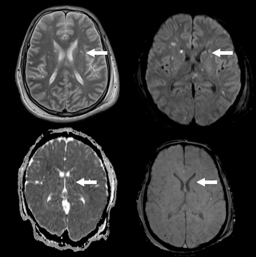
Fig. 1. Brain magnetic resonance images show extensive bilateral, symmetrical punctate foci of nearly the same size and abnormal signal intensities affecting both cerebral and cerebellar hemispheres with diffusion restriction and decreased susceptibility-weighted imaging (microbleeds) and mottled fluid-attenuated inversion recovery signals. Arrows indicate one of the foci as an example.
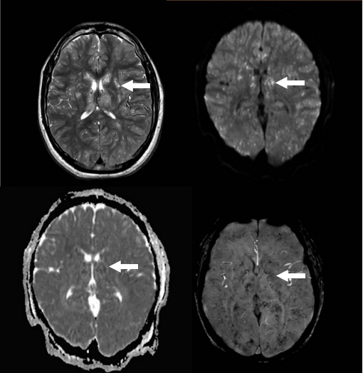
Fig. 2. Brain magnetic resonance images show extensive small high-signal intensity lesions involving the whole brain giving the appearance of a starfield pattern on T2-weighted images with restricted diffusion and profuse microhemorrhages in susceptibility-weighted imaging. Arrows indicate one of the lesions as an example.

Fig. 3. Brain magnetic resonance images show innumerable deep-white-matter, high-signal foci on T2-weighted images and low-signal foci on T1-weighted images showing restricted diffusion on diffusion-weighted imaging. Some images show blooming artifacts on susceptibility-weighted imaging consistent with multiple small infarctions and some show hemorrhagic components consistent with fat embolism. Arrows indicate one of the foci as an example.
Cited by 1 articles
-
Fat embolism syndrome: a review in cosmetic surgery
Hongil Kim, Bommie Florence Seo, Gregory Randolph Dean Evans
Kosin Med J. 2024;39(3):169-178. doi: 10.7180/kmj.24.126.
Reference
-
References
1. Rothberg DL, Makarewich CA. Fat embolism and fat embolism syndrome. J Am Acad Orthop Surg. 2019; 27:e346–55.2. Adeyinka A, Pierre L. Fat embolism. In: StatPearls [Internet]. Treasure Island (FL): StatPearls Publishing;2022. [cited 2022 Jul 23]. https://www.ncbi.nlm.nih.gov/books/NBK499885/.3. Tzioupis CC, Giannoudis PV. Fat embolism syndrome: what have we learned over the years? Trauma. 2011; 13:259–81.4. Newbigin K, Souza CA, Torres C, Marchiori E, Gupta A, Inacio J, et al. Fat embolism syndrome: state-of-the-art review focused on pulmonary imaging findings. Respir Med. 2016; 113:93–100.5. Morales-Vidal SG. Neurologic complications of fat embolism syndrome. Curr Neurol Neurosci Rep. 2019; 19:14.6. Scarpino M, Lanzo G, Lolli F, Grippo A. From the diagnosis to the therapeutic management: cerebral fat embolism, a clinical challenge. Int J Gen Med. 2019; 12:39–48.7. Godoy DA, Di Napoli M, Rabinstein AA. Cerebral fat embolism: recognition, complications, and prognosis. Neurocrit Care. 2018; 29:358–65.8. Fernández-Torre JL, Burgueño P, Ballesteros MA, Hernández-Hernández MA, Villagrá-Terán N, de Lucas EM. Super-refractory nonconvulsive status epilepticus secondary to fat embolism: a clinical, electrophysiological, and pathological study. Epilepsy Behav. 2015; 49:184–8.9. Kuo KH, Pan YJ, Lai YJ, Cheung WK, Chang FC, Jarosz J. Dynamic MR imaging patterns of cerebral fat embolism: a systematic review with illustrative cases. AJNR Am J Neuroradiol. 2014; 35:1052–7.10. Sen RK, Tripathy SK, Krishnan V. Role of corticosteroid as a prophylactic measure in fat embolism syndrome: a literature review. Musculoskelet Surg. 2012; 96:1–8.
- Full Text Links
-
- Actions
-
Cited
- CITED
-
- Close
- Share
-
- Similar articles
-
- Cerebral Fat Embolism as a Rare Complication of Postgastrectomy: Case Report
- Cardiac arrest occurred by cerebral fat embolism
- Gradient-Echo MRI in Defining the Severity of Cerebral Fat Embolism
- Cerebral fat embolism after bilateral total knee replacement arthroplasty: A case report
- Cerebral Fat Embolism That Was Initially Negative on DiffusionWeighted Magnetic Resonance Imaging
