Case 9: A 62-Year-Old Woman With Jaundice and General Weakness
- Affiliations
-
- 1The Catholic University Liver Research Center, College of Medicine, The Catholic University of Korea, Seoul, Korea
- 2Division of Hepatology, Department of Internal Medicine, College of Medicine, Seoul St. Mary’s Hospital, The Catholic University of Korea, Seoul, Korea
- 3Division of Hepatology, Department of Internal Medicine, College of Medicine, Uijeongbu St. Mary’s Hospital, The Catholic University of Korea, Seoul, Korea
- 4Division of Hepatology, Department of Internal Medicine, College of Medicine, Eunpyeong St. Mary’s Hospital, The Catholic University of Korea, Seoul, Korea
- KMID: 2541545
- DOI: http://doi.org/10.3346/jkms.2023.38.e105
Figure
-
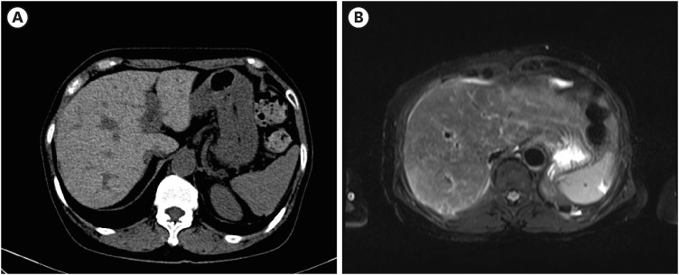
Fig. 1 Imaging findings at the hospitalization. (A) Non-contrast phase imaging of computed tomography showing high density of the liver (B) T2-weighted imaging of magnetic resonance imaging showing the low signal intensity of the liver.
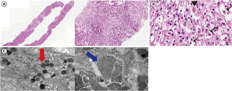
Fig. 2 Histologic findings obtained from the initial liver biopsy. For the confirmation of diagnosis, liver biopsy was performed. (A-C) Histological findings of the initial liver biopsy. Hematoxylin and eosin stain was done. ×20 (left) and ×100 (right) slides showing severe lobular, portal, and periportal inflammation with hepatocyte necrosis (A). ×400 slide shows a red granule (arrowhead) among the necrotic cells. Ballooned hepatocytes and Mallory bodies are seen. (black narrow arrows) (B) Electron microscopy shows phospholipid laden lysosomal lamellar bodies (left, red arrow) and fatty deposition in the lysosome (right, blue arrow) (C).
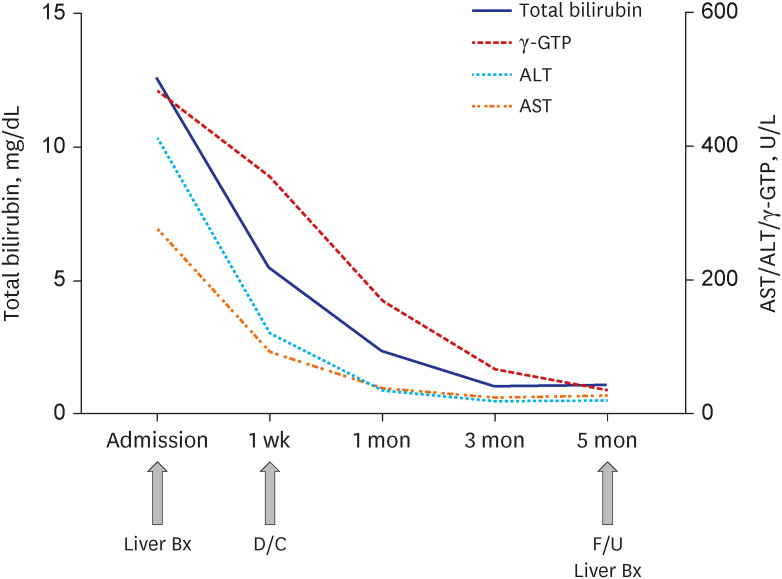
Fig. 3 Serial laboratory findings from admission to 5 months after cessation of amiodarone. Serum levels of total bilirubin, AST, ALT, and γ-GTP at admission and 1 week, 1 month, 3 months, 5 months after the cessation of amiodarone are presented.γ-GTP = γ-glutamyl transpeptidase, ALT = alanine aminotransferase, AST = aspartate aminotransferase, Bx = biopsy, D/C = discharge, F/U = follow up.
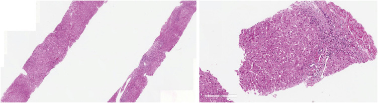
Fig. 4 Histologic findings obtained from liver biopsy 5 month after amiodarone withdrawal. H&E stain was done. ×20 (left) and ×100 (right) slides H&E staining from the liver biopsy samples at 5 months after the cessation of amiodarone showing significant recovery from the liver injury.H&E = hematoxylin and eosin.
Reference
-
1. Hennes EM, Zeniya M, Czaja AJ, Parés A, Dalekos GN, Krawitt EL, et al. Simplified criteria for the diagnosis of autoimmune hepatitis. Hepatology. 2008; 48(1):169–176. PMID: 18537184.
Article2. Komori A. Recent updates on the management of autoimmune hepatitis. Clin Mol Hepatol. 2021; 27(1):58–69. PMID: 33291862.
Article3. Lee JY, Yoo KH, Hahn SH. HFE gene mutation, C282Y causing hereditary hemochromatosis in Caucasian is extremely rare in Korean population. J Korean Med Sci. 2000; 15(2):179–182. PMID: 10803694.
Article4. Bacon BR, Adams PC, Kowdley KV, Powell LW, Tavill AS. American Association for the Study of Liver Diseases. Diagnosis and management of hemochromatosis: 2011 practice guideline by the American Association for the Study of Liver Diseases. Hepatology. 2011; 54(1):328–343. PMID: 21452290.
Article5. Mahroum N, Alghory A, Kiyak Z, Alwani A, Seida R, Alrais M, et al. Ferritin - from iron, through inflammation and autoimmunity, to COVID-19. J Autoimmun. 2022; 126:102778. PMID: 34883281.6. Fontana RJ, Liou I, Reuben A, Suzuki A, Fiel MI, Lee W, et al. AASLD practice guidance on drug, herbal, and dietary supplement-induced liver injury. Hepatology. 2023; 77(3):1036–1065. PMID: 35899384.7. LiverTox: Clinical and Research Information on Drug-Induced Liver Injury. Bethesda, MD, USA: National Institute of Diabetes and Digestive and Kidney Diseases;2012.8. Low EX, Zheng Q, Chan E, Lim SG. Drug induced liver injury: East versus West - a systematic review and meta-analysis. Clin Mol Hepatol. 2020; 26(2):142–154. PMID: 31816676.
Article9. Lee HA, Chang Y, Sung PS, Yoon EL, Lee HW, Yoo JJ, et al. Therapeutic mechanisms and beneficial effects of non-antidiabetic drugs in chronic liver diseases. Clin Mol Hepatol. 2022; 28(3):425–472. PMID: 35850495.
Article10. Buggey J, Kappus M, Lagoo AS, Brady CW. Amiodarone-induced liver injury and cirrhosis. ACG Case Rep J. 2015; 2(2):116–118. PMID: 26157932.
Article11. Gregory SA, Webster JB, Chapman GD. Acute hepatitis induced by parenteral amiodarone. Am J Med. 2002; 113(3):254–255. PMID: 12208392.
Article12. Gayam V, Khalid M, Dahal S, Garlapati P, Gill A, Alex R, et al. Fatal acute liver failure with intravenous amiodarone: a case report and literature review. Gastroenterol Res. 2018; 11(1):62–63.
Article13. Jaiswal P, Attar BM, Yap JE, Devani K, Jaiswal R, Wang Y, et al. Acute liver failure with amiodarone infusion: a case report and systematic review. J Clin Pharm Ther. 2018; 43(1):129–133. PMID: 28714083.14. Sung PS, Yoon SK. Amiodarone hepatotoxicity. Hepatology. 2012; 55(1):325–326. PMID: 21898482.
Article
- Full Text Links
-
- Actions
-
Cited
- CITED
-
- Close
- Share
-
- Similar articles
-
- A Case of Hyperkalemia discovered immediately after the Induction of General Anesthesia
- A Case of Obstructive Jaundice Caused by Extrinsic Compresson of Biliary Cystadenoma of the Common Hepatic Duct
- A Case of Acute Fatty Liver of Pregnancy
- A Case of Leiomyoma in the Common Bile Duct
- Ultrasonography of jaundice
