Cancer Res Treat.
2023 Apr;55(2):506-512. 10.4143/crt.2022.1008.
The Additive Impact of Transbronchial Cryobiopsy Using a 1.1-mm Diameter Cryoprobe on Conventional Biopsy for Peripheral Lung Nodules
- Affiliations
-
- 1Department of Internal Medicine, Pusan National University School of Medicine, Busan, Korea
- 2Biomedical Research Institute, Pusan National University Hospital, Busan, Korea
- KMID: 2541237
- DOI: http://doi.org/10.4143/crt.2022.1008
Abstract
- Purpose
The diagnostic yield of transbronchial biopsy (TBB) using radial probe endobronchial ultrasound (RP-EBUS) is 71%, which is lower than that of transthoracic needle biopsy. We investigated the performance and safety of sequential transbronchial cryobiopsy (TBC) using a novel 1.1-mm diameter cryoprobe, after conventional TBB using RP-EBUS for the diagnosis of peripheral lung lesions (PLLs).
Materials and Methods
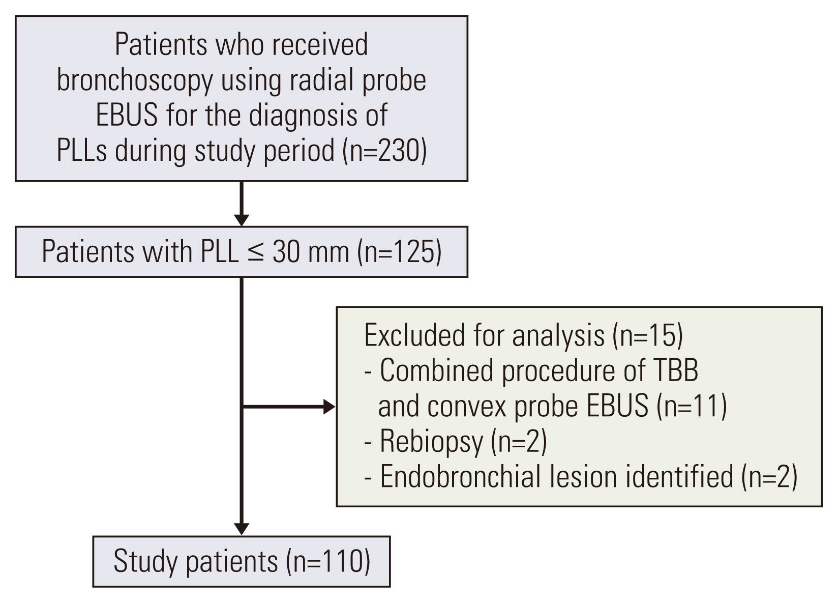
From April 2021 to November 2021, 110 patients who underwent bronchoscopy using RP-EBUS for the diagnosis of PLL ≤ 30 mm were retrospectively included in our study. All records were followed until June 2022.
Results
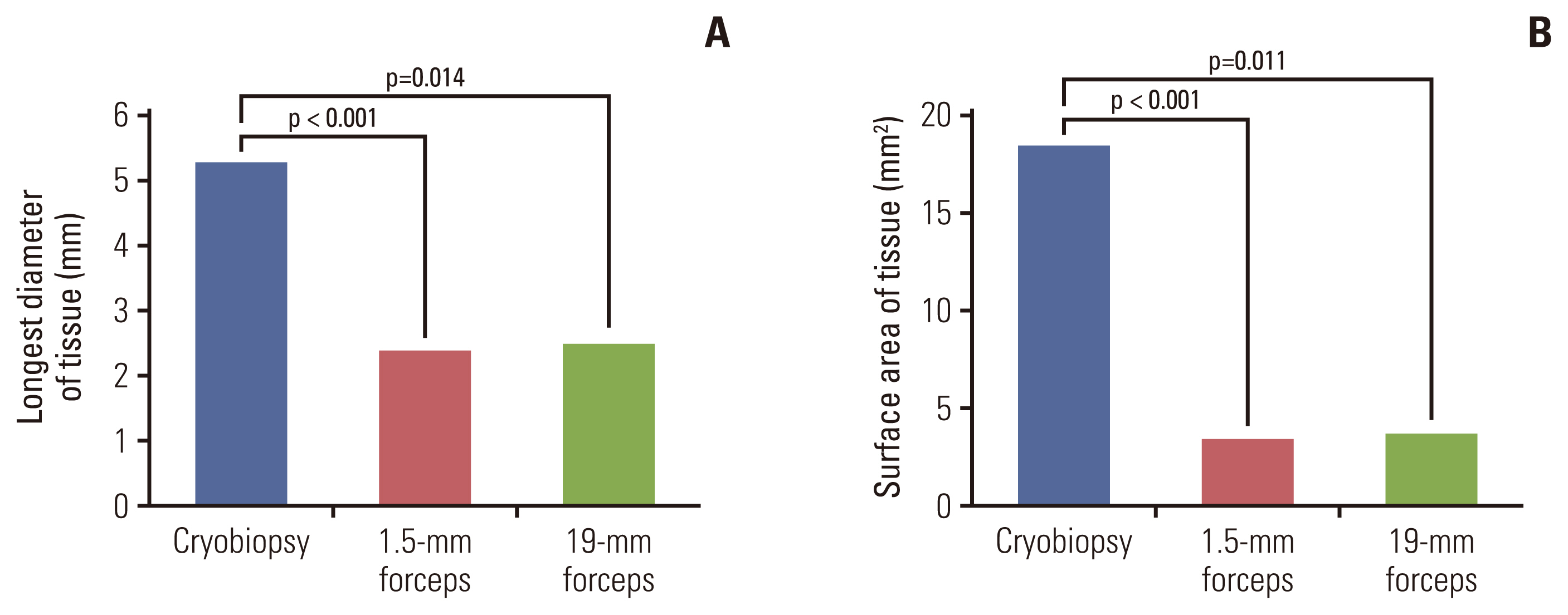
The overall diagnostic yield of combined TBB and TBC was 79.1%, which was higher than 60.9% of TBB alone (p=0.005). The diagnostic yield of sequential TBC was 65.5%, which increased the overall diagnostic yield by 18.2%. The surface area of tissues by TBC (mean area, 18.5 mm2) was significantly larger than those of TBB by 1.5-mm forceps (3.4 mm2, p < 0.001) and 1.9-mm forceps (3.7 mm2, p=0.011). In the multivariate analysis, PLLs with the longest diameter of ≤ 22 mm were found to be related to additional diagnostic benefits from sequential TBC (odds ratio, 3.51; 95% confidence interval, 1.043 to 11.775; p=0.042). Complications were found in 10.5% of the patients: pneumothorax (1.0%), infection (1.0%), and significant bleeding (8.6%). None of the patients developed any life-threatening complications.
Conclusion
Sequential TBC with a 1.1-mm cryoprobe improved the performance of conventional TBB using RP-EBUS without serious complications.
Keyword
Figure
Cited by 1 articles
-
Development of the Korean Association for Lung Cancer Clinical Practice Guidelines: Recommendations on Radial Probe Endobronchial Ultrasound for Diagnosing Lung Cancer - An Updated Meta-Analysis
Soo Han Kim, Hyun Sung Chung, Jinmi Kim, Mi-Hyun Kim, Min Ki Lee, Insu Kim, Jung Seop Eom
Cancer Res Treat. 2024;56(2):464-483. doi: 10.4143/crt.2023.749.
Reference
-
References
1. National Lung Screening Trial Research Team, Church TR, Black WC, Aberle DR, Berg CD, Clingan KL, et al. Results of initial low-dose computed tomographic screening for lung cancer. N Engl J Med. 2013; 368:1980–91.
Article2. Nanavaty P, Alvarez MS, Alberts WM. Lung cancer screening: advantages, controversies, and applications. Cancer Control. 2014; 21:9–14.
Article3. Gould MK, Donington J, Lynch WR, Mazzone PJ, Midthun DE, Naidich DP, et al. Evaluation of individuals with pulmonary nodules: when is it lung cancer? Diagnosis and management of lung cancer, 3rd ed: American College of Chest Physicians evidence-based clinical practice guidelines. Chest. 2013; 143:e93S–120S.4. Kurimoto N, Miyazawa T, Okimasa S, Maeda A, Oiwa H, Miyazu Y, et al. Endobronchial ultrasonography using a guide sheath increases the ability to diagnose peripheral pulmonary lesions endoscopically. Chest. 2004; 126:959–65.
Article5. Hayama M, Izumo T, Matsumoto Y, Chavez C, Tsuchida T, Sasada S. Complications with endobronchial ultrasound with a guide sheath for the diagnosis of peripheral pulmonary lesions. Respiration. 2015; 90:129–35.
Article6. Eom JS, Mok JH, Kim I, Lee MK, Lee G, Park H, et al. Radial probe endobronchial ultrasound using a guide sheath for peripheral lung lesions in beginners. BMC Pulm Med. 2018; 18:137.
Article7. Ali MS, Trick W, Mba BI, Mohananey D, Sethi J, Musani AI. Radial endobronchial ultrasound for the diagnosis of peripheral pulmonary lesions: a systematic review and meta-analysis. Respirology. 2017; 22:443–53.
Article8. Schuhmann M, Bostanci K, Bugalho A, Warth A, Schnabel PA, Herth FJ, et al. Endobronchial ultrasound-guided cryobiopsies in peripheral pulmonary lesions: a feasibility study. Eur Respir J. 2014; 43:233–9.
Article9. Sryma PB, Mittal S, Madan NK, Tiwari P, Hadda V, Mohan A, et al. Efficacy of radial endobronchial ultrasound (R-EBUS) guided transbronchial cryobiopsy for peripheral pulmonary lesions (PPL’s): a systematic review and meta-analysis. Pulmonology. 2021.
Article10. Yarmus LB, Semaan RW, Arias SA, Feller-Kopman D, Ortiz R, Bosmuller H, et al. A randomized controlled trial of a novel sheath cryoprobe for bronchoscopic lung biopsy in a porcine model. Chest. 2016; 150:329–36.
Article11. Matsumoto Y, Nakai T, Tanaka M, Imabayashi T, Tsuchida T, Ohe Y. Diagnostic outcomes and safety of cryobiopsy added to conventional sampling methods: an observational study. Chest. 2021; 160:1890–901.
Article12. Kuiper JL, Heideman DA, Thunnissen E, Paul MA, van Wijk AW, Postmus PE, et al. Incidence of T790M mutation in (sequential) rebiopsies in EGFR-mutated NSCLC-patients. Lung Cancer. 2014; 85:19–24.
Article13. Burgers JA, Herth F, Becker HD. Endobronchial ultrasound. Lung Cancer. 2001; 34(Suppl 2):S109–13.
Article14. Kurimoto N, Morita K. Bronchial branch tracing. Singapore: Sprinter;2020.15. Ishida T, Asano F, Yamazaki K, Shinagawa N, Oizumi S, Moriya H, et al. Virtual bronchoscopic navigation combined with endobronchial ultrasound to diagnose small peripheral pulmonary lesions: a randomised trial. Thorax. 2011; 66:1072–7.
Article16. Eberhardt R, Kahn N, Gompelmann D, Schumann M, Heussel CP, Herth FJ. LungPoint: a new approach to peripheral lesions. J Thorac Oncol. 2010; 5:1559–63.17. Yamada N, Yamazaki K, Kurimoto N, Asahina H, Kikuchi E, Shinagawa N, et al. Factors related to diagnostic yield of transbronchial biopsy using endobronchial ultrasonography with a guide sheath in small peripheral pulmonary lesions. Chest. 2007; 132:603–8.
Article18. Hetzel M, Hetzel J, Schumann C, Marx N, Babiak A. Cryorecanalization: a new approach for the immediate management of acute airway obstruction. J Thorac Cardiovasc Surg. 2004; 127:1427–31.
Article19. Yoshikawa M, Sukoh N, Yamazaki K, Kanazawa K, Fukumoto S, Harada M, et al. Diagnostic value of endobronchial ultrasonography with a guide sheath for peripheral pulmonary lesions without X-ray fluoroscopy. Chest. 2007; 131:1788–93.
Article20. Kikuchi E, Yamazaki K, Sukoh N, Kikuchi J, Asahina H, Imura M, et al. Endobronchial ultrasonography with guide-sheath for peripheral pulmonary lesions. Eur Respir J. 2004; 24:533–7.
Article21. Lee J, Kim C, Seol HY, Chung HS, Mok J, Lee G, et al. Safety and diagnostic yield of radial probe endobronchial ultrasound-guided biopsy for peripheral lung lesions in patients with idiopathic pulmonary fibrosis: a multicenter cross-sectional study. Respiration. 2022; 101:401–7.
Article22. Oki M, Saka H, Asano F, Kitagawa C, Kogure Y, Tsuzuku A, et al. Use of an ultrathin vs thin bronchoscope for peripheral pulmonary lesions: a randomized trial. Chest. 2019; 156:954–64.
Article23. Steinfort DP, Khor YH, Manser RL, Irving LB. Radial probe endobronchial ultrasound for the diagnosis of peripheral lung cancer: systematic review and meta-analysis. Eur Respir J. 2011; 37:902–10.
Article
- Full Text Links
- Actions
-
Cited
- CITED
-
- Close
- Share
- Similar articles
-
- Advanced Bronchoscopic Diagnostic Techniques in Lung Cancer
- The Effects of Bronchoscope Diameter on the Diagnostic Yield of Transbronchial Lung Biopsy of Peripheral Pulmonary Nodules
- The diagnosis of peripheral lung lesions: transbronchial biopsy using a radial probe endobronchial ultrasound
- Diagnostic Approaches for Idiopathic Pulmonary Fibrosis
- Usefulness of CT-Guided Automatic Needle Biopsy of Solitary Pulmonary Nodule Smaller than 15 mm


