Anat Cell Biol.
2023 Mar;56(1):9-15. 10.5115/acb.22.118.
The clinical anatomy of the accessory submandibular gland: a comprehensive review
- Yazbeck A
 1
1 - Iwanaga J2,3,4,5,6
- Walocha JA7
- Olewnik 8
- Tubbs RS1,4,5,9,10
- Affiliations
-
- 1Department of Structural & Cellular Biology, Tulane University School of Medicine, New Orleans, LA, USA
- 2Division of Gross and Clinical Anatomy, Department of Anatomy, Kurume University School of Medicine, Kurume, Fukuoka
- 3Dental and Oral Medical Center, Kurume University School of Medicine, Kurume, Fukuoka, Japan
- 4Department of Neurosurgery, Tulane Center for Clinical Neurosciences, Tulane University School of Medicine, New Orleans, LA
- 5Department of Neurology, Tulane Center for Clinical Neurosciences, Tulane University School of Medicine, New Orleans, LA, USA
- 6Department of Oral and Maxillofacial Anatomy, Graduate School of Medical and Dental Sciences, Tokyo Medical and Dental University, Tokyo, Japan
- 7Department of Anatomy, Jagiellonian University Medical College, Krakow
- 8Department of Anatomical Dissection and Donation, Medical University of Lodz, Lodz, Poland
- 9Department of Anatomical Sciences, St. George’s University, St. George’s, Grenada, West Indies
- 10Department of Neurosurgery and Ochsner Neuroscience Institute, Ochsner Health System, New Orleans, LA
- 11Department of Surgery, Tulane University School of Medicine, New Orleans, LA, USA
- 12University of Queensland, Brisbane, Australia
- KMID: 2540979
- DOI: http://doi.org/10.5115/acb.22.118
Abstract
- An accessory submandibular gland is a rare variation. As such, there is limited literature regarding the embryology, anatomy, variations, clinical imaging, and pathology of the accessory submandibular gland. In this article, we review the existing literature on the accessory submandibular gland from clinical and anatomical perspectives. The goal of this review is to provide comprehensive knowledge of this variation which can be useful for oral and maxillofacial/head and neck surgeons, radiologists, and anatomists. Within this review, the embryologic origin as well as the anatomy of the accessory submandibular gland is detailed. Several imaging modalities which can be used to visualize the accessory submandibular gland are outlined as well as its variations. Lastly, this review investigates several reported clinical considerations regarding the accessory submandibular gland including sialoliths, Wharton’s duct obstruction, and pleomorphic adenoma.
Figure
-

Fig. 1 The accessory submandibular gland (arrow) was noted on left side in 85 years-old at death male cadaver. The gland was located superior to the mylohyoid muscle (the main submandibular gland is removed).
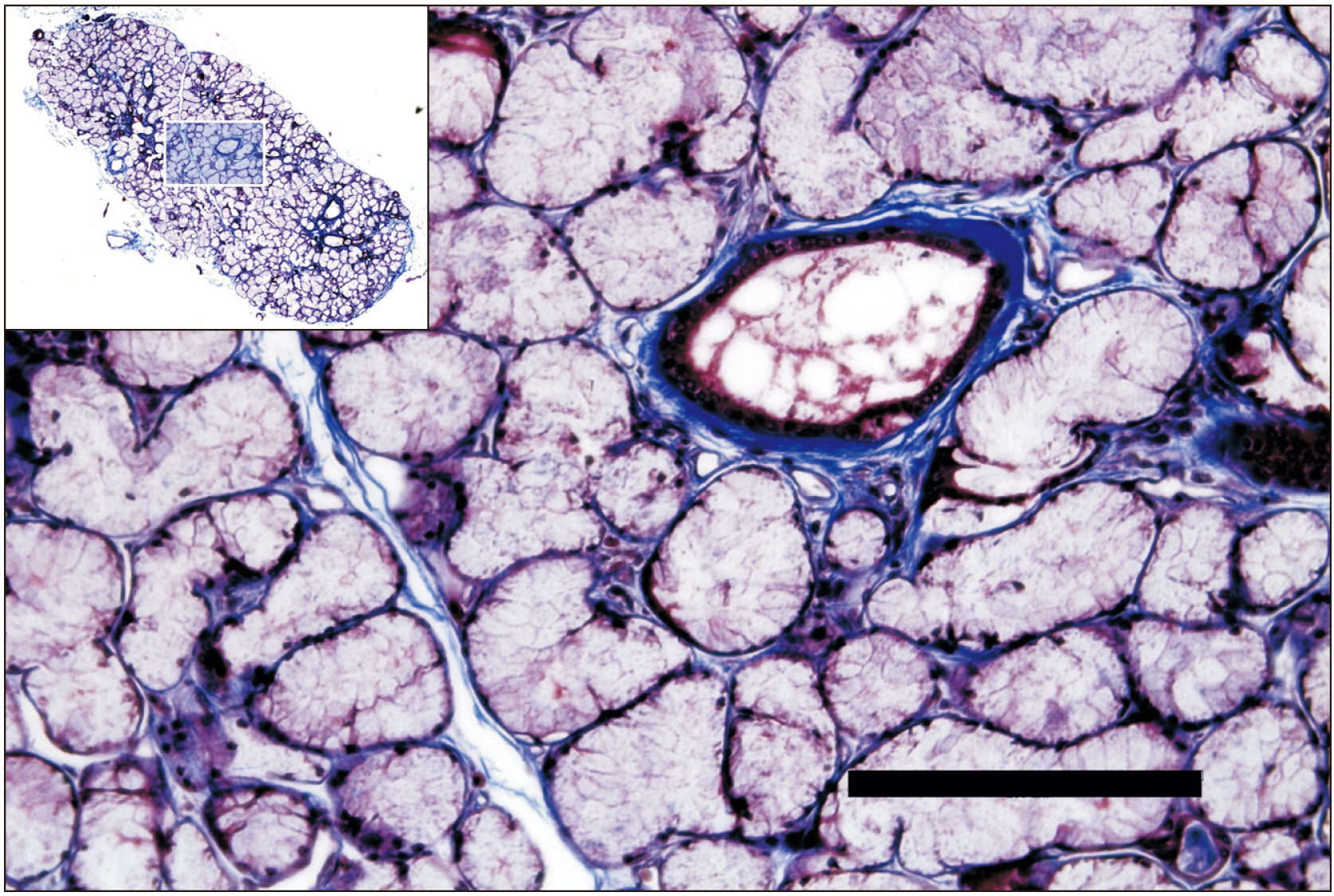
Fig. 2 Masson-trichrome staining of the accessory submandibular gland shown in Fig. 1. The rectangular area in the upper left corner is magnified. Note that the gland tissue is filled with mucinous gland. Scale bar: 200 μm.
Reference
-
References
1. Iwanaga J, Haikata Y, Nakamura K, Kusukawa J, Watanabe K, Tubbs RS. 2021; An anatomical and histological study of mental nerve branches to the inferior labial glands. Surg Radiol Anat. 43:1801–4. DOI: 10.1007/s00276-021-02795-6. PMID: 34232370.
Article2. Iwanaga J, Nakamura K, Alonso F, Kirkpatrick C, Oskouian RJ, Watanabe K, Tubbs RS. 2018; Anatomical study of the so-called "retromolar gland": distinguishing normal anatomy from oral cavity pathology. Clin Anat. 31:462–5. DOI: 10.1002/ca.23047. PMID: 29349817.
Article3. Kamijo Y. 1969. Oral Anatomy. Anatom;Tokyo:4. Shen D, Ono K, Do Q, Ohyama H, Nakamura K, Obata K, Ibaragi S, Watanabe K, Tubbs RS, Iwanaga J. 2021; Clinical anatomy of the inferior labial gland: a narrative review. Gland Surg. 10:2284–92. DOI: 10.21037/gs-21-143. PMID: 34422599. PMCID: PMC8340335.
Article5. Wang Z, Li W, Hong X, Su JZ, Hua H, Peng X, Lv L, Yu GY. 2016; Minor salivary glands function is decreased in hyposalivation-related diseases. Arch Oral Biol. 69:63–70. DOI: 10.1016/j.archoralbio.2016.05.012. PMID: 27243418.
Article6. Iorgulescu G. 2009; Saliva between normal and pathological. Important factors in determining systemic and oral health. J Med Life. 2:303–7. PMID: 20112475. PMCID: PMC5052503.7. Grewal JS, Jamal Z, Ryan J. 2022. Anatomy, head and neck, submandibular gland [Internet]. StatPearls Publishing;Treasure Island, FL: Available from: http://www.ncbi.nlm.nih.gov/books/NBK542272/. cited 2022 May 11.8. Ryu EJ, Kim DH. 2021; Anatomical insights of the mylohyoid for clinical procedures in dentistry. Clin Anat. 34:461–9. DOI: 10.1002/ca.23675. PMID: 32893917.
Article9. Gadodia A, Seith A, Neyaz Z, Sharma R, Thakkar A. 2007; Magnetic resonance identification of an accessory submandibular duct and gland: an unusual variant. J Laryngol Otol. 121:e18. DOI: 10.1017/S0022215107008602. PMID: 17517164.
Article10. Sanchez Barrueco A, Santillan Coello J, Sobrino Guijarro B, Villacampa Aubá JM, Cenjor Español C. 2016; Sialolithiasis in an accessory submandibular gland identified by magnetic resonance sialography. Ann Otol Rhinol Laryngol. 125:603–6. DOI: 10.1177/0003489416636128. PMID: 26961009.
Article11. Jafek BW, Strife JL. 1973; Accessory lobe of the submandibular gland. Radiology. 109:75–7. DOI: 10.1148/109.1.75. PMID: 4783131.
Article12. Priya. 2020; P DS, Anitha DN, Rajesh DE, Masthan DKMK. Embryology and development of salivary gland. Eur J Mol Clin Med. 7:764–70.13. Quirós-Terrón L, Arráez-Aybar LA, Murillo-González J, De-la-Cuadra-Blanco C, Martínez-Álvarez MC, Sanz-Casado JV, Mérida-Velasco JR. 2019; Initial stages of development of the submandibular gland (human embryos at 5.5-8 weeks of development). J Anat. 234:700–8. DOI: 10.1111/joa.12955. PMID: 30740679. PMCID: PMC6481420.
Article14. Batsakis JG. 1986; Heterotopic and accessory salivary tissues. Ann Otol Rhinol Laryngol. 95(4 Pt 1):434–5. DOI: 10.1177/000348948609500422. PMID: 3740723.
Article15. Bryan S, Bodner L, Manor E, Brennan PA. 2013; Pleomorphic adenoma occurring outside the submandibular gland: a case report of an accessory submandibular gland. J Oral Maxillofac Surg. 71:1703–5. DOI: 10.1016/j.joms.2013.04.014. PMID: 23769461.
Article16. Singer MI, Applebaum EL, Loy KD. 1979; Heterotopic salivary tissue in the neck. Laryngoscope. 89:1772–8. DOI: 10.1288/00005537-197911000-00009. PMID: 502698.
Article17. Brazen B, Dyer J. 2022. Histology, salivary glands [Internet]. StatPearls Publishing;Treasure Island, FL: Available from: http://www.ncbi.nlm.nih.gov/books/NBK551688/. cited 2022 May 12.18. Drake RL, Vogl W, Mitchell AWM, Gray H. 2020. Gray's anatomy for students. 4th ed. Elsevier;Philadelphia: p. 1102–5. DOI: 10.1288/00005537-197911000-00009.19. Atamaz Pinar Y, Govsa F, Bilge O. 2005; The anatomical features and surgical usage of the submental artery. Surg Radiol Anat. 27:201–5. DOI: 10.1007/s00276-005-0317-8. PMID: 16003485.
Article20. Lettau J, Bordoni B. 2022. Anatomy, head and neck, lingual artery [Internet]. StatPearls Publishing;Treasure Island, FL: Available from: http://www.ncbi.nlm.nih.gov/books/NBK554513/. cited 2022 May 11.21. Ono K, Yoshioka N, Hage D, Ibaragi S, Tubbs RS, Iwanaga J. 2021; Duplication of the external jugular vein: a language barrier of database search in classic anatomical studies. Surg Radiol Anat. 43:1721–8. Erratum in: Surg Radiol Anat 2021;43:1729-30. DOI: 10.1007/s00276-021-02717-6. PMID: 33620594.
Article22. Yeh CK, Johnson DA, Dodds MW. 1998; Impact of aging on human salivary gland function: a community-based study. Aging (Milano). 10:421–8. DOI: 10.1007/BF03339889. PMID: 9932146.
Article23. Nayak SB. 2018; Accessory submandibular salivary gland forming a "horseshoe" with the main submandibular salivary gland: a unique variation. J Craniofac Surg. 29:1376–7. DOI: 10.1097/SCS.0000000000004537. PMID: 29570527.24. Hasson O. 2010; Modern sialography for screening of salivary gland obstruction. J Oral Maxillofac Surg. 68:276–80. DOI: 10.1016/j.joms.2009.09.044. PMID: 20116695.
Article25. Sobrino-Guijarro B, Cascarini L, Lingam RK. 2013; Advances in imaging of obstructed salivary glands can improve diagnostic outcomes. Oral Maxillofac Surg. 17:11–9. DOI: 10.1007/s10006-012-0327-8. PMID: 22562281.
Article26. Köybaşioğlu A, Ileri F, Gençay S, Poyraz A, Uslu S, Inal E. 2000; Submandibular accessory salivary gland causing Warthin's duct obstruction. Head Neck. 22:717–21. DOI: 10.1002/1097-0347(200010)22:7<717::AID-HED12>3.0.CO;2-1. PMID: 11002328.
Article27. Bokhari MR, Greene J. 2022. Pleomorphic adenoma [Internet]. StatPearls Publishing;Treasure Island, FL: Available from: http://www.ncbi.nlm.nih.gov/books/NBK430829/. cited 2022 May 12.28. Nayak SB, Vasudeva SK, Pamidi N, Sirasanagandla SR. 2020; Anomalous course of facial artery through the submandibular gland and its redundant loop at the base of mandible. J Craniofac Surg. 31:2015–6. DOI: 10.1097/SCS.0000000000006539. PMID: 32472879.
Article29. Nayak SB, Kumar N. 2018; Lingual nerve entrapment in fused submandibular and sublingual salivary glands: a unique finding. J Craniofac Surg. 29:e677–9. DOI: 10.1097/SCS.0000000000004842. PMID: 30106809.
Article30. Iwanaga J, Singh V, Ohtsuka A, Hwang Y, Kim HJ, Moryś J, Ravi KS, Ribatti D, Trainor PA, Sañudo JR, Apaydin N, Şengül G, Albertine KH, Walocha JA, Loukas M, Duparc F, Paulsen F, Del Sol M, Adds P, Hegazy A, Tubbs RS. 2021; Acknowledging the use of human cadaveric tissues in research papers: recommendations from anatomical journal editors. Clin Anat. 34:2–4. DOI: 10.1002/ca.23671. PMID: 32808702.
Article31. Iwanaga J, Singh V, Takeda S, Ogeng'o J, Kim HJ, Moryś J, Ravi KS, Ribatti D, Trainor PA, Sañudo JR, Apaydin N, Sharma A, Smith HF, Walocha JA, Hegazy AMS, Duparc F, Paulsen F, Del Sol M, Adds P, Louryan S, Fazan VPS, Boddeti RK, Tubbs RS. 2022; Standardized statement for the ethical use of human cadaveric tissues in anatomy research papers: recommendations from Anatomical Journal Editors-in-Chief. Clin Anat. 35:526–8. DOI: 10.1002/ca.23849. PMID: 35218594.
- Full Text Links
-
- Actions
-
Cited
- CITED
-
- Close
- Share
-
- Similar articles
-
- Bilateral Plunging Ranula Arising from Accessory Submandibular Gland
- Localization of Nerves Innervating Sublingual and Submandibular Gland in the CNS Using Cholera Toxin B Subnit and Pseudorabies Virus
- A Study on the Diverticular Enlargement of the Rat's Submandibular Duct (II)
- A Study on the Diverticular Enlargement of the Rat's Submandibular Duct
- A Case of Unilateral Absence of the Submandibular Gland Secondary to Sialolithiasis
