Korean J Orthod.
2023 Mar;53(2):106-115. 10.4041/kjod22.179.
Clinical predictors of potentially impacted canines in low-risk patients: A retrospective study in mixed dentition
- Affiliations
-
- 1Division of Orthodontics, Faculty of Dentistry, Federal University of Rio Grande do Sul. Porto Alegre, RS, Brazil
- KMID: 2540950
- DOI: http://doi.org/10.4041/kjod22.179
Abstract
Objective
To evaluate the null hypothesis that there is no difference in a set of clinical predictors of potentially impacted canines between low-risk patients with and without displaced canines.
Methods
The normal canine position group consisted of 30 patients with 60 normally erupting canines ranked in sector I (age, 9.30 ± 0.94 years). The displaced canine group comprised 30 patients with 41 potentially impacted canines ranked in sectors II to IV (age, 9.46 ± 0.78 years). Maxillary lateral incisor crown angulation, inclination, rotation, width, height, and shape, as well as palatal depth, arch length, width, and perimeter composed a set of clinical predictors, which were evaluated on digital dental casts. Statistical analyses consisted of group comparisons and variable correlations (p < 0.05).
Results
There was a significant association between sex and mesially displaced canines. Unilateral canine displacement was more prevalent than bilateral displacement. The crown of the maxillary lateral incisors was significantly angulated more mesially and rotated mesiolabially in low-risk patients with displaced canines, who also had a shallower palate and shorter anterior dental arch length. Lateral incisor crown angulation and rotation, as well as palatal depth and arch length, were significantly correlated with the canine displacement severity.
Conclusions
The null hypothesis was rejected. Maxillary lateral incisor angulation inconsistent with the “ugly duckling” stage as well as a shallow palate and short arch length are clinical predictors that can significantly contribute to the early screening of ectopic canines in low-risk patients.
Figure
-

Figure 1 Positional and dimensional characteristics of the clinical crown of the maxillary lateral incisor. A, Angulation. B, Inclination. C, Rotation. D, Mesiodistal width. E, Cervico-occlusal height. F, Buccolingual width.
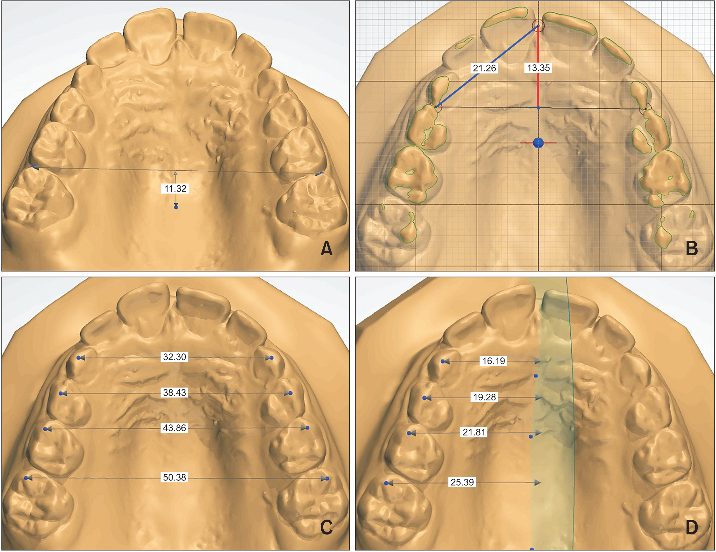
Figure 2 Dimensional characteristics of the maxillary dental arch. A, Palatal depth. B, Anterior arch length (red line) and anterior hemiarch perimeter (blue line). C, Transverse dimensions of the dental arch. D, Transverse dimensions of the dental hemiarch.
Reference
-
1. Garib DG, Lancia M, Kato RM, Oliveira TM, Neves LT. 2016; Risk of developing palatally displaced canines in patients with early detectable dental anomalies: a retrospective cohort study. J Appl Oral Sci. 24:549–54. DOI: 10.1590/1678-775720150535. PMID: 28076458. PMCID: PMC5161253.2. Mercuri E, Cassetta M, Cavallini C, Vicari D, Leonardi R, Barbato E. 2013; Dental anomalies and clinical features in patients with maxillary canine impaction. Angle Orthod. 83:22–8. DOI: 10.2319/021712-149.1. PMID: 22639824. PMCID: PMC8805528.3. Sacerdoti R, Baccetti T. 2004; Dentoskeletal features associated with unilateral or bilateral palatal displacement of maxillary canines. Angle Orthod. 74:725–32. DOI: 10.1043/0003-3219(2004)074<0725:DFAWUO>2.0.CO;2. PMID: 15673132.4. Alessandri Bonetti G, Zanarini M, Danesi M, Parenti SI, Gatto MR. 2009; Percentiles relative to maxillary permanent canine inclination by age: a radiologic study. Am J Orthod Dentofacial Orthop. 136:486.e1–6. discussion 486–7. DOI: 10.1016/j.ajodo.2009.01.022. PMID: 19815142.5. Lindauer SJ, Rubenstein LK, Hang WM, Andersen WC, Isaacson RJ. 1992; Canine impaction identified early with panoramic radiographs. J Am Dent Assoc. 123:91–2. 95–7. Erratum in: J Am Dent Assoc 1992;123:16. DOI: 10.14219/jada.archive.1992.0069. PMID: 1545064.6. Sajnani AK, King NM. 2012; Early prediction of maxillary canine impaction from panoramic radiographs. Am J Orthod Dentofacial Orthop. 142:45–51. DOI: 10.1016/j.ajodo.2012.02.021. PMID: 22748989.7. Ericson S, Kurol J. 1988; Early treatment of palatally erupting maxillary canines by extraction of the primary canines. Eur J Orthod. 10:283–95. DOI: 10.1093/ejo/10.1.283. PMID: 3208843.8. Heimisdottir K, Bosshardt D, Ruf S. 2005; Can the severity of root resorption be accurately judged by means of radiographs? A case report with histology. Am J Orthod Dentofacial Orthop. 128:106–9. DOI: 10.1016/j.ajodo.2004.11.028. PMID: 16027634.9. Mucedero M, Ricchiuti MR, Cozza P, Baccetti T. 2013; Prevalence rate and dentoskeletal features associated with buccally displaced maxillary canines. Eur J Orthod. 35:305–9. DOI: 10.1093/ejo/cjr133. PMID: 22084202.10. Becker A, Smith P, Behar R. 1981; The incidence of anomalous maxillary lateral incisors in relation to palatally-displaced cuspids. Angle Orthod. 51:24–9. DOI: 10.1043/0003-3219(1981)051<0024:TIOAML>2.0.CO;2. PMID: 6939351.11. Ericson S, Kurol J. 1987; Radiographic examination of ectopically erupting maxillary canines. Am J Orthod Dentofacial Orthop. 91:483–92. DOI: 10.1016/0889-5406(87)90005-9. PMID: 3473928.12. Ericson S, Kurol J. 1986; Longitudinal study and analysis of clinical supervision of maxillary canine eruption. Community Dent Oral Epidemiol. 14:172–6. DOI: 10.1111/j.1600-0528.1986.tb01526.x. PMID: 3459617.13. Jacobs SG. 1996; The impacted maxillary canine. Further observations on aetiology, radiographic localization, prevention/interception of impaction, and when to suspect impaction. Aust Dent J. 41:310–6. DOI: 10.1111/j.1834-7819.1996.tb03139.x. PMID: 8961604.14. Leifert S, Jonas IE. 2003; Dental anomalies as a microsymptom of palatal canine displacement. J Orofac Orthop. 64:108–20. DOI: 10.1007/s00056-003-0222-x. PMID: 12649707.15. Perillo L, Masucci C, Ferro F, Apicella D, Baccetti T. 2010; Prevalence of orthodontic treatment need in southern Italian schoolchildren. Eur J Orthod. 32:49–53. DOI: 10.1093/ejo/cjp050. PMID: 19706641.16. Becker A, Chaushu S. 2015; Etiology of maxillary canine impaction: a review. Am J Orthod Dentofacial Orthop. 148:557–67. DOI: 10.1016/j.ajodo.2015.06.013. PMID: 26432311.17. Kim Y, Hyun HK, Jang KT. 2017; Morphological relationship analysis of impacted maxillary canines and the adjacent teeth on 3-dimensional reconstructed CT images. Angle Orthod. 87:590–7. DOI: 10.2319/071516-554.1. PMID: 28156127. PMCID: PMC8366694.18. Liuk IW, Olive RJ, Griffin M, Monsour P. 2013; Maxillary lateral incisor morphology and palatally displaced canines: a case-controlled cone-beam volumetric tomography study. Am J Orthod Dentofacial Orthop. 143:522–6. DOI: 10.1016/j.ajodo.2012.11.023. PMID: 23561414.19. Barros SE, Hoffelder L, Araújo F, Janson G, Chiqueto K, Ferreira E. 2018; Short-term impact of rapid maxillary expansion on ectopically and normally erupting canines. Am J Orthod Dentofacial Orthop. 154:524–34. DOI: 10.1016/j.ajodo.2018.01.011. PMID: 30268263.20. Nolla CM. 1960; The development of the permanent teeth. J Dent Child. 27:254–66.21. Ericson S, Kurol J. 1988; Resorption of maxillary lateral incisors caused by ectopic eruption of the canines. A clinical and radiographic analysis of predisposing factors. Am J Orthod Dentofacial Orthop. 94:503–13. DOI: 10.1016/0889-5406(88)90008-X. PMID: 3195514.22. Koutzoglou SI, Kostaki A. 2013; Effect of surgical exposure technique, age, and grade of impaction on ankylosis of an impacted canine, and the effect of rapid palatal expansion on eruption: a prospective clinical study. Am J Orthod Dentofacial Orthop. 143:342–52. DOI: 10.1016/j.ajodo.2012.10.017. PMID: 23452968.23. Arriola-Guillén LE, Aliaga-Del Castillo A, Ruíz-Mora GA, Rodríguez-Cárdenas YA, Dias-Da Silveira HL. 2019; Influence of maxillary canine impaction characteristics and factors associated with orthodontic treatment on the duration of active orthodontic traction. Am J Orthod Dentofacial Orthop. 156:391–400. DOI: 10.1016/j.ajodo.2018.10.018. PMID: 31474269.24. Bazargani F, Magnuson A, Dolati A, Lennartsson B. 2013; Palatally displaced maxillary canines: factors influencing duration and cost of treatment. Eur J Orthod. 35:310–6. DOI: 10.1093/ejo/cjr143. PMID: 22275512.25. Alqerban A, Jacobs R, Lambrechts P, Loozen G, Willems G. 2009; Root resorption of the maxillary lateral incisor caused by impacted canine: a literature review. Clin Oral Investig. 13:247–55. DOI: 10.1007/s00784-009-0262-8. PMID: 19277728.26. Abbing A, Koretsi V, Eliades T, Papageorgiou SN. 2020; Duration of orthodontic treatment with fixed appliances in adolescents and adults: a systematic review with meta-analysis. Prog Orthod. 21:37. DOI: 10.1186/s40510-020-00334-4. PMID: 33015719. PMCID: PMC7533275. PMID: b426baf33a9e45cda8928373bb860e0d.27. Liuk IW, Olive RJ, Griffin M, Monsour P. 2013; Associations between palatally displaced canines and maxillary lateral incisors. Am J Orthod Dentofacial Orthop. 143:622–32. DOI: 10.1016/j.ajodo.2012.11.025. PMID: 23631964.28. Sajnani AK, King NM. 2014; Prevalence and characteristics of impacted maxillary canines in Southern Chinese children and adolescents. J Investig Clin Dent. 5:38–44. DOI: 10.1111/jicd.12027. PMID: 23355390.29. Schindel RH, Duffy SL. 2007; Maxillary transverse discrepancies and potentially impacted maxillary canines in mixed-dentition patients. Angle Orthod. 77:430–5. DOI: 10.2319/0003-3219(2007)077[0430:MTDAPI]2.0.CO;2. PMID: 17465649.30. Lövgren ML, Dahl O, Uribe P, Ransjö M, Westerlund A. 2019; Prevalence of impacted maxillary canines-an epidemiological study in a region with systematically implemented interceptive treatment. Eur J Orthod. 41:454–9. DOI: 10.1093/ejo/cjz056. PMID: 31369665.31. Camilleri S, Lewis CM, McDonald F. 2008; Ectopic maxillary canines: segregation analysis and a twin study. J Dent Res. 87:580–3. DOI: 10.1177/154405910808700606. PMID: 18502969. PMCID: PMC2572751.32. Rutledge MS, Hartsfield JK Jr. 2010; Genetic factors in the etiology of palatally displaced canines. Semin Orthod. 16:165–71. DOI: 10.1053/j.sodo.2010.05.001.33. Yan B, Sun Z, Fields H, Wang L, Luo L. 2013; Etiologic factors for buccal and palatal maxillary canine impaction: a perspective based on cone-beam computed tomography analyses. Am J Orthod Dentofacial Orthop. 143:527–34. DOI: 10.1016/j.ajodo.2012.11.021. PMID: 23561415.34. Becker A, Gillis I, Shpack N. 1999; The etiology of palatal displacement of maxillary canines. Clin Orthod Res. 2:62–6. DOI: 10.1111/ocr.1999.2.2.62. PMID: 10534981.35. Olive RJ. 2005; Factors influencing the non-surgical eruption of palatally impacted canines. Aust Orthod J. 21:95–101. PMID: 16429864.36. Shapira Y, Kuftinec MM. 1998; Early diagnosis and interception of potential maxillary canine impaction. J Am Dent Assoc. 129:1450–4. DOI: 10.14219/jada.archive.1998.0080. PMID: 9787542.37. Basdra EK, Kiokpasoglou M, Stellzig A. 2000; The class II division 2 craniofacial type is associated with numerous congenital tooth anomalies. Eur J Orthod. 22:529–35. DOI: 10.1093/ejo/22.5.529. PMID: 11105409.38. Jacobs SG. 1999; Localization of the unerupted maxillary canine: how to and when to. Am J Orthod Dentofacial Orthop. 115:314–22. DOI: 10.1016/S0889-5406(99)70335-5. PMID: 10066981.39. Peck S, Peck L, Kataja M. 1994; The palatally displaced canine as a dental anomaly of genetic origin. Angle Orthod. 64:249–56. DOI: 10.1043/0003-3219(1994)064<0249:WNID>2.0.CO;2. PMID: 7978519.40. Anic-Milosevic S, Varga S, Mestrovic S, Lapter-Varga M, Slaj M. 2009; Dental and occlusal features in patients with palatally displaced maxillary canines. Eur J Orthod. 31:367–73. DOI: 10.1093/ejo/cjp014. PMID: 19401354.41. Becker A, Sharabi S, Chaushu S. 2002; Maxillary tooth size variation in dentitions with palatal canine displacement. Eur J Orthod. 24:313–8. DOI: 10.1093/ejo/24.3.313. PMID: 12143095.42. Chaushu S, Bongart M, Aksoy A, Ben-Bassat Y, Becker A. 2009; Buccal ectopia of maxillary canines with no crowding. Am J Orthod Dentofacial Orthop. 136:218–23. DOI: 10.1016/j.ajodo.2007.10.047. PMID: 19651351.43. Bizzarro M, Generali C, Maietta S, Martorelli M, Ferrillo M, Flores-Mir C, et al. 2018; Association between 3D palatal morphology and upper arch dimensions in buccally displaced maxillary canines early in mixed dentition. Eur J Orthod. 40:592–6. DOI: 10.1093/ejo/cjy023. PMID: 29726936.44. Jacoby H. 1983; The etiology of maxillary canine impactions. Am J Orthod. 84:125–32. DOI: 10.1016/0002-9416(83)90176-8. PMID: 6576636.45. D' Oleo-Aracena MF, Arriola-Guillén LE, Rodríguez-Cárdenas YA, Ruíz-Mora GA. 2017; Skeletal and dentoalveolar bilateral dimensions in unilateral palatally impacted canine using cone beam computed tomography. Prog Orthod. 18:7. DOI: 10.1186/s40510-017-0160-6. PMID: 28164257. PMCID: PMC5316518. PMID: 65270d0a6d0e4fe99a189d8435742860.46. Aznar T, Galán AF, Marín I, Domínguez A. 2006; Dental arch diameters and relationships to oral habits. Angle Orthod. 76:441–5. DOI: 10.1043/0003-3219(2006)076[0441:DADART]2.0.CO;2. PMID: 16637724.47. Ogaard B, Larsson E, Lindsten R. 1994; The effect of sucking habits, cohort, sex, intercanine arch widths, and breast or bottle feeding on posterior crossbite in Norwegian and Swedish 3-year-old children. Am J Orthod Dentofacial Orthop. 106:161–6. DOI: 10.1016/S0889-5406(94)70034-6. PMID: 8059752.48. Naoumova J. 2014; Interceptive treatment of palatally displaced canines. Swed Dent J Suppl. 234:7–118.
- Full Text Links
-
- Actions
-
Cited
- CITED
-
- Close
- Share
-
- Similar articles
-
- Evaluation of Impacted Maxillary Canine Position Using Panoramic Radiographs and Cone-beam Computed Tomography
- Retrospective Analysis of Incisor Root Resorption Associated with Impacted Maxillary Canines
- Factors Influencing the Duration of Forced Eruption in Impacted Maxillary Canines
- Evaluation of potency of panoramic radiography for estimating the position of maxillary impacted canines using 3D CT
- Pressure Root Resorption of the Second Molar Caused by Third Molar Impaction: A Case Report of Severely Resorbed Root with Vital Pulp
