Evaluation of potency of panoramic radiography for estimating the position of maxillary impacted canines using 3D CT
- Affiliations
-
- 1Department of Orthodontics, School of Dentistry, Kyungpook National University, Korea. parkhs@knu.ac.kr
- KMID: 2273367
- DOI: http://doi.org/10.4041/kjod.2008.38.4.265
Abstract
OBJECTIVE
The aim of this study was to evaluate the potency of panoramic radiography for the detection of maxillary impacted canines. METHODS: Twenty-five patients were selected, comprised of 7 males (mean age: 10.9 years, range: 8.5 - 14.5 years) and 18 females (mean age: 10.9 years, range: 8.2 - 15.7 years). In total, thirty-five maxillary impacted canines were estimated. The position of the canine and root resorption of adjacent teeth were evaluated on panoramic radiography and 3D CT. RESULTS: Except for angulation to the occlusal plane, the other parameters, such as tooth length, crown width, vertical distance and lateral shift showed larger values on panoramic radiography compared to 3D CT. In palatally impacted cases, the angulation of canine was smaller, and the vertical distance to the occlusal place was larger on panoramic radiography than 3D CT. For labially impacted canines, tooth length, crown width, and angulation to the occlusal plane were similar for the two methods. The sensitivity for detecting root resorption on panoramic radiography was calculated as being 33.3% of 3D CT. CONCLUSIONS: The position of labially impacted canines can be effectively estimated using panoramic radiography, but palatally impacted canines need further investigation such as 3D CT for proper diagnosis.
MeSH Terms
Figure
-
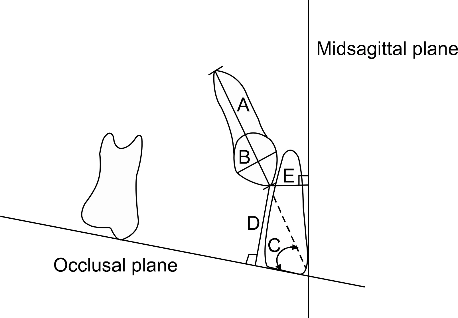
Fig 1. Panoramic measurements. A, tooth length; B, crown width; C, angulation related to the occlusal plane; D, vertical distance related to the occlusal plane; E, lateral shift related to the midsagittal plane.

Fig 2. Three dimensional reconstruction image of impacted canine from CT scanning.

Fig 3. Three dimensional segmented image of impacted canine.
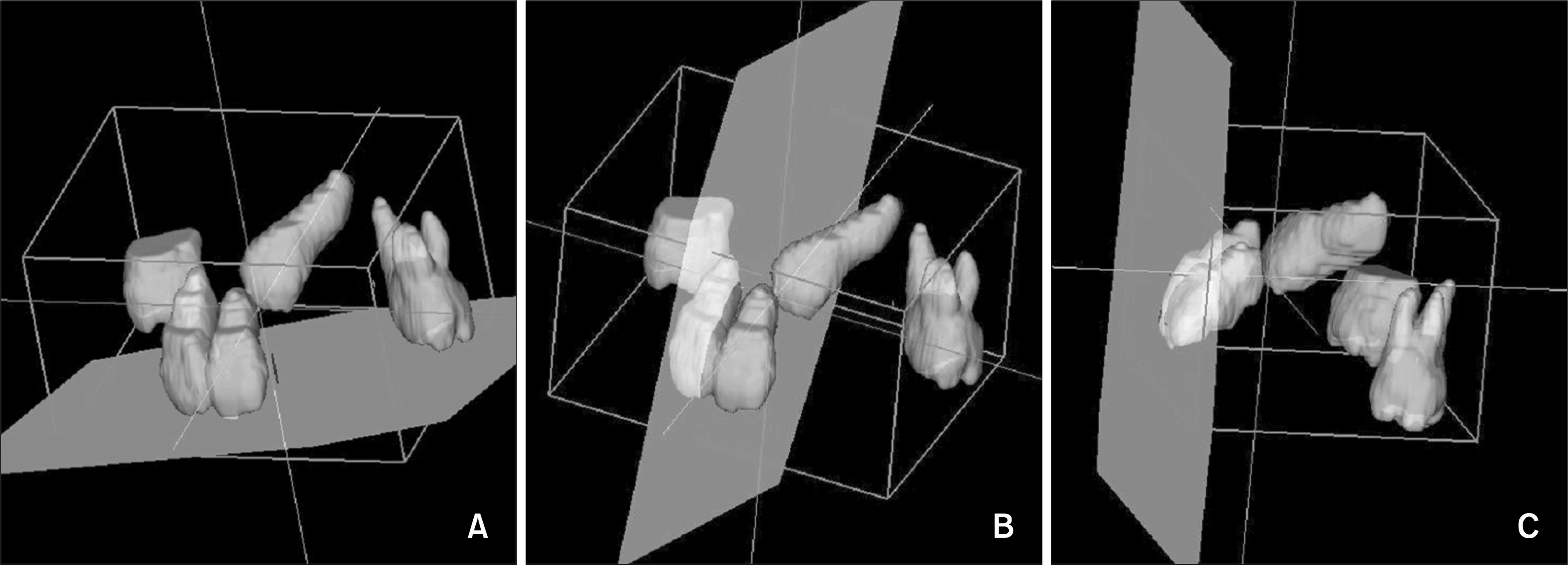
Fig 4. Angular measurements at reconstructed image from CT scanning. A, Angulation of impacted canine to occlusal plane; B, angulation of impacted canine to midsagittal plane; C, angulation of impacted canine to coronal plane.
Cited by 2 articles
-
Pharyngeal airway analysis of different craniofacial morphology using cone-beam computed tomography (CBCT)
Yong-Il Kim, Seong-Sik Kim, Woo-Sung Son, Soo-Byung Park
Korean J Orthod. 2009;39(3):136-145. doi: 10.4041/kjod.2009.39.3.136.The structural change in the hyoid bone and upper airway after orthognathic surgery for skeletal class III anterior open bite patients using 3-dimensional computed tomography
Yoon-seob Lee, Hyoung-seon Baik, Kee-joon Lee, Hyung-seog Yu
Korean J Orthod. 2009;39(2):72-82. doi: 10.4041/kjod.2009.39.2.72.
Reference
-
1.Kung SH., Hwang CJ. Diagnosis and treatment plan of maxillary impacted canine. Korean J Orthod. 1993. 23:165–77.2.Dachi SF., Howel FV. A survey of 3, 874 routine full-month radiographs. II. A study of impacted teeth. Oral Surg Oral Med Oral Pathol. 1961. 14:1165–9.3.Thilander B., Jakobsson SO. Local factors in impaction of maxillary canines. Acta Odontol Scand. 1968. 26:145–68.
Article4.Ericson S., Kurol J. Radiographic assement of maxillary canine eruption in children with clinical signs of eruption disturbance. Eur J Orthod. 1986. 8:133–40.5.Rayne J. The unerupted maxillary canine. Dent Pract Dent Rec. 1969. 19:194–204.6.Hitchein AD. The impacted maxillary canine. Dent Pract Dent Rec. 1951. 2:100–3.7.Dewel BF. The upper cuspid: its development and impaction. Angle Orthod. 1949. 19:79–90.8.Miller BH. The influence of congenitally missing teeth on the eruption of the upper canine. Dent Practit. 1963. 50:17–24.9.Becker A., Smith P., Behar R. The incidence of anomalous maxillary lateral incisors in relation to palatally-displaced cuspids. Angle Orthod. 1981. 51:24–9.10.Bass TB. Observations on the misplaced upper canine tooth. Dent Pract Dent Rec. 1967. 18:25–33.11.Ericson S., Kurol J. Resorption of maxillary lateral incisors caused by ectopic eruption of the canines. A clinical and radiographic analysis of predisposing factors. Am J Orthod Dentofacial Orthop. 1988. 94:503–13.12.Becker A., Chaushu S. Success rate and duration of orthodontic treatment for adult patients with palatally impacted maxillary canines. Am J Orthod Dentofacial Orthop. 2003. 124:509–14.
Article13.Spoor CF., Zonneveld FW., Macho GA. Linear measurements of cortical bone and dental enamel by computed tomography: applications and problems. Am J Phys Anthropol. 1993. 91:469–84.
Article14.Gavel V., Dermaut L. The effect of tooth position on the image of unerupted canines on panoramic radiographs. Eur J Orthod. 1999. 21:551–60.
Article15.Warford JH Jr., Grandhi RK., Tira DE. Prediction of maxillary canine impaction using sectors and angular measurement. Am J Orthod Dentofacial Orthop. 2003. 124:651–5.
Article16.Dahlberg G. Statistical methods for medical and biological students. London: George Allen and Unwin;1940.17.Langland OE. Panoramic radiology. 2nd ed.Philadelphia: Lea & Febiger;1989. 192.18.Otto RL. Early and unusual incisor resorption due to impacted maxillary canines. Am J Orthod Dentofacial Orthop. 2003. 124:446–9.
Article19.Ericson S., Kurol J. Resorption of incisors after ectopic eruption of maxillary canines: a CT study. Angle Orthod. 2000. 70:415–23.20.Ericson S., Kurol J. Radiographic examination of ectopically erupting maxillary canines. Am J Orthod Dentofacial Orthop. 1987. 91:483–92.
Article
- Full Text Links
-
- Actions
-
Cited
- CITED
-
- Close
- Share
-
- Similar articles
-
- Evaluation of Impacted Maxillary Canine Position Using Panoramic Radiographs and Cone-beam Computed Tomography
- Panoramic radiological study to identify locally displaced maxillary canines in Bangladeshi population
- The radiographic localization of unerupted maxillary incisors and supernumeraries
- Factors Influencing the Duration of Forced Eruption in Impacted Maxillary Canines
- Retrospective Analysis of Incisor Root Resorption Associated with Impacted Maxillary Canines
